Podcast
Questions and Answers
What is the primary role of the alpha motor neuron in the context of muscle contraction?
What is the primary role of the alpha motor neuron in the context of muscle contraction?
- To transmit sensory information from the muscle to the spinal cord.
- To carry action potentials from the spinal cord to skeletal muscle fibers, initiating contraction. (correct)
- To inhibit muscle contraction by releasing inhibitory neurotransmitters.
- To regulate blood flow to the muscle tissue during physical activity.
Which of the following accurately describes the process of depolarization in the axon terminal?
Which of the following accurately describes the process of depolarization in the axon terminal?
- Influx of sodium ions, leading to a more positive charge inside the cell. (correct)
- Efflux of chloride ions, maintaining the resting membrane potential.
- Influx of potassium ions, making the inside of the cell more negative.
- Efflux of sodium ions, causing the inside of the cell to become less positive.
What role does calcium influx into the synaptic bulb play in neuromuscular transmission?
What role does calcium influx into the synaptic bulb play in neuromuscular transmission?
- It directly binds to actin and myosin filaments, initiating muscle contraction.
- It hyperpolarizes the synaptic bulb membrane, preventing further neurotransmitter release.
- It facilitates the fusion of synaptic vesicles with the presynaptic membrane, releasing acetylcholine. (correct)
- It triggers the synthesis of acetylcholine within the synaptic bulb.
Where are synaptic vesicles formed, and how do they reach the axon terminal?
Where are synaptic vesicles formed, and how do they reach the axon terminal?
What two primary components combine to synthesize acetylcholine (ACh), and where does this synthesis primarily occur?
What two primary components combine to synthesize acetylcholine (ACh), and where does this synthesis primarily occur?
How is choline transported into the synaptic bulb for acetylcholine synthesis?
How is choline transported into the synaptic bulb for acetylcholine synthesis?
What role does choline acetyltransferase play in the synthesis of acetylcholine (ACh)?
What role does choline acetyltransferase play in the synthesis of acetylcholine (ACh)?
How is acetylcholine (ACh) transported into synaptic vesicles, and what is the significance of maintaining a proton gradient?
How is acetylcholine (ACh) transported into synaptic vesicles, and what is the significance of maintaining a proton gradient?
What are SNARE proteins, and how do they facilitate neurotransmitter release at the neuromuscular junction?
What are SNARE proteins, and how do they facilitate neurotransmitter release at the neuromuscular junction?
In the context of SNARE proteins, what is the difference between v-SNAREs and t-SNAREs, and where are they located?
In the context of SNARE proteins, what is the difference between v-SNAREs and t-SNAREs, and where are they located?
What role does calcium play in the interaction between v-SNAREs and t-SNAREs during neurotransmitter release?
What role does calcium play in the interaction between v-SNAREs and t-SNAREs during neurotransmitter release?
What best describes the process of repolarization in the axon terminal, and which ion is primarily involved?
What best describes the process of repolarization in the axon terminal, and which ion is primarily involved?
What is the equilibrium potential of potassium, and how is it related to repolarization?
What is the equilibrium potential of potassium, and how is it related to repolarization?
What is the order of the following events?
- Action potential stimulates calcium channels.
- Neurotransmitter is released by exocytosis.
- Acetylcholine is synthesized.
- Action potential reaches axon terminal.
- SNARE proteins facilitate fusion.
What is the order of the following events?
- Action potential stimulates calcium channels.
- Neurotransmitter is released by exocytosis.
- Acetylcholine is synthesized.
- Action potential reaches axon terminal.
- SNARE proteins facilitate fusion.
Following the release of acetylcholine into the synaptic cleft, what is the immediate next step?
Following the release of acetylcholine into the synaptic cleft, what is the immediate next step?
Flashcards
Alpha Motor Neuron
Alpha Motor Neuron
Neuron connecting the spinal cord to skeletal muscles.
Anterior Gray Horn
Anterior Gray Horn
The anterior part of the spinal cord containing motor neuron cell bodies.
Action Potential
Action Potential
A rapid change in the electrical potential of a neuron or muscle fiber.
Synaptic Bulb (Axon Terminal)
Synaptic Bulb (Axon Terminal)
Signup and view all the flashcards
Depolarization
Depolarization
Signup and view all the flashcards
Calcium
Calcium
Signup and view all the flashcards
Acetylcholine (ACh)
Acetylcholine (ACh)
Signup and view all the flashcards
Choline Acetyltransferase
Choline Acetyltransferase
Signup and view all the flashcards
Synaptic Vesicles
Synaptic Vesicles
Signup and view all the flashcards
SNARE Proteins
SNARE Proteins
Signup and view all the flashcards
Synaptobrevin
Synaptobrevin
Signup and view all the flashcards
Syntaxin
Syntaxin
Signup and view all the flashcards
Exocytosis
Exocytosis
Signup and view all the flashcards
Repolarization
Repolarization
Signup and view all the flashcards
Potassium (K+) Efflux
Potassium (K+) Efflux
Signup and view all the flashcards
Study Notes
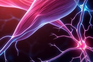
Neuromuscular Junction and Neuromuscular Transmission
- Focus is on the neuromuscular junction and neuromuscular transmission.
- A long motor neuron extends from the spinal cord to the muscle.
Long Motor Neuron Origin
- The anterior grey horn (also called the ventral grey horn) of the spinal cord contains the cell bodies of somatic motor neurons.
- Somatic nervous system controls skeletal muscles.
- Somatic nervous system is under voluntary control.
Alpha Motor Neuron
- The cerebral cortex sends impulses down to stimulate muscle contraction.
- These impulses travel via motor neurons, specifically alpha motor neurons, to the skeletal muscles.
- A muscle cell requires stimulation to contract.
- Muscles are excitable, responding to neural stimulus with changes in membrane potential.
Action Potential
- Stimulation of cell bodies generates an action potential that travels down the axon.
- Action potentials are caused by a depolarizing wave.
- Voltage sensitive sodium channels open at a specific threshold voltage.
- Sodium flows into the axon, causing depolarization.
- Depolarization makes the inside of the cell more positive.
Synaptic Bulb
- The action potential reaches the axon terminal (synaptic bulb).
- Depolarization to approximately +30 mV stimulates voltage-gated calcium channels in the synaptic bulb.
- Calcium ions influx into the synaptic bulb.
Acetylcholine Vesicles
- Synaptic vesicles containing the neurotransmitter acetylcholine (ACh) are present in the synaptic bulb.
- Vesicles are made in the cell body by the Golgi apparatus and transported to the synaptic bulb.
- Acetylcholine (ACh) is NOT a protein.
- It is synthesized in the synaptic bulb.
- Acetylcholine synthesized from choline (obtained from dietary sources) and acetate (derived from acetyl-CoA from mitochondria).
Acetylcholine Synthesis
- Choline is transported into the synaptic bulb via a choline transporter.
- Acetyl-CoA, from mitochondrial Krebs cycle activity, provides acetate.
- Choline and acetyl-CoA react, releasing CoA and forming acetylcholine.
- Choline acetyltransferase is the enzyme that stimulates the reaction.
Transporting Acetylcholine
- Acetylcholine is transported into synaptic vesicles via protein transporters.
- Protons are actively pumped into the synaptic vesicle using ATP (primary active transport), creating a proton gradient.
- The proton gradient facilitates secondary active transport of acetylcholine into the vesicle.
Role of Calcium and SNARE Proteins
- Calcium influx is crucial for neurotransmitter release.
- SNARE proteins (v-SNAREs on the vesicle and t-SNAREs on the target membrane) mediate vesicle fusion.
- v-SNAREs: synaptobrevin and synaptotagmin.
- t-SNAREs: SNAP-25 and syntaxin.
- Calcium acts as a cross-link, stimulating v-SNAREs and t-SNAREs to interact and intertwine.
Acetylcholine Release
- SNARE protein interaction pulls the synaptic vesicle toward the cell membrane.
- Fusion of the vesicle membrane with the cell membrane releases acetylcholine into the synaptic cleft via exocytosis.
- Acetylcholine diffuses across the synaptic cleft.
Steps of Neuromuscular Transmission Recap
- Stimulation of somatic motor neuron cell bodies in the anterior ventral grey horn.
- Action potential propagation down the alpha motor neuron, involving sodium influx and depolarization.
- Choline uptake from the diet via choline transporters into the synaptic bulb.
- Acetyl-CoA (acetate) combines with choline via choline acetyltransferase to form acetylcholine.
- Acetylcholine transported into vesicles using proton pumps (primary active transport) and secondary active transport.
- Interaction of v-SNAREs (synaptobrevin, synaptotagmin) and t-SNAREs (SNAP-25, syntaxin) triggered by calcium influx.
- Exocytosis: Acetylcholine release into the synaptic cleft.
Repolarization
- Voltage-gated potassium channels open at approximately +30 mV.
- Potassium efflux causes repolarization.
- Repolarization makes the cell more electronegative.
- Potassium moves out until the inside of the cell reaches approximately -70 mV.
- The potassium moving out causes inactivation gates to close, therefore calcium cannot come in.
- Acetylcholine cannot get released when calcium cannot come in because the synap proteins are not able to link up together during the resting period.
- Potassium is crucial for repolarizing the membrane, which makes the axon terminal rest and prepare for subsequent stimulation.
Studying That Suits You
Use AI to generate personalized quizzes and flashcards to suit your learning preferences.
Description
Overview of the neuromuscular junction and neuromuscular transmission. A long motor neuron extends from the spinal cord to the muscle. Stimulation of cell bodies generates an action potential that travels down the axon.