Podcast
Questions and Answers
What is the primary role of the sarcoplasmic reticulum in skeletal muscle contraction?
What is the primary role of the sarcoplasmic reticulum in skeletal muscle contraction?
- Provides ATP for myosin head re-cocking
- Functions as the synapse between motor neurons and muscle fibers
- Releases calcium ions to initiate contraction (correct)
- Transmits action potentials to muscle fibers
During which phase of muscle contraction does the myosin head detach from the actin filament?
During which phase of muscle contraction does the myosin head detach from the actin filament?
- Cross-Bridge Formation
- Cross-Bridge Detachment (correct)
- Power Stroke
- Resetting Myosin Head
Which component of the muscle fiber is responsible for transmitting the action potential into the muscle fiber?
Which component of the muscle fiber is responsible for transmitting the action potential into the muscle fiber?
- Neuromuscular Junction
- T-Tubules (correct)
- Motor Neuron
- Sarcoplasm
What happens to calcium ions after the termination of muscle contraction?
What happens to calcium ions after the termination of muscle contraction?
Which process links the electrical signal to mechanical contraction in muscle fibers?
Which process links the electrical signal to mechanical contraction in muscle fibers?
How does the sliding filament theory describe muscle contraction?
How does the sliding filament theory describe muscle contraction?
What role does tropomyosin play in muscle contraction?
What role does tropomyosin play in muscle contraction?
What initiates the release of acetylcholine at the neuromuscular junction?
What initiates the release of acetylcholine at the neuromuscular junction?
Flashcards are hidden until you start studying
Study Notes
Overview of Skeletal Muscle Contraction
- Skeletal muscle contraction involves the shortening of muscle fibers to produce movement.
- The process is initiated by neural signals and involves several key components.
Key Components
-
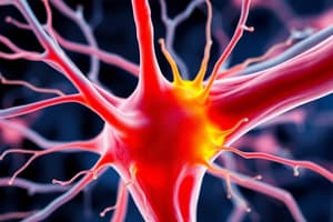
Motor Neuron
- Transmits action potentials from the central nervous system to muscle fibers.
-
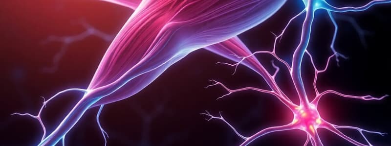
Neuromuscular Junction
- The synapse between a motor neuron and a muscle fiber.
- Release of neurotransmitter acetylcholine (ACh) triggers muscle contraction.
-
Sarcolemma
- The muscle fiber cell membrane that depolarizes in response to ACh, generating an action potential.
-
T-Tubules
- Extensions of the sarcolemma that penetrate into the muscle fiber, helping to transmit the action potential into the fiber.
-
Sarcoplasmic Reticulum (SR)
- Stores calcium ions (Ca²⁺).
- Releases Ca²⁺ in response to the action potential, initiating contraction.
-
Myofibrils
- Rod-like structures within muscle fibers containing the contractile units.
Mechanism of Contraction
-
Excitation-Contraction Coupling
- The process that links the electrical signal (action potential) to mechanical contraction.
- Ca²⁺ is released from the SR into the cytoplasm, binding to troponin.
-
Sliding Filament Theory
- Actin and myosin filaments slide past each other during contraction.
- Myosin heads form cross-bridges with actin, pulling actin filaments toward the center of the sarcomere.
-
Muscle Shortening
- Shortening of the muscle occurs as sarcomeres contract, reducing the distance between Z discs.
- When multiple sarcomeres contract simultaneously, the entire muscle shortens.
Cycle of Muscle Contraction
-
Cross-Bridge Formation
- Myosin heads attach to actin binding sites.
-
Power Stroke
- Myosin head pivots, pulling actin filaments inward (contraction).
- ATP is used to re-cock the myosin head.
-
Cross-Bridge Detachment
- New ATP molecule binds, causing myosin to detach from actin.
-
Resetting Myosin Head
- Energy from ATP hydrolysis repositions the myosin head for another cycle.
Termination of Contraction
- Calcium ions are pumped back into the SR, decreasing Ca²⁺ concentration.
- Tropomyosin blocks binding sites on actin, halting the contraction cycling.
Key Terms
- Sarcomere: The functional unit of muscle contraction.
- Troponin: A protein that regulates muscle contraction in response to Ca²⁺.
- Tropomyosin: A protein that blocks actin binding sites in a relaxed muscle.
Summary
- Skeletal muscle contraction is a highly coordinated process involving neural stimulation, calcium signaling, and the interaction of myosin and actin filaments.
- Contraction terminates when calcium ions are reabsorbed and myosin can no longer bind to actin.
Overview of Skeletal Muscle Contraction
- Skeletal muscle contraction is the shortening of muscle fibers to produce movement.
- This process is initiated by neural signals and involves a sequence of steps.
Key Components
- Motor Neuron: Transmits action potentials from the central nervous system to muscle fibers.
- Neuromuscular Junction: The synapse between a motor neuron and a muscle fiber. Acetylcholine (ACh) is released, triggering muscle contraction.
- Sarcolemma: The muscle fiber cell membrane. It depolarizes in response to ACh, generating an action potential.
- T-Tubules: Extensions of the sarcolemma that penetrate into the muscle fiber, transmitting the action potential into the fiber.
- Sarcoplasmic Reticulum (SR): Stores calcium ions (Ca²⁺). Releases Ca²⁺ in response to the action potential, initiating contraction.
- Myofibrils: Rod-like structures within muscle fibers containing the contractile units (sarcomeres).
Mechanism of Contraction
- Excitation-Contraction Coupling: Links the electrical signal (action potential) to mechanical contraction. Ca²⁺ is released from the SR into the cytoplasm and binds to troponin.
- Sliding Filament Theory: Actin and myosin filaments slide past each other during contraction. Myosin heads form cross-bridges with actin, pulling actin filaments toward the center of the sarcomere.
- Muscle Shortening: The sarcomeres contract, reducing the distance between Z discs. Multiple sarcomeres contracting simultaneously shortens the entire muscle.
Cycle of Muscle Contraction
- Cross-Bridge Formation: Myosin heads attach to actin binding sites.
- Power Stroke: Myosin head pivots, pulling actin filaments inward (contraction). ATP is used to re-cock the myosin head.
- Cross-Bridge Detachment: A new ATP molecule binds, causing myosin to detach from actin.
- Resetting Myosin Head: Energy from ATP hydrolysis repositions the myosin head for another cycle.
Termination of Contraction
- Calcium ions are pumped back into the SR, decreasing Ca²⁺ concentration.
- Tropomyosin blocks binding sites on actin, halting the contraction cycling.
Key Terms
- Sarcomere: The functional unit of muscle contraction.
- Troponin: A protein that regulates muscle contraction in response to Ca²⁺.
- Tropomyosin: A protein that blocks actin binding sites in a relaxed muscle.
Summary
- Skeletal muscle contraction is a complex process that involves neural stimulation, calcium signaling, and the interaction of myosin and actin filaments.
- Contraction terminates when calcium ions are reabsorbed, preventing myosin from binding to actin.
Studying That Suits You
Use AI to generate personalized quizzes and flashcards to suit your learning preferences.