Podcast
Questions and Answers
Which of the following is the MOST accurate description of the relationship between a neurological deficit and neuroanatomic localization?
Which of the following is the MOST accurate description of the relationship between a neurological deficit and neuroanatomic localization?
- Neurological deficits are solely determined by the specific location of the lesion, irrespective of the functional systems involved.
- Understanding the pattern of neurological deficits is crucial for determining the most likely neuroanatomic location of the lesion. (correct)
- Neuroanatomic localization is primarily based on the severity of the neurological deficit, with more severe deficits indicating larger lesions.
- Neuroanatomic localization is independent of the observed neurological deficits; therefore, diagnostic imaging is always required for accurate diagnosis.
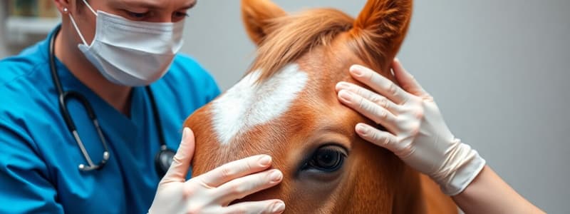
In equine neurological examinations, what is the significance of observing a horse in unfamiliar surroundings?
In equine neurological examinations, what is the significance of observing a horse in unfamiliar surroundings?
- It is essential for determining the horse's pain threshold.
- It mainly serves to evaluate the horse's respiratory capacity under varying environmental conditions.
- It primarily helps in assessing the horse's cardiovascular response to stress.
- It allows for a more accurate evaluation of the horse's mentation and vision by challenging their perception and responses. (correct)
What is the MOST appropriate method for assessing cortical function in horses during a neurological examination?
What is the MOST appropriate method for assessing cortical function in horses during a neurological examination?
- Evaluating pupillary light reflexes.
- Assessing muscle tone.
- Observing mentation and consciousness. (correct)
- Testing limb reflexes.
In evaluating cranial nerve function in horses, asymmetry of the face is MOST indicative of a lesion affecting which cranial nerve?
In evaluating cranial nerve function in horses, asymmetry of the face is MOST indicative of a lesion affecting which cranial nerve?
Which of the following BEST describes the afferent and efferent components of the menace response in horses?
Which of the following BEST describes the afferent and efferent components of the menace response in horses?
Which cranial nerve(s) is/are involved in physiologic nystagmus (Doll's eye response)?
Which cranial nerve(s) is/are involved in physiologic nystagmus (Doll's eye response)?
In Horner's syndrome, disruption of the sympathetic pathway affects several structures. Which of the following is NOT directly innervated by the sympathetic nerve in the head and neck?
In Horner's syndrome, disruption of the sympathetic pathway affects several structures. Which of the following is NOT directly innervated by the sympathetic nerve in the head and neck?
When assessing a horse for ataxia during a neurological exam, which of the following techniques is MOST effective for detecting subtle proprioceptive deficits?
When assessing a horse for ataxia during a neurological exam, which of the following techniques is MOST effective for detecting subtle proprioceptive deficits?
In the grading system for neurological deficits in horses, a grade of '2' for ataxia is typically defined as:
In the grading system for neurological deficits in horses, a grade of '2' for ataxia is typically defined as:
Which statement accurately reflects the appropriate technique and interpretation of cerebrospinal fluid (CSF) collection in horses with suspected neurologic disease?
Which statement accurately reflects the appropriate technique and interpretation of cerebrospinal fluid (CSF) collection in horses with suspected neurologic disease?
What does xanthochromia in cerebrospinal fluid (CSF) indicate>
What does xanthochromia in cerebrospinal fluid (CSF) indicate>
What is the MOST likely cause of iatrogenic hemorrhage during CSF collection?
What is the MOST likely cause of iatrogenic hemorrhage during CSF collection?
A horse exhibits homogenous distribution of blood during aspiration, does not clear with subsequent aspiration, yellow supernatant after centrifugation and no platelets are identified on cytologic examination. Which of the following is the MOST likely cause?
A horse exhibits homogenous distribution of blood during aspiration, does not clear with subsequent aspiration, yellow supernatant after centrifugation and no platelets are identified on cytologic examination. Which of the following is the MOST likely cause?
When evaluating unusual gaits in horses during a neurological examination, what is the primary characteristic of stringhalt?
When evaluating unusual gaits in horses during a neurological examination, what is the primary characteristic of stringhalt?
A horse is presented with muscle fasciculations of the hind limbs when asked to back or pick up its hooves. Which of the following conditions is MOST likely responsible for these clinical signs?
A horse is presented with muscle fasciculations of the hind limbs when asked to back or pick up its hooves. Which of the following conditions is MOST likely responsible for these clinical signs?
Which of the following best describes the underlying pathophysiology of Sweeny in horses?
Which of the following best describes the underlying pathophysiology of Sweeny in horses?
A horse presents with neurogenic atrophy of the supraspinatus and infraspinatus muscles. Which of the following additional clinical signs would MOST strongly support a diagnosis of Sweeny?
A horse presents with neurogenic atrophy of the supraspinatus and infraspinatus muscles. Which of the following additional clinical signs would MOST strongly support a diagnosis of Sweeny?
After performing a complete neurological examination, you suspect a horse has a compressive lesion in the cervical spinal cord. Which diagnostic imaging modality would be MOST appropriate to confirm the location and nature of the compression?
After performing a complete neurological examination, you suspect a horse has a compressive lesion in the cervical spinal cord. Which diagnostic imaging modality would be MOST appropriate to confirm the location and nature of the compression?
When performing a myelogram on a horse with suspected spinal cord compression, where is the positive contrast material injected?
When performing a myelogram on a horse with suspected spinal cord compression, where is the positive contrast material injected?
What is the primary indication for performing skull radiographs in a horse with neurological signs?
What is the primary indication for performing skull radiographs in a horse with neurological signs?
Flashcards
Neurologic Dysfunction
Neurologic Dysfunction
Impairment of neurological function in horses.
Neuroanatomic Localization
Neuroanatomic Localization
Localization based on loss of neurologic function.
Cortical Dysfunction
Cortical Dysfunction
Abnormal mentation or altered consciousness.
Brainstem Dysfunction
Brainstem Dysfunction
Signup and view all the flashcards
Cerebellar Dysfunction
Cerebellar Dysfunction
Signup and view all the flashcards
Spinal Cord dysfunction
Spinal Cord dysfunction
Signup and view all the flashcards
Interest in Feed
Interest in Feed
Signup and view all the flashcards
Facial Symmetry
Facial Symmetry
Signup and view all the flashcards
Menace Response
Menace Response
Signup and view all the flashcards
Pupillary Light Reflex
Pupillary Light Reflex
Signup and view all the flashcards
Palpebral Response
Palpebral Response
Signup and view all the flashcards
Strabismus
Strabismus
Signup and view all the flashcards
Nystagmus
Nystagmus
Signup and view all the flashcards
Gag Reflex
Gag Reflex
Signup and view all the flashcards
Assessment of limbs
Assessment of limbs
Signup and view all the flashcards
Cerebrospinal Fluid
Cerebrospinal Fluid
Signup and view all the flashcards
Xanthochromic CSF
Xanthochromic CSF
Signup and view all the flashcards
Myelographic Examination
Myelographic Examination
Signup and view all the flashcards
Stringhalt
Stringhalt
Signup and view all the flashcards
Sweeny
Sweeny
Signup and view all the flashcards
Study Notes
Neurologic Examination in Horses
- Recognise neurologic dysfunction and perform relevant examinations
- Describe the relationship between neurologic deficit and neuroanatomic localization with emphasis on differentiating cortical, brainstem, cerebellar, and spinal cord dysfunction
- Distinguish between upper and lower motor neuron deficits
- Identify focal, multifocal, and diffuse lesions
History and Observation
- Key information includes age, breed, gender, disease progression, and environment
- Observe the horse in unfamiliar surroundings to assess mentation and vision
Neuroanatomic Localization
- Neuroanatomic localization is based upon loss of neurologic function
- Cortical dysfunction can manifest as abnormal mentation (altered consciousness), seizures, head pressing, and blindness with a normal pupillary light reflex
- Brainstem issues may present as cranial nerve deficits, altered consciousness, and gait deficits
- Cerebellar dysfunction affects depth and direction of motion, potentially causing intention tremor, opisthotonos, and absent menace response, though strength is typically preserved
- Spinal cord issues typically cause ataxia, weakness, and spasticity of the limbs
- Determining if lesions are diffuse, focal, or multifocal is key for differential diagnoses
Cranial Nerve Examination
- CN1 (olfactory): Assess interest in feed
- CN5 (trigeminal): Check for facial symmetry, as it innervates muscles of mastication
- CN7 (facial): Responsible for muscles of facial expression
- Menace Response: Involves CN2 (optic) as the afferent and CN7 (facial) as the efferent
- Pupillary Light Reflex: Involves CN2 (optic) as the afferent and CN3 (oculomotor) as the efferent
- Palpebral Response: Involves CN5 (trigeminal) as the afferent and CN7 (facial) as the efferent
- Strabismus: Indicates abnormal ocular position
- Elevation of the head can improve detection of certain deficits
- CN3 (Oculomotor) function is ventrolateral
- CN4 (Trochlear) function is dorsal deviation of the medial angle
- CN6 (Abducens) function is medial
- CN8 (Vestibulocochlear) function is ventrolateral and has UMN input
- Nystagmus involves rapid, rhythmic eye movements
- Physiologic nystagmus (Doll's eye response) requires normal function of cranial nerves III, IV, VI, and VIII
- Pathologic nystagmus indicates CN8 (vestibulocochlear) or cerebellar dysfunction
- Peripheral damage leads to horizontal nystagmus with a fast phase away from the lesion
- Central damage can cause horizontal, vertical, or rotary nystagmus, with the direction changing based on head position
- Facial sensation is mediated by CN5 (trigeminal) in the nasal passages
- Gag reflex involves CN9 (glossopharyngeal) and CN10 (vagus)
- Tongue tone is assessed via CN12 (hypoglossal)
Horner's Syndrome
- Sympathetic disruption can cause miosis, ptosis, 3rd eyelid prolapse, enophthalmus, sweating of the head and neck, and poor airflow through the nostril on the affected side
- The sympathetic nerve pathway includes the intermediate grey column T1 to T3, vagosympathetic trunk, cervical thoracic ganglion, cranial cervical ganglion, traveling caudomedial to the tympanic bulla and with the ophthalmic branch of CN5
- The nerve innervates smooth muscle of the periorbital area, eyelids (Mueller's muscle), third eyelid, iris, blood vessels, and sweat glands of the head and neck
Limb Assessment
- Assess limbs for weakness, spasticity, and ataxia
- Manipulation is used to test proprioceptive positioning like crossing limbs, placing limbs wide, or on the dorsal hoof wall
- Circling can indicate ataxia through circumduction of limbs or posting of a pivotal limb
- Tail pull assesses weakness when inclining
- Weakness, spasticity, and ataxia are graded from 0 to 5, noting symmetry
- Grade 0: no deficits detected
- Grade 1: mild deficits detected by a trained eye
- Grade 2: deficits detected by most observers
- Grade 3: prominent deficits detected by all observers
- Grade 4: marked deficits, potentially causing the horse to fall during examination
- Grade 5: The horse is unable to rise
Diagnostic Evaluation through CSF Analysis
- CSF should be obtained from the lumbosacral (LS) site in horses with spinal cord dysfunction
- CSF flows from cranial to caudal, a lumbosacral sample accurately reflects spinal cord pathology better than an atlanto-occipital (A-O) sample
- LS CSF taps are performed in standing, sedated animals
- If CSF flow is poor, the handler can occlude the jugular veins to increase CSF pressure (Quakenstat maneuver)
- If brain/brainstem dysfunction is observed, an A-O tap is indicated, requiring general anesthesia
- Damaging the spinal cord during an A-O tap is possible if you insert the needle too far
CSF Analysis Specifics
- Normal nucleated cell count is 0-5 cells/µl (small lymphocytes), and normal CSF protein concentration is < 80 mg/dl
- Hemorrhagic CSF can be iatrogenic or pathologic
- Iatrogenic hemorrhage shows blood swirling with CSF during aspiration, clears with subsequent aspiration, clear CSF after centrifugation, and platelets identified on cytologic examination
- Pathologic hemorrhage shows a homogenous distribution of blood during aspiration, doesn't clear with subsequent aspiration, presents a yellow supernatant after centrifugation, and no platelets are identified on cytologic examination
- Xanthochromic (yellow) CSF indicates bilirubin in CSF from RBC destruction, CNS hemorrhage happened 1 to 10 days prior
- Pandy test is a qualitative assessment of CSF immunoglobulin, layering CSF over phenol causes proteins to precipitate at the interface
Radiographic Examination
- Cervical spine radiographs assess malformation of the cervical vertebrae and vertebral canal diameter, vertebral trauma, vertebral body osteomyelitis, or tumors
- Myelographic examination uses positive contrast to identify spinal cord compression and is injected at the A-O site under general anesthesia
- Skull radiographs can detect fractures or tumors
- CT scans are used to detect fractures, tumors, or abscesses
Unusual Gaits
- Stringhalt involves exaggerated hindlimb advancement, unilaterally or bilaterally
- Etiology is unknown in the US, but in Australia, it is related to a plant toxin
- Pathophysiology is unknown
- Shivers involves mm fasciculation of hindlimbs when asked to back or pick up a hoof, common in draft horses and warmblood breeds, pathophysiology is unknown
- Sweeny involves suprascapular nerve damage
- Pathophysiology is suprascapular nerve trauma at the cranial aspect of the scapula, where the nerve traverses medial to lateral to innervate the supraspinatus and infraspinatus mm
- Neurologic examination reveals neurogenic atrophy of the supraspinatus and infraspinatus mm, a prominent scapular spine, subluxation of the scapulohumeral joint during weight-bearing (loss of rotator cuff function), and circumduction of the limb (decreased anterior phase)
Studying That Suits You
Use AI to generate personalized quizzes and flashcards to suit your learning preferences.


