Podcast
Questions and Answers
What regulates secretions from endocrine glands controlling reproduction, sexual behavior, stress response, and growth?
What regulates secretions from endocrine glands controlling reproduction, sexual behavior, stress response, and growth?
- Epithalamus
- Hypothalamus
- Subthalamus
- Anterior Pituitary (correct)
The lateral hypothalamus acts as a satiety center.
The lateral hypothalamus acts as a satiety center.
False (B)
What part of the hypothalamus triggers the release of vasopressin?
What part of the hypothalamus triggers the release of vasopressin?
Thirst center
The __________ is known as the body clock that keeps the brain on a day-night cycle.
The __________ is known as the body clock that keeps the brain on a day-night cycle.
What areas does the habenular trigone regulate?
What areas does the habenular trigone regulate?
What is the primary hormone secreted by the pineal body that is involved in circadian rhythms?
What is the primary hormone secreted by the pineal body that is involved in circadian rhythms?
Which of the following lobes is responsible for auditory processing?
Which of the following lobes is responsible for auditory processing?
Match the following lobes with their primary functions:
Match the following lobes with their primary functions:
What are the three components of the basal ganglia?
What are the three components of the basal ganglia?
The caudate nucleus is located on the superficial layer of the cerebral hemispheres.
The caudate nucleus is located on the superficial layer of the cerebral hemispheres.
What separates the putamen and globis pallidus from caudate and thalamus?
What separates the putamen and globis pallidus from caudate and thalamus?
Which type of tracts are contained in the internal capsule?
Which type of tracts are contained in the internal capsule?
The __________ is a thin layer of gray matter beneath the insular cortex.
The __________ is a thin layer of gray matter beneath the insular cortex.
The pia mater is the outermost layer of the meninges.
The pia mater is the outermost layer of the meninges.
What is the role of the meninges?
What is the role of the meninges?
Match the fibers in the internal capsule with their respective functions:
Match the fibers in the internal capsule with their respective functions:
Which structures are included in the diencephalon?
Which structures are included in the diencephalon?
What is the primary function of the thalamus?
What is the primary function of the thalamus?
Which of the following is a component of thalamic radiations?
Which of the following is a component of thalamic radiations?
What hormone is associated with water retention?
What hormone is associated with water retention?
What types of nuclei are found in the thalamus?
What types of nuclei are found in the thalamus?
Match the following thalamic nuclei with their functions:
Match the following thalamic nuclei with their functions:
The hypothalamus is located beneath the thalamus.
The hypothalamus is located beneath the thalamus.
What regulatory functions is the hypothalamus involved in?
What regulatory functions is the hypothalamus involved in?
The hypothalamus communicates with the net of blood vessels surrounding the ______.
The hypothalamus communicates with the net of blood vessels surrounding the ______.
Which hormone stimulates corticotrophs in the anterior pituitary?
Which hormone stimulates corticotrophs in the anterior pituitary?
What is a fissure?
What is a fissure?
What is a sulcus?
What is a sulcus?
Which fissure separates the temporal lobe from the frontal and parietal lobes?
Which fissure separates the temporal lobe from the frontal and parietal lobes?
Which fissure separates the frontal and parietal lobes?
Which fissure separates the frontal and parietal lobes?
Which structure connects the two cerebral hemispheres?
Which structure connects the two cerebral hemispheres?
What is the function of the motor cortex?
What is the function of the motor cortex?
Where is the primary visual cortex located?
Where is the primary visual cortex located?
What does the term 'basal ganglia' refer to?
What does the term 'basal ganglia' refer to?
Which part of the brain is primarily associated with judgment and abstract reasoning?
Which part of the brain is primarily associated with judgment and abstract reasoning?
The __________ fissure separates the parietal lobe from the occipital lobe.
The __________ fissure separates the parietal lobe from the occipital lobe.
Match the following fissures with their correct functions:
Match the following fissures with their correct functions:
Flashcards are hidden until you start studying
Study Notes
Diencephalon Overview
- Composed of the thalamus, hypothalamus, subthalamus, and epithalamus, acting as a crucial brain region.
- Fornix and commissures (anterior and posterior) are notable landmarks.
Thalamus
- Central sensory hub; large gray mass situated bilaterally.
- Acts as a waystation for sensory signals before they travel to the cerebral cortex.
- Contains thalamic radiations: axons that connect thalamus to various cortical areas.
- Thalamic White Matter includes:
- Superior Radiation: Connects to motor and sensory cortices.
- Anterior Radiation: Connects to prefrontal cortex.
- Posterior Radiation: Serves occipital, temporal, and parietal cortices.
- Inferior Radiation: Targets auditory cortex in temporal lobe.
- Major nuclei categorized into five groups:
- Anterior Nuclei: Connects to limbic system and prefrontal cortex.
- Midline and Medial Nuclei: Links to hypothalamus and frontal cortex.
- Lateral Nuclei: Involved in motor (VA, VL) and sensory (VP) processing.
- Posterior Nuclei: Includes:
- Medial Geniculate Nucleus (MGN): Auditory relay to temporal cortex.
- Lateral Geniculate Nucleus (LGN): Visual relay to occipital cortex.
- Pulvinar Nucleus: Integrates sensory information to posterior cortical areas.
Hypothalamus
- Central hub below the thalamus, regulating autonomic and appetitive functions.
- Divided into regions:
- Preoptic Region: Contains medial and lateral preoptic nuclei.
- Anterior Hypothalamus: Involves supraoptic and suprachiasmatic nuclei, among others.
- Central Hypothalamus: Features dorsomedial and arcuate nuclei.
- Posterior Hypothalamus: Includes mammillary bodies.
- Utilizes afferent and efferent connections to regulate the body, interfacing with the reticular formation of the brainstem.
Hypothalamus and Pituitary Gland Interaction
- Posterior Pituitary Signaling:
- Neural structure releasing hormones like vasopressin (ADH) for water retention and oxytocin for childbirth and lactation directly into the bloodstream.
- Anterior Pituitary Signaling:
- Peptide hormones synthesized in pituicytes (anterior pituitary cells) controlled by hypothalamic neurons, regulating a variety of endocrine functions:
- CRH stimulates ACTH release influencing stress response.
- GnRH controls reproductive hormone release (FSH and LH).
- Peptide hormones synthesized in pituicytes (anterior pituitary cells) controlled by hypothalamic neurons, regulating a variety of endocrine functions:
Homeostasis and Negative Feedback
- Hormonal feedback loops maintain body functions, such as the HPA axis controlling cortisol levels.
- Abnormal regulation can result in conditions like depression due to elevated cortisol.
Hypothalamic Functions
- Eating: Lateral hypothalamus promotes feeding, while the ventromedial area induces satiety.
- Body Temperature: Regulates heat loss and production through autonomic responses.
- Water Balance: The thirst center triggers vasopressin release based on osmotic pressure.
- Circadian Rhythms: Suprachiasmatic nucleus maintains the day-night cycle.
- Emotional Regulation: Influenced by inputs from the limbic system, affects emotions like fear and pleasure.
Subthalamus
- Contains the subthalamic nucleus, instrumental in extrapyramidal motor functions linked to basal ganglia.
Epithalamus
- Habenular Trigone: Connects neural systems of forebrain to midbrain, regulating dopamine and serotonin pathways.
- Pineal Body: Associated with circadian rhythms and sometimes referred to as the "seat of the soul," per Descartes's philosophy.### Epithalamus
- Habenular Trigone (Habenula): A small triangular area above and in front of the superior colliculus; regulates dopamine and serotonin systems between the forebrain and midbrain.
- Pineal Body: Located between superior colliculi, attaches via the pineal stalk; historically referred to as the "Seat of the Soul" in Rene Descartes’s 1600s philosophy.
- Melatonin Secretion: Critical for regulating circadian rhythms; also involved in stress and sex hormone secretion.
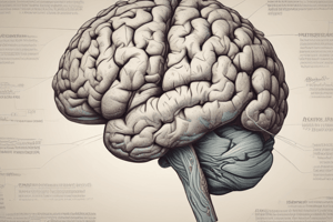
Cerebral Cortex
- Structure: Divided into two hemispheres, each containing six lobes: frontal, parietal, temporal, occipital, insular, and limbic; assists in various functions.
- Cortical Anatomy: Features gyri (folds) and sulci (valleys) that expand the surface area of the cortex to approximately 2.5 square feet when unfolded; about 50% of the cortex is hidden within sulci and fissures.
Functional Areas of the Cerebral Cortex
- Frontal Lobe: Contains the motor cortex; involved in ideation and cognition.
- Parietal Lobe: Houses the somatosensory cortex and associative cortex.
- Occipital Lobe: Responsible for visual processing through the visual cortex.
- Temporal Lobe: Contains the auditory cortex, crucial for hearing.
- Insular Lobe: Functions in sensory processing, motor control, risk assessment, decision-making, self-awareness, and emotion.
- Limbic Lobe: Important for motivation, emotion, learning, and memory; specific sections of each lobe pertain to these functions.
Cerebral White Matter
- Comprises myelinated fiber tracts that connect inputs to and outputs from the cerebral cortex.
Deep Subcortical Gray Areas
- Contains clusters of brain nuclei, such as the basal ganglia, which are essential for motor control and other functions.### Fissures and Sulci
- Fissures are deep grooves that separate lobes; sulci are grooves that delineate gyri within a lobe.
Main Sulci and Fissures
- Lateral Cerebral Fissure (Sylvian Fissure): Divides the temporal lobe from the frontal and parietal lobes.
- Central Sulcus (Fissure of Rolando): Separates the frontal lobe from the parietal lobe.
- Longitudinal Cerebral Fissure: Divides the left and right cerebral hemispheres.
- Circuminsular Fissure (Circular Sulcus): Distinguishes the insula from the surrounding frontal, parietal, and temporal lobes.
- Parieto-occipital Fissure: Located on the medial surface, separating parietal and occipital lobes.
- Calcarine Fissure: Runs horizontally in the occipital lobe, part of the visual cortex.
Corpus Callosum
- Large structure composed of myelinated and non-myelinated fibers connecting the two hemispheres.
- Parts include:
- Body: Arched central portion.
- Genu: Curved front section.
- Rostrum: Anterior part extending downward.
- Splenium: Thick back section.
Frontal Lobe Anatomy
- Motor Cortex: Controls voluntary movements, located in the precentral gyrus.
- Contains pyramidal upper motor neurons, mirroring a somatotropic map.
- Premotor Area: Involved in motor planning, located anterior to the central sulcus.
- Frontal Association Areas: Responsible for complex functions such as judgment and creativity, located in the prefrontal cortex.
- Divided by sulci:
- Precentral Sulcus: In front of the primary motor cortex.
- Superior and Inferior Frontal Sulci: Separate the lateral frontal lobe into gyrus regions.
Parietal Lobe Anatomy
- Located between the central sulcus and the parieto-occipital fissure.
- Postcentral Gyrus: Primary somatosensory cortex.
- Intraparietal Sulcus: May connect with the postcentral sulcus.
- Supramarginal and Angular Gyri: Adjacent to each other in the parietal lobe.
Occipital Lobe Anatomy
- Situated behind the parieto-occipital fissure.
- Contains the primary visual cortex along the occipital gyri.
- Divided by the calcarine sulcus into:
- Cuneus: Above the fissure.
- Lingual Gyrus: Below the fissure.
Temporal Lobe Anatomy
- Located below the lateral fissure, extending to the parieto-occipital fissure.
- Contains three main gyri:
- Superior, Middle, and Inferior Temporal Gyri.
- Sulci include the Superior and Inferior Temporal Sulci, which separate the gyri.
Insular Lobe Anatomy
- Found in a sunken area of the cortex within the lateral cerebral fissure.
Limbic System
- Comprises structures such as the cingulate gyrus and parahippocampal gyrus.
- Known as the archicortex, reflecting its ancient evolutionary origin.
Basal Ganglia Anatomy
- Masses of gray matter located deep within the cerebral hemispheres.
- Major components include:
- Caudate Nucleus: A C-shaped structure involved in movement regulation.
- Putamen and Globus Pallidus: Together known as the corpus striatum, with the putamen serving as the principal input and the globus pallidus as the main output.
Subcortical White Matter
- Commissural Fibers: Connect the two hemispheres; includes:
- Corpus Callosum: Main connector between cortex areas.
- Anterior and Hippocampal Commissures: Connect specific brain regions.
- Projection Fibers: Connect the cortex with lower brain areas and the spinal cord, encompassing optic and acoustic radiations.
- Association Fibers: Connect different regions of the same hemisphere, including the uncinate and arcuate fasciculus.
Summary of Key Areas
- Brain structures are distinguished by specific anatomical features and neural functions that are essential for integrating and processing sensory, motor, and cognitive information.
Studying That Suits You
Use AI to generate personalized quizzes and flashcards to suit your learning preferences.