Podcast
Questions and Answers
Which of the following is NOT a function of neuroglia?
Which of the following is NOT a function of neuroglia?
- Support for neurons
- Insulation for neurons
- Transmitting nerve impulses (correct)
- Protection for neurons
Which type of neuroglia is responsible for monitoring the health of nearby neurons and disposing of debris?
Which type of neuroglia is responsible for monitoring the health of nearby neurons and disposing of debris?
- Oligodendrocytes
- Ependymal cells
- Astrocytes
- Microglia (correct)
What is the primary function of ependymal cells?
What is the primary function of ependymal cells?
- Lining the central cavities of the brain and spinal cord. (correct)
- Forming myelin sheaths in the CNS
- Controlling the chemical environment around neurons
- Anchoring neurons to blood capillaries
Oligodendrocytes serve what critical function in the central nervous system (CNS)?
Oligodendrocytes serve what critical function in the central nervous system (CNS)?
Which of the following is a characteristic that distinguishes glial cells from neurons?
Which of the following is a characteristic that distinguishes glial cells from neurons?
What role do Schwann cells play in the peripheral nervous system (PNS)?
What role do Schwann cells play in the peripheral nervous system (PNS)?
What is a key characteristic of neurons that contributes to their function?
What is a key characteristic of neurons that contributes to their function?
Which region of the neuron is primarily responsible for receiving incoming signals?
Which region of the neuron is primarily responsible for receiving incoming signals?
Where on a neuron is the axon hillock located and what is its significance?
Where on a neuron is the axon hillock located and what is its significance?
Which structural component of a neuron contains neurotransmitters?
Which structural component of a neuron contains neurotransmitters?
What characterizes the myelin sheath that surrounds axons?
What characterizes the myelin sheath that surrounds axons?
What are the gaps between Schwann cells along a myelinated axon called and what is their function?
What are the gaps between Schwann cells along a myelinated axon called and what is their function?
Which of the following best describes 'nuclei' in the context of nervous tissue?
Which of the following best describes 'nuclei' in the context of nervous tissue?
What is the primary difference between white matter and gray matter?
What is the primary difference between white matter and gray matter?
How are neurons functionally classified?
How are neurons functionally classified?
What is the role of motor (efferent) neurons?
What is the role of motor (efferent) neurons?
How are interneurons characterized in terms of their function?
How are interneurons characterized in terms of their function?
What structural feature differentiates multipolar neurons from bipolar or unipolar neurons?
What structural feature differentiates multipolar neurons from bipolar or unipolar neurons?
A neuron with a single process emerging from the cell body is classified as:
A neuron with a single process emerging from the cell body is classified as:
What is the main function of sensory receptors?
What is the main function of sensory receptors?
Which type of receptor is primarily responsible for detecting pain and temperature?
Which type of receptor is primarily responsible for detecting pain and temperature?
What stimulus does a Meissner’s corpuscle primarily detect?
What stimulus does a Meissner’s corpuscle primarily detect?
What kind of stimulus does a lamellar corpuscle detect?
What kind of stimulus does a lamellar corpuscle detect?
What type of receptor are Golgi tendon organs and muscle spindles?
What type of receptor are Golgi tendon organs and muscle spindles?
What information do proprioceptors provide?
What information do proprioceptors provide?
Flashcards
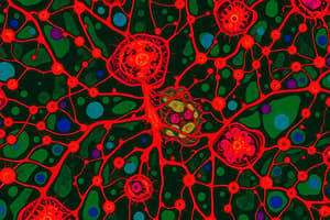
Neuroglia Function
Neuroglia Function
Support, insulate, and protect delicate neurons.
Nervous Tissue
Nervous Tissue
Nervous tissue is highly cellular, densely packed, and intertwined.
Astrocytes
Astrocytes
Abundant, star-shaped neuroglia in the CNS that anchor neurons to capillaries, control the chemical environment, and exchange between neurons.
Microglia
Microglia
Signup and view all the flashcards
Ependymal cells
Ependymal cells
Signup and view all the flashcards
Oligodendrocytes
Oligodendrocytes
Signup and view all the flashcards
Schwann Cells
Schwann Cells
Signup and view all the flashcards
Satellite Cells
Satellite Cells
Signup and view all the flashcards
Neurons
Neurons
Signup and view all the flashcards
Three Functional Regions of Most Neurons
Three Functional Regions of Most Neurons
Signup and view all the flashcards
Cell Body of a Neuron
Cell Body of a Neuron
Signup and view all the flashcards
Dendrites
Dendrites
Signup and view all the flashcards
Axons
Axons
Signup and view all the flashcards
Myelin Sheaths
Myelin Sheaths
Signup and view all the flashcards
Nodes of Ranvier
Nodes of Ranvier
Signup and view all the flashcards
Nuclei (CNS)
Nuclei (CNS)
Signup and view all the flashcards
Ganglia (PNS)
Ganglia (PNS)
Signup and view all the flashcards
Tracts
Tracts
Signup and view all the flashcards
White Matter
White Matter
Signup and view all the flashcards
Gray Matter
Gray Matter
Signup and view all the flashcards
Sensory Neurons
Sensory Neurons
Signup and view all the flashcards
Motor Neurons
Motor Neurons
Signup and view all the flashcards
Interneurons
Interneurons
Signup and view all the flashcards
Multipolar Neurons
Multipolar Neurons
Signup and view all the flashcards
Bipolar Neurons
Bipolar Neurons
Signup and view all the flashcards
Study Notes
- Nervous tissue is characterized by being highly cellular, densely packed, and intricately intertwined.
- There are two main types of cells within nervous tissue: supporting cells and neurons.
Supporting Cells
- Commonly referred to as neuroglia, glia, or glial cells.
- Six types exist: four are located in the central nervous system (CNS), and two are in the peripheral nervous system (PNS).
- Neuroglia functions include supporting, insulating, and safeguarding delicate neurons.
Neuroglia in the CNS
- Four types are present
- These types are: Astrocytes, Microglia, Ependymal cells, and Oligodendrocytes.
Astrocytes
- They are abundant and have a star-like shape.
- Astrocytes account for almost half of the neural tissue.
- Numerous projections with swollen ends are used to anchor neurons to blood capillaries.
- They contribute to the blood-brain barrier through determining capillary permeability and making exchanges.
- Astrocytes protect neurons against harmful substances found in blood.
- They maintain the brains chemical environment through removal of excess K+ ions.
Microglia
- They are spider-like phagocytes.
- They monitor the health of nearby neurons and dispose of debris, including dead brain cells and bacteria.
Ependymal Cells
- These range from squamous to columnar in shape and are often ciliated
- They line the central cavities of the brain and spinal cord.
- Cilia aids in the circulation of cerebrospinal fluid
- They form a protective cushion around the CNS.
Oligodendrocytes
- These glial cells have fewer processes than astrocytes.
- Oligodendrocytes wrap tightly around nerve fibers, forming insulating myelin sheaths.
- They serve an insulation role in the nervous system.
- Glial cells and neurons share structural similarities but differ in function, glial cells do not transmit impulses nor divide.
- Most brain tumors are gliomas derived from glial cells.
Neuroglia in the PNS
- There are two types: Schwann cells and satellite cells.
Schwann cells
- Schwann cells form myelin sheaths around larger nerve fibers in the PNS.
- They are similar to oligodendrocytes in function.
- These cells are vital for the regeneration of damaged peripheral nerve fibers.
Satellite cells
- Satellite cells surround neuron cell bodies in the PNS.
- These cells have protective and cushioning functions.
Neurons
- Also known as nerve cells.
- Specialized in transmitting messages as nerve impulses from one area of the body to another.
- Neurons consist of a cell body with a nucleus and slender processes.
- Special characteristics include extreme longevity, being amitotic, and having a high metabolic rate.
- Neurons are complex cells that possess three functional components: an input region, a conducting component, and an output region.
Structure of a Neuron
- Consist of a cell body, which is the metabolic center of the neuron.
- A nucleus containing a nucleolus is contained in cell body
- Surrounded by cytoplasm containing organelles, excluding centrioles.
- Nissl bodies and neurofibrils within the rough ER are important for maintaining cell shape.
- Armlike extensions, or fibers, that vary in length constitute neuron processes.
- Processes that convey incoming messages toward the cell body are dendrites.
- Axons conduct impulses away from the cell body.
- Neurons can have hundreds of dendrites but only one axon, which originates from the axon hillock.
- Axons occasionally branch into collateral branches, and all axons profusely branch at their terminal end into axon terminals.
- Neurotransmitters are chemicals contained within tiny vesicles or membranous sacs inside the axon terminals.
- A synaptic cleft, which is a tiny gap, separates each axon terminal from the next neuron.
- The functional junction between neurons has a name: synapse.
- Most long nerve fibers are enveloped by myelin, a whitish, fatty material and has a waxy appearance.
- An axon outside the CNS are myelinated by Schwann cells.
- It protects and insulates nerve fibers as well as increases the transmission rate of nerve impulses.
- The myelin sheath is formed as Schwann cells wrap around the axon in a jelly-roll fashion, squeezing out cytoplasm.
- The myelin sheath encloses the axon when the wrapping process is complete, leaving most of the Schwann cell cytoplasm beneath the plasma membrane.
- The outer part of the Schwann cell around the myelin sheath is called the neurolemma.
- Nodes of Ranvier are gaps or indentations between Schwann cells.
- Multiple sclerosis is a homeostatic imbalance (SELF STUDY).
Terminology
- Nuclei: clusters of cell bodies within the CNS
- Ganglia: small groups of cell bodies
- Tracts: bundles of nerve fibers within the CNS.
- White matter is composed of myelinated regions of the CNS.
- Gray matter is composed of unmyelinated regions of the CNS.
Functional Classification of Neurons
- Neurons are grouped based on the direction that the nerve impulse travels relative to the CNS.
Sensory (afferent) neurons
- They transmit impulses from sensory receptors to the CNS.
Motor (efferent) neurons
- They transmit impulses from the CNS to the viscera, muscles, or glands.
Interneurons (association) neurons
- Interneurons connect sensory and motor neurons in neural pathways.
Structural Classification of Neurons
- Classification is based on the number of processes extending from the cell body.
Multipolar neurons
- They have several processes extending from the cell body.
Bipolar neurons
- They have two processes.
Unipolar neurons
- These neurons have a single process emerging from the cell body.
- Sensory receptors are activated via specific changes nearby.
Studying That Suits You
Use AI to generate personalized quizzes and flashcards to suit your learning preferences.