Podcast
Questions and Answers
Which of the following best describes the primary function of the central nervous system?
Which of the following best describes the primary function of the central nervous system?
- Sensing the environment
- Directing reactions to incoming information
- Processing information and directing actions (correct)
- Carrying messages to and from the peripheral nervous system
What primarily forms ganglia throughout the trunk?
What primarily forms ganglia throughout the trunk?
- Ependymal cells
- Neural crest cells (correct)
- Neural plate
- Neural tube
Which structures are products of the neural tube?
Which structures are products of the neural tube?
- Ganglia
- Schwann cells
- Neurons of the PNS
- Brain and spinal cord (correct)
Which glial cells are responsible for myelination within the central nervous system (CNS)?
Which glial cells are responsible for myelination within the central nervous system (CNS)?
What is the main function of the peripheral nervous system?
What is the main function of the peripheral nervous system?
Which of the following is a primary function of neurons?
Which of the following is a primary function of neurons?
Which characteristic is unique to the axolemma?
Which characteristic is unique to the axolemma?
Which of the following best describes the function of Nissl substance in neurons?
Which of the following best describes the function of Nissl substance in neurons?
Which type of glial cell is the primary immune defense in the central nervous system?
Which type of glial cell is the primary immune defense in the central nervous system?
What is the main role of satellite glial cells?
What is the main role of satellite glial cells?
What is a key function of ependymal cells in the nervous system?
What is a key function of ependymal cells in the nervous system?
Where are bipolar neurons most commonly found?
Where are bipolar neurons most commonly found?
Which type of neuron is most abundant in the human body?
Which type of neuron is most abundant in the human body?
Which type of neuron is responsible for carrying signals from the central nervous system to muscles or glands?
Which type of neuron is responsible for carrying signals from the central nervous system to muscles or glands?
What is the primary function of interneurons?
What is the primary function of interneurons?
What is the main role of receptors in the nervous system concerning information?
What is the main role of receptors in the nervous system concerning information?
What is the primary means of synaptic communication between neurons?
What is the primary means of synaptic communication between neurons?
Which of the following describes the presynaptic component of a chemical synapse?
Which of the following describes the presynaptic component of a chemical synapse?
Which of the following are found in the synaptic cleft?
Which of the following are found in the synaptic cleft?
Which of the following characterizes an electrical synapse?
Which of the following characterizes an electrical synapse?
What is the critical function of astrocytes in relation to the blood-brain barrier?
What is the critical function of astrocytes in relation to the blood-brain barrier?
What is the term for the outermost nucleated cytoplasmic layer of Schwann cells?
What is the term for the outermost nucleated cytoplasmic layer of Schwann cells?
What is a primary difference between oligodendrocytes and Schwann cells?
What is a primary difference between oligodendrocytes and Schwann cells?
Which of the following is a function of microglia?
Which of the following is a function of microglia?
In the context of myelinated axons, what are the uninsulated regions between myelin sheaths called?
In the context of myelinated axons, what are the uninsulated regions between myelin sheaths called?
What is the functional significance of the nodes of Ranvier?
What is the functional significance of the nodes of Ranvier?
Which process best describes saltatory conduction?
Which process best describes saltatory conduction?
Which of the following is most closely associated with multiple sclerosis (MS)?
Which of the following is most closely associated with multiple sclerosis (MS)?
Which glial cells are derived from neural crest cells?
Which glial cells are derived from neural crest cells?
Which of the following best defines a synapse?
Which of the following best defines a synapse?
In a synapse, what is the function of the postsynaptic neuron?
In a synapse, what is the function of the postsynaptic neuron?
Which of the following does NOT describe a function of Astrocytes?
Which of the following does NOT describe a function of Astrocytes?
What name is given to motor neurons that connect to skeletal muscle at a neuromuscular junction?
What name is given to motor neurons that connect to skeletal muscle at a neuromuscular junction?
What is the key distinction between protoplasmic and fibrous astrocytes concerning their location?
What is the key distinction between protoplasmic and fibrous astrocytes concerning their location?
Which of the central nervous tissues, in addition to the ependyma lining the ventricles and central canal, did Ramon y Cajal and del Rio-Hortega contribute to discovering?
Which of the central nervous tissues, in addition to the ependyma lining the ventricles and central canal, did Ramon y Cajal and del Rio-Hortega contribute to discovering?
Which of the following is a functional characteristic of unipolar neurons?
Which of the following is a functional characteristic of unipolar neurons?
Which of the central neurotransmitter molecules is MOST involved in synaptic communication?
Which of the central neurotransmitter molecules is MOST involved in synaptic communication?
Which of the following would MOST influence a drug's ability to affect the brain?
Which of the following would MOST influence a drug's ability to affect the brain?
How would disrupting the function of microglia MOST affect the brain?
How would disrupting the function of microglia MOST affect the brain?
Flashcards
Nervous System
Nervous System
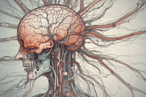
The nervous system consists of the brain, spinal cord, sensory organs, and all of the nerves that connect these organs with the rest of the body.
Central Nervous System (CNS)
Central Nervous System (CNS)
The CNS (brain and spinal cord) processes information and directs actions.
Peripheral Nervous System (PNS)
Peripheral Nervous System (PNS)
The PNS consists of all nerves that are not encased in bone.
Neural Crest Cells
Neural Crest Cells
Signup and view all the flashcards
Neural Tube
Neural Tube
Signup and view all the flashcards
Neural Crest
Neural Crest
Signup and view all the flashcards
Telencephalon
Telencephalon
Signup and view all the flashcards
Nervous Tissue Composition
Nervous Tissue Composition
Signup and view all the flashcards
Neurons
Neurons
Signup and view all the flashcards
Neuroglia
Neuroglia
Signup and view all the flashcards
Cell Body (Soma)
Cell Body (Soma)
Signup and view all the flashcards
Processes of Neurons
Processes of Neurons
Signup and view all the flashcards
Axon Hillock
Axon Hillock
Signup and view all the flashcards
Initial Segment
Initial Segment
Signup and view all the flashcards
Axon Collaterals
Axon Collaterals
Signup and view all the flashcards
Axon Terminals
Axon Terminals
Signup and view all the flashcards
Nissl Substance
Nissl Substance
Signup and view all the flashcards
Axolemma
Axolemma
Signup and view all the flashcards
Microtubules (Neurons)
Microtubules (Neurons)
Signup and view all the flashcards
Neurofilaments
Neurofilaments
Signup and view all the flashcards
Microfilaments (Neurons)
Microfilaments (Neurons)
Signup and view all the flashcards
Sensory Neurons
Sensory Neurons
Signup and view all the flashcards
Motor Neurons
Motor Neurons
Signup and view all the flashcards
Interneurons
Interneurons
Signup and view all the flashcards
Receptors
Receptors
Signup and view all the flashcards
Multipolar Neuron
Multipolar Neuron
Signup and view all the flashcards
Bipolar Neuron
Bipolar Neuron
Signup and view all the flashcards
Unipolar Neuron
Unipolar Neuron
Signup and view all the flashcards
Synapses Definition
Synapses Definition
Signup and view all the flashcards
Synapse Function
Synapse Function
Signup and view all the flashcards
Presynaptic Neuron
Presynaptic Neuron
Signup and view all the flashcards
Postsynaptic Neuron
Postsynaptic Neuron
Signup and view all the flashcards
Synaptic Communication
Synaptic Communication
Signup and view all the flashcards
The motor endplate
The motor endplate
Signup and view all the flashcards
Astrocytes
Astrocytes
Signup and view all the flashcards
Blood-Brain Barrier
Blood-Brain Barrier
Signup and view all the flashcards
Microglia as Macrophages.
Microglia as Macrophages.
Signup and view all the flashcards
Ependymal Cells
Ependymal Cells
Signup and view all the flashcards
Oligodendrocytes
Oligodendrocytes
Signup and view all the flashcards
Oligodendrocytes myelin
Oligodendrocytes myelin
Signup and view all the flashcards
Schwann cells : PNS
Schwann cells : PNS
Signup and view all the flashcards
Study Notes
- Nervous tissue consists of the brain, spinal cord, sensory organs, and connecting nerves.
- It senses the environment and processes incoming information.
- It influences and directs reactions to incoming information.
- Nerves encased in bone form the central nervous system, i.e. the brain and spinal cord.
- The central nervous system is responsible for processing information and directing actions.
- All nerves not encased in bone make up the peripheral nervous system
- The peripheral nervous systems main function is to carry messages to and from the central nervous system.
- Neural crest cells migrate to form ganglia throughout the trunk and in a variety of other tissues.
- The neural tube forms the brain and spinal cord, which makes up the central nervous system.
- The neural crest gives rise to neurons with cell bodies in the PNS and to Schwann cells, among other tissues.
- The nervous system includes the peripheral and central nervous systems.
- The peripheral nervous system is split into the autonomic and somatic nervous systems.
- The autonomic nervous system is split into sympathetic and parasympathetic divisions.
- The central nervous system contains the brain and spinal cord.
- The brain consists of the forebrain, midbrain, and hindbrain.
- The forebrain includes the telencephalon and diencephalon.
- The telencephalon includes the cerebral cortex, basal ganglia, hippocampus, and amygdala.
- The diencephalon includes the thalamus and hypothalamus.
- The midbrain consists of the mesencephalon.
- The mesencephalon includes the tectum and the cerebellum.
- The hindbrain consists of the metencephalon and myelencephalon.
- The metencephalon includes the pons and cerebellum.
- The myelencephalon includes the medulla.
- Nervous tissue is composed of neurons and neuroglia (nerve glue).
- Neurons generate and transduce electric impulses and are responsible for the nervous systems receptive, integrative, and motor functions.
- Neuroglia supports and protects neurons
Neuroglia cells:
- Astrocytes
- Schwann cells
- Oligodendrocytes
- Satellite Cells
- Microglia
- Ependymal cells
Neurons:
- All have a cell body which contains a cytoplasm, organelles, and is the metabolic and integrative center of neuron.
- All neurons have processes specialized for communication, including an axon and dendrites.
- Axon hillock is the origin of the axon.
- The initial segment is the part of the axon where action potential is initiated.
- Axon collaterals are branches of axons.
- Axon terminals are the effector portion, synaptic terminals, and terminal boutons.
Organelles in Neurons
- Cell membrane: Neuron's plasma membrane (plasmalemma), axon’s cell membrane is the axolemma.
- Nucleus: Prominent, centrally located, and the site of ribosomal synthesis.
- Nissl substance: Stacks of RER (rough endoplasmic reticulum) which are the site of protein synthesis, abundant in the cell body and proximal parts of dendrites, but absent in axon hillock and axons.
- Smooth endoplasmic reticulum.
- Golgi apparatus: Site of glycosylation, abundant, perinuclear location.
- Mitochondria: Abundant.
- Lysosomes.
- Pigments: Lipofuscin (aging pigment); melanin
- Microtubules (25 µm): Part of cytoskeleton that gives neurons their shape and is abundant in axons and dendrites and is responsible for axon transport.
- Neurofilaments (intermediate filaments, 10 um): Part of the cytoskeleton responsible for the shape of neurons and are abundant in axons and dendrites; also known as "Neurofibrils" and are bundles of neurofilaments seen with the light microscope; they are argyrophilic and visualized with silver stains.
- Microfilaments (f-actin, 5 µm): Part of the cytoskeleton, abundant in growth cones where they give contractile properties
- Vesicles: Vesicles in synaptic endings that contain neurotransmitters and are called synaptic vesicles.
Functional Classification of Neurons
- Sensory neurons (afferent nerves) carry signals from the outer parts of your body (periphery) into the central nervous system.
- Motor neurons (efferent nerves) carry signals from the central nervous system to the outer parts (muscles or glands) of your body.
- Interneurons connect various neurons within the brain and spinal cord.
- Interneurons facilitate or inhibit a motor response to a sensory stimulus.
- Receptors sense the environment (chemicals, light, sound, touch) and encode that information into electrochemical messages.
- These transformed messages are then transmitted by sensory neurons.
Morphological classification based on the number of processes:
- Multipolar neurons have one axon and two or more dendrites.
- Bipolar neurons have one axon and one dendrite.
- Unipolar (pseudounipolar) neurons have one process that divides close to the cell body into two long processes, an axon and a dendrite.
- 99% of neurons in the body are multipolar.
- Bipolar neurons are rare and occur in special sense organs of ear, nose and eye.
- Unipolar neurons begin as bipolar but processes fuse.
- They are primarily sensory neurons such as dorsal root ganglion neurons.
- Neurons communicate with other neurons and effector cells by means of synapses.
- Synapses are specialized junctions between neurons that transmit impulses from one neuron to another or to effector cells like muscle or gland cells
- Synapse is the site where two nerves communicate with each other.
- The presynaptic neuron is the neuron generating information toward the next neuron
- The postsynaptic neuron transmits information away from synapse
- Most synaptic communication is via chemical messengers, e.g. acetylcholine, serotonin, norepinephrine, dopamine, endorphins, GABA, glycine, glutamic acid, etc.
Synapse Components:
- Presynaptic component consists of synaptic vesicles (~40 nm) which are flat or round, clear or dense, and contain neurotransmitters or neuropeptides; and mitochondria. May contain pre-synaptic membrane densities (= active zones = sites of release).
- Synaptic cleft: (20-40 nm) May contain carbohydrate-containing material that serves to tightly adhere pre- to post-synaptic component. Postsynaptic component: membrane densities representing receptor sites may be present, can be dendrites or dendritic spines, cell body (soma), axon, or other target cells.
- Neurons communicate via chemical and electrical transmissions
- Chemical neurotransmission which makes up 99% of transmissions utilize nerve impulses traveling down the axon of the pre-synaptic cleft.
- Electrical neurotransmission makesup 1% of transmissions.
- synapses are classified by their position
The Three Types of Synapses
- Axosomatic synapse: an axon terminal ending on the soma of a neuron.
- Axoaxonic synapse: an axon terminal contacting another axon terminal.
- Axodendritic synapse: An axon ending on a dendrite.
- Axospinous synapse: An axon terminal facing a dendritic spine.
- The dendritic shaft contains microtubules.
- Dendritic spine cytoskeleton consists of actin filaments, and neurotransmitter receptors and ion channels localize in the postsynaptic density.
- Telodendria are the end branches of an axon
- The motor endplate (neuromuscular junction) is a specialized synapse.
- The motor endplate joins a motoneuron's axon terminal and a skeletal muscle fiber.
- It has the features of a synapse, but the postsynaptic component consists of muscle fiber
- There are deep invaginations of the sarcolemma (muscle membrane) at the junction
Neuromuscular Junction Diagram key:
- Presynaptic terminal
- Sarcolemma
- Synaptic vesicle
- Nicotinic acetylcholine receptor
- Mitochondrion
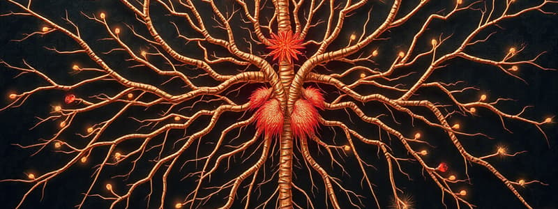
- Neuroglia are nerve glue and are made up of different cell types
- The term neuroglia, or "nerve glue," was coined in 1859 by Rudolph Virchow.
- Virchow conceived of the neuroglia as an inactive "connective tissue" holding neurons together in the central nervous system.
- The metallic staining techniques developed by Ramón y Cajal and del Rio-Hortega allowed the identification of the types of supporting cells in the CNS: oligodendrocytes, astrocytes, and microglia.
- The Schwann cell is the major neuroglial component in the peripheral nervous system (PNS).
- Astrocytes are star-shaped glial cells in the brain and spinal cord, that have small cell bodies and radial branched processes.
- They perform biochemical control of endothelial cells to form the blood-brain barrier
Astrocyte Functions:
- Clearing excess neurotransmitters.
- Stabilizing and regulating the blood-brain barrier.
- Promoting synapse formation.
- Provision of nutrients to the nervous tissue.
- Maintenance of extracellular ion balance.
- Regulation of cerebral blood flow.
- Repair and scarring the brain and spinal cord following infection and traumatic injuries.
- Astrocytes fence in neurons and oligodendrocytes.
- Astrocytes help isolate the brain parenchyma.
- They achieve this via long processes that project to the pia mater and the ependyma
- Glia limitans are formed through the connection of pia mater and ependyma.
- They enmesh synapses and dendrites and project processes to cell somas.
- Astrocytes form a syncytium via gap junctions and enables ion transfer and also allows for small molecules to diffuse across the brain parenchyma.
- Protoplasmic astrocytes have many processes and are large and star-shaped, attach to blood vessels
- Fibrous astrocytes are similar to protoplasmic astrocytes but have more filaments and glycogen. And lie in the white matter.
- Capillaries of the CNS form the blood–brain barrier, a protective layer that prevents exchange of solutes between the blood and brain.
- The difference with systematic circulation is the presence in certain brain interendothelial tight junctions that form the barrier.
- Finally, astrocytes induce and maintain these junctions.
- Microglia (Hortega cells) are macrophages of the CNS.
- Ramified microglia may transform into migratory, ameboid, and phagocytic cells.
- Microglia help phagocytose degenerating cells that undergo programmed cell death as part of normal development.
- They retain the ability to divide and have the immunophenotypic properties of monocytes and macrophages.
- Microglia secrete cytokines and growth factors for fiber tract development, gliogenesis, and angiogenesis.
- They also present antigens to T lymphocytes.
- Ependymal cells line ventricles and central cavities of brain and spinal cord and secrete cerebrospinal fluid (CSF)
- Their cilia help to circulate CSF
- Cell types include ependymocytes, choroid plexus epithelial cells, tanycytes, and, within the retina, Müller cells and retinal pigment epithelial cells.
- Oligodendrocytes have plump cell bodies with fairly dense cytoplasm and a darker nucleus and fewer, shorter processes than an astrocyte.
- Oligodendrocytes accomplish myelination in the CNS.
- They are the myelinating cells of the central nervous system (CNS)
- Myelination of the spinal cord begins in the ventral (motor) roots during month 4.
- Myelination of the association neocortex extends to 30 years of age.
- Myelin is an electrically insulating material to form the myelin sheath around the axon of a neuron
- Oligodendrocytes myelinate CNS axons while Schwann cells myelinate PNS axons.
- Nodes of Ranvier are uninsulated portions of axonal membrane between myelin sheaths
- Myelin segments are called internodes.
- Myelination increases speed of impulse propagation by saltatory conduction.
- Schwann cells accomplish myelination in the PNS, individual myelinating Schwann cells form a single internode
- Neurolemma (aka neurilemma or Schwann's Sheath) is the outermost nucleated cytoplasm layer of Schwann cells that encircle the axon of the neuron.
- It forms outermost layer of the nerve fiber in the peripheral nervous system.
- The neurolemma serves a protective function for peripheral nerve fibers.
- Non-myelinating Schwann cells are involved in maintenance of axons and are crucial for neuronal survival.
- Satellite glial cells are glial cells that cover the surface of nerve cell bodies in sensory, sympathetic and parasympathetic ganglia.
- Both satellite glial cells and Schwann cells are derived from the neural crest cells.
- Satellite Cells provide nutrients, remove metabolites, and have structural role (similar to astrocytes).
Studying That Suits You
Use AI to generate personalized quizzes and flashcards to suit your learning preferences.