Podcast
Questions and Answers
Which of the following accurately describes a ganglion in the nervous system?
Which of the following accurately describes a ganglion in the nervous system?
- A collection of myelinated axons within the spinal cord
- A protective layer around a nerve root
- A type of neuron that conducts impulses away from the brain
- A cluster of nerve cell bodies located outside the central nervous system (correct)
Which type of neuron is primarily responsible for transmitting signals from sensory receptors to the central nervous system?
Which type of neuron is primarily responsible for transmitting signals from sensory receptors to the central nervous system?
- Efferent neurons
- Motor neurons
- Interneurons
- Sensory neurons (correct)
What is the primary function of glial cells in the nervous system?
What is the primary function of glial cells in the nervous system?
- Storing neurotransmitters
- Regulating blood flow to the brain
- Transmitting electrical impulses
- Providing structural and metabolic support to neurons (correct)
Which part of the spinal cord is primarily responsible for processing and relaying efferent (motor) signals?
Which part of the spinal cord is primarily responsible for processing and relaying efferent (motor) signals?
Which of the following structures is involved in the production and flow of cerebrospinal fluid (CSF)?
Which of the following structures is involved in the production and flow of cerebrospinal fluid (CSF)?
Which of the following correctly defines an effector in the nervous system?
Which of the following correctly defines an effector in the nervous system?
What is the primary difference between gray matter and white matter in the central nervous system?
What is the primary difference between gray matter and white matter in the central nervous system?
In the context of autonomic nervous system pathways, which neurotransmitter is primarily associated with the sympathetic nervous system?
In the context of autonomic nervous system pathways, which neurotransmitter is primarily associated with the sympathetic nervous system?
Which of the following statements correctly describes a reflex arc?
Which of the following statements correctly describes a reflex arc?
What role do glial cells play in nerve regeneration?
What role do glial cells play in nerve regeneration?
Flashcards are hidden until you start studying
Study Notes
Nervous System Divisions
- Central Nervous System (CNS): Brain and spinal cord
- Peripheral Nervous System (PNS): Nerves that connect the CNS to the rest of the body
Definitions
- Ganglion: a cluster of neuron cell bodies located outside the CNS
- Nerve: a bundle of axons that transmit signals between the CNS and other parts of the body
- Nucleus: a cluster of neuron cell bodies located within the CNS
- Receptor: a specialized cell or structure that detects a specific stimulus
- Effector: a muscle or gland that carries out a response to a stimulus
Neuron Types
- Sensory neurons: transmit signals from receptors to the CNS
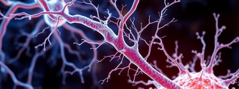
- Interneurons: connect neurons within the CNS, responsible for processing and integrating information
- Motor neurons: transmit signals from the CNS to effectors
Neuron Anatomy
- Cell body (soma): contains the nucleus and other organelles
- Axon: a long, slender extension that transmits signals away from the cell body
- Dendrites: branched extensions that receive signals from other neurons
Glial Cells
- Astrocytes: support and nourish neurons, regulate the blood-brain barrier
- Oligodendrocytes (CNS) and Schwann cells (PNS): produce myelin, insulating the axons and speeding up signal transmission
- Microglia: phagocytic cells that remove cellular debris and pathogens
Myelination
- Myelin: a fatty substance that insulates axons, increasing the speed of signal conduction
- Nodes of Ranvier: gaps in the myelin sheath, allowing for saltatory conduction, where the signal jumps from node to node
Nerve Regeneration
- PNS: limited regeneration possible due to Schwann cells guiding axonal growth
- CNS: regeneration is very limited due to the lack of Schwann cells and the presence of inhibitory factors
Synapse Anatomy
- Synapse: the junction between two neurons where communication occurs
- Presynaptic neuron: releases neurotransmitters into the synaptic cleft
- Postsynaptic neuron: receives neurotransmitters
- Synaptic cleft: the space between the presynaptic and postsynaptic neurons
Neuronal Circuits
- Converging circuits: multiple neurons converge onto a single neuron, allowing integration of information
- Diverging circuits: a single neuron branches to multiple neurons, allowing for amplification of a signal
- Reverberating circuits: neurons are organized in a loop, creating repetitive patterns to amplify the signal or create a rhythmic pattern.
Spinal Cord Gross Anatomy
- Cervical, Thoracic, Lumbar, Sacral, Coccygeal regions
- Central canal: filled with cerebrospinal fluid (CSF)
- Gray matter: contains neuron cell bodies, dendrites, and unmyelinated axons
- White matter: contains myelinated axons
Gray vs. White Matter
- Gray matter: involved in processing information and forming connections between neurons
- White matter: responsible for communicating information between different parts of the CNS
Nerve Anatomy
- Endoneurium: surrounds individual axons
- Perineurium: surrounds bundles of axons (fascicles)
- Epineurium: surrounds the entire nerve
Rootlets to Rami
- Rootlets: small bundles of axons that emerge from the spinal cord
- Dorsal roots: contain sensory axons
- Ventral roots: contain motor axons
- Spinal nerve: formed by the fusion of dorsal and ventral roots
- Rami: branches of spinal nerves that innervate specific regions of the body
- Dorsal rami: innervate the back
- Ventral rami: innervate the front and limbs
Nerve Plexuses
- Cervical plexus: innervates the neck, shoulders, and diaphragm
- Brachial plexus: innervates the upper limbs
- Lumbar plexus: innervates the lower limbs
- Sacral plexus: innervates the lower limbs and pelvic region
Meninges
- Dura mater: outer, tough layer
- Arachnoid mater: middle, web-like layer
- Pia mater: inner, delicate layer that adheres to the surface of the brain and spinal cord
- Subarachnoid space: space between the arachnoid mater and pia mater, filled with CSF
Spinal Tracts
- Ascending tracts: carry sensory information from the body to the brain
- Descending tracts: carry motor commands from the brain to the body
Neuron Types
- First-order neuron: receives sensory input from receptors
- Second-order neuron: receives information from the first-order neuron and transmits it to the thalamus
- Third-order neuron: receives information from the thalamus and transmits it to the cerebral cortex
Upper vs. Lower Motor Neurons
- Upper motor neurons (UMN): located in the cerebral cortex, control voluntary movement
- Lower motor neurons (LMN): located in the brainstem or spinal cord, directly innervate muscle fibers
Reflex Arcs
- Reflex: an involuntary, rapid response to a stimulus
- Reflex arc: the neural pathway involved in a reflex
- Components of a reflex arc: receptor, sensory neuron, interneuron, motor neuron, effector
Nerve Innervation and Spinal Cord Injuries
- Spinal nerves: can be damaged by injury, causing loss of sensation or motor function
- Complete spinal cord injury: all function below the level of the injury is lost
- Incomplete spinal cord injury: some function below the level of the injury is preserved
Major Brain Landmarks
- Cerebrum: the largest part of the brain, responsible for higher cognitive functions
- Cerebellum: located at the back of the brain, responsible for coordination and balance
- Brainstem: connects the cerebrum and cerebellum to the spinal cord, responsible for vital functions like breathing and heart rate
Dural Venous Sinuses
- Dural venous sinuses: spaces between the layers of dura mater that collect venous blood from the brain
- Superior sagittal sinus: a large sinus that runs along the top of the brain
- Transverse sinus: connects the superior sagittal sinus to the sigmoid sinus
- Sigmoid sinus: drains into the jugular vein
Cerebrospinal Fluid (CSF) Production & Flow
- CSF: a clear, colorless fluid that circulates through the CNS, providing cushioning, nutrient supply, and waste removal
- Produced by: choroid plexuses, vascular structures located in the ventricles of the brain
- Flow: ventricles (lateral, third, fourth) – subarachnoid space – absorbed into venous blood
Brainstem Functions
- Midbrain: controls eye movements, auditory reflexes, and motor control
- Pons: relays signals between the cerebrum and cerebellum, controls breathing, and sleep
- Medulla oblongata: controls heart rate, blood pressure, and breathing
Cerebellar Anatomy
- Two cerebellar hemispheres
- Vermis: the narrow strip of tissue that connects the hemispheres
- Folia: folds on the surface of the cerebellum
- Arbor vitae: the tree-like pattern of white matter
Cerebellum Function
- Coordination and balance: receives information from the cerebrum, inner ear, and muscles, and adjusts motor output to ensure smooth, coordinated movements
- Motor learning and memory: plays a role in learning and refining motor skills
- Cognitive functions: may also be involved in higher cognitive functions such as language and attention
Diencephalon
- Thalamus: relays sensory information to the cerebral cortex
- Hypothalamus: controls body temperature, hunger, thirst, and other homeostatic functions
- Epithalamus: includes the pineal gland, which secretes melatonin
Cerebral Lobes and Major Functions
- Frontal lobe: responsible for planning, decision-making, language, and voluntary movement
- Parietal lobe: responsible for sensory perception, spatial awareness, and motor control
- Temporal lobe: responsible for hearing, memory, and language comprehension
- Occipital lobe: responsible for visual processing
Limbic System
- Hippocampus: involved in memory formation and consolidation
- Amygdala: involved in emotional responses, especially fear and aggression
- Hypothalamus: controls basic drives and emotions
- Cingulate gyrus: involved in emotional processing and behavior regulation
Primary Sensory Cortices
- Somatosensory cortex: receives sensory input from the body
- Visual cortex: receives visual input
- Auditory cortex: receives auditory input
- Gustatory cortex: receives taste input
- Olfactory cortex: receives smell input
White Matter Tract Types
- Association fibers: connect areas within the same hemisphere
- Commissural fibers: connect areas between hemispheres
- Projection fibers: connect the cerebrum to lower brain structures and the spinal cord
Motor Function, Basal Nuclei, and Substantia Nigra
- Basal nuclei: a group of nuclei deep within the cerebrum that help control voluntary movements, posture, and muscle tone
- Substantia nigra: a brain structure that produces dopamine, a neurotransmitter involved in movement control
Cranial Nerve Number & Function
- 12 pairs of cranial nerves: numbered I-XII
- Functions: sensory (vision, hearing, taste, smell), motor (eye, jaw, facial, tongue movements), and mixed (sensory and motor)
Hemispheric Lateralization and Language
- Cerebral hemispheres: specialized for different functions
- Left hemisphere: typically dominant for language, speech, and logic
- Right hemisphere: typically dominant for spatial processing, music, and artistic ability
Autonomic vs. Somatic NS
- Somatic nervous system: controls voluntary movements of skeletal muscles
- Autonomic nervous system (ANS): regulates involuntary functions such as heart rate, breathing, digestion, and blood pressure
Sympathetic vs. Parasympathetic Structure and Function
- Sympathetic nervous system (fight-or-flight): prepares the body for stressful situations
- Parasympathetic nervous system (rest-and-digest): promotes relaxation and conserves energy
ANS Pathways
- Sympathetic: preganglionic neurons are short, postganglionic neurons are long
- Parasympathetic: preganglionic neurons are long, postganglionic neurons are short
ANS Neurotransmitters
- Sympathetic: norepinephrine and epinephrine
- Parasympathetic: acetylcholine
ANS Antagonism, Coopertivity, and Autonomic Tone
- Antagonism: sympathetic and parasympathetic systems often have opposing effects
- Cooperativity: systems can work together to achieve a common goal
- Autonomic tone: the balance between sympathetic and parasympathetic activity always has a slight activation of both systems, but one is usually dominant
Sensory Receptor Classification
- Mechanoreceptors: respond to mechanical stimuli (pressure, touch, sound, vibration)
- Chemoreceptors: respond to chemical stimuli (taste, smell, pH)
- Photoreceptors: respond to light stimuli (vision)
- Thermoreceptors: respond to temperature stimuli
- Nociceptors: respond to painful stimuli
Receptive Field
- Receptive field: the area of the body or environment that, when stimulated, will activate a particular sensory receptor
Studying That Suits You
Use AI to generate personalized quizzes and flashcards to suit your learning preferences.


