Podcast
Questions and Answers
What is the primary function of the Sympathetic Division of the Autonomic Nervous System?
What is the primary function of the Sympathetic Division of the Autonomic Nervous System?
- Monitors internal organ health
- Promotes rest and digest functions
- Facilitates fight or flight response (correct)
- Regulates voluntary muscle movement
Which part of a neuron is responsible for transmitting electrical signals away from the cell body?
Which part of a neuron is responsible for transmitting electrical signals away from the cell body?
- Soma
- Myelin sheath
- Dendrites
- Axon (correct)
What distinguishes the Parasympathetic Division from the Sympathetic Division in the Autonomic Nervous System?
What distinguishes the Parasympathetic Division from the Sympathetic Division in the Autonomic Nervous System?
- Both divisions manage voluntary muscle control
- Parasympathetic only regulates respiratory functions
- Parasympathetic leads to rest and digest responses, while Sympathetic leads to fight or flight (correct)
- Sympathetic promotes digestion while Parasympathetic increases heart rate
What is the role of myelin sheath in neurons?
What is the role of myelin sheath in neurons?
Which division of the Peripheral Nervous System is primarily responsible for voluntary movements?
Which division of the Peripheral Nervous System is primarily responsible for voluntary movements?
Which component of the nervous system primarily monitors internal stimuli like hunger and pain?
Which component of the nervous system primarily monitors internal stimuli like hunger and pain?
What are the basic building blocks of the nervous system?
What are the basic building blocks of the nervous system?
In which part of the Peripheral Nervous System would you find the Enteric Nervous System?
In which part of the Peripheral Nervous System would you find the Enteric Nervous System?
What is the primary function of the myelin sheath?
What is the primary function of the myelin sheath?
What is the role of the nodes of Ranvier?
What is the role of the nodes of Ranvier?
Which part of the neuron is responsible for receiving signals from other neurons?
Which part of the neuron is responsible for receiving signals from other neurons?
What surrounds each individual axonal fiber and Schwann cells in a nerve?
What surrounds each individual axonal fiber and Schwann cells in a nerve?
Which layer encases the entire nerve?
Which layer encases the entire nerve?
What is the purpose of the paraneurium in nerve anatomy?
What is the purpose of the paraneurium in nerve anatomy?
Which structure assists in the formation of myelin in the peripheral nervous system?
Which structure assists in the formation of myelin in the peripheral nervous system?
What is a fascicle in nerve anatomy?
What is a fascicle in nerve anatomy?
What is the primary role of the autonomic nervous system?
What is the primary role of the autonomic nervous system?
Which branch of the autonomic nervous system is primarily responsible for 'fight or flight' responses?
Which branch of the autonomic nervous system is primarily responsible for 'fight or flight' responses?
Which pathway in the sympathetic nervous system involves preganglionic neurons bypassing the sympathetic chain ganglia?
Which pathway in the sympathetic nervous system involves preganglionic neurons bypassing the sympathetic chain ganglia?
Where do sympathetic preganglionic neurons originate?
Where do sympathetic preganglionic neurons originate?
Which of the following statements is true regarding the structure of sympathetic neurons?
Which of the following statements is true regarding the structure of sympathetic neurons?
What is the primary function of the parasympathetic nervous system?
What is the primary function of the parasympathetic nervous system?
Which structure is involved in the direct pathway to the adrenal medulla in the sympathetic nervous system?
Which structure is involved in the direct pathway to the adrenal medulla in the sympathetic nervous system?
What type of tissue surrounds the epineurium as a protective layer?
What type of tissue surrounds the epineurium as a protective layer?
Flashcards
Myelin Sheath
Myelin Sheath
Fatty layer covering axons; increases speed of nerve impulse conduction.
Nodes of Ranvier
Nodes of Ranvier
Gaps in the myelin sheath that speed up signal transmission.
Schwann Cells
Schwann Cells
Glial cells that myelinate axons in the peripheral nervous system, supporting neurons and nerve repair.
Endoneurium
Endoneurium
Signup and view all the flashcards
Perineurium
Perineurium
Signup and view all the flashcards
Fascicle
Fascicle
Signup and view all the flashcards
Epineurium
Epineurium
Signup and view all the flashcards
Nerve
Nerve
Signup and view all the flashcards
Peripheral Nervous System (PNS)
Peripheral Nervous System (PNS)
Signup and view all the flashcards
Sensory Division (Afferent)
Sensory Division (Afferent)
Signup and view all the flashcards
Somatic Sensory
Somatic Sensory
Signup and view all the flashcards
Visceral Sensory
Visceral Sensory
Signup and view all the flashcards
Motor Division (Efferent)
Motor Division (Efferent)
Signup and view all the flashcards
Autonomic Nervous System (ANS)
Autonomic Nervous System (ANS)
Signup and view all the flashcards
Neuron (Nerve cell)
Neuron (Nerve cell)
Signup and view all the flashcards
Neuron Soma
Neuron Soma
Signup and view all the flashcards
Autonomic Nervous System
Autonomic Nervous System
Signup and view all the flashcards
Sympathetic Nervous System
Sympathetic Nervous System
Signup and view all the flashcards
Parasympathetic Nervous System
Parasympathetic Nervous System
Signup and view all the flashcards
Sympathetic Nervous System (SNS) Pathways
Sympathetic Nervous System (SNS) Pathways
Signup and view all the flashcards
Sympathetic Chain Ganglia
Sympathetic Chain Ganglia
Signup and view all the flashcards
Splanchnic Nerves
Splanchnic Nerves
Signup and view all the flashcards
Adrenal Medulla
Adrenal Medulla
Signup and view all the flashcards
Parasympathetic Nervous System Anatomy
Parasympathetic Nervous System Anatomy
Signup and view all the flashcards
Study Notes
Peripheral Nervous System (PNS) Overview
- The PNS connects the CNS (brain and spinal cord) to the rest of the body
- It consists of nerves outside the brain and spinal cord
- It has two main divisions: Sensory (afferent) and Motor (efferent)
Sensory Division (Afferent)
- Carries signals from the body to the CNS
- Divided into:
- Somatic Sensory: Detects external stimuli (skin, muscles, joints, special senses) and is consciously perceived (touch, temperature, pain)
- Visceral Sensory: Monitors internal stimuli in organs (heart, lungs, stomach), and is usually unconsciously perceived (hunger, internal pain)
Motor Division (Efferent)
- Carries signals from the CNS to the body
- Divided into:
- Somatic Nervous System: Controls voluntary movements (skeletal muscles)
- Autonomic Nervous System (ANS): Controls involuntary functions (smooth muscle, cardiac muscle, glands)
Autonomic Nervous System (ANS)
- Part of the PNS governing involuntary functions
- Divided into:
- Sympathetic Division: "Fight or Flight" response, activates during stress. Increases heart rate, dilates pupils, inhibits digestion
- Parasympathetic Division: "Rest and Digest" response, calms the body during relaxation. Slows heart rate, constricts pupils, stimulates digestion
- The ANS also has specialized nervous pathways.
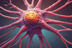
Neuron Anatomy
- Soma (cell body): Contains the nucleus and organelles. It maintains neuron structure and provides energy.
- Dendrites: Branching fibers that receive signals from other neurons. They can form multiple dendritic trees.
- Axon: A long projection transmitting electrical signals away from the cell body and joins the cell body at the axon hillock. Insulated by the myelin sheath for faster signal conduction
- Myelin sheath: Fatty layer insulating axons, speeding up impulse conduction.
- Nodes of Ranvier: Gaps in the myelin sheath that facilitate saltatory conduction, speeding up signal transmission.
- Schwann cells: Myelinate axons of peripheral neurons and support nerve function and repair.
Anatomy of a Nerve
- Endoneurium surrounds each individual axonal fiber and Schwann cells
- Perineurium surrounds groups of axons, forming fascicles (bundles)
- Fascicle is a bundle of nerve fibers organized for efficient signal transmission
- Epineurium is the outermost layer encasing the entire nerve
- Paraneurium (Mesoneurium) is the loose areolar tissue surrounding the epineurium for added support
Cranial Nerves
- There are 12 pairs of cranial nerves, numbered I through XII. Each plays specific roles in the body, including sensory (e.g. Olfaction), vision (e.g. Optic), eye movement, face sensation and expression, hearing and balance, and motor (e.g. tongue movements).
Forelimb Nerves
- Radial Nerve: controls extension of the elbow, carpus, digits, supination of the paw. Provides sensation.
- Deep branch: Deep branch innervates extensor muscles of the forelimb.
- Superficial Branch: Provides sensory information to the skin.
- Median Nerve: Travels deep, flexes muscles of the forearm, flexes carpal and digital joints. Provides sensory information to the palmar side of the forepaw and dorsal surface.
- Ulnar Nerve: flexes wrist and digits, sensation to forepaw and 5th digit.
Hindlimb Nerves
- Femoral Nerve: Straightens leg and moves the hip (innervates sartorius, iliacus and quadriceps femoris muscles)
- Tibial Nerve: Supplies movement to the lower leg (innervates gastrocnemius, superficial digital flexor, deep digital flexor, and popliteus muscles)
- Common Peroneal Nerve: Supplies movement to the lower leg.
- Sural Nerve: Detect foot position, purely sensory nerve functioning
- Saphenous Nerve: Provides sensation to the medial aspect of lower leg and medial foot, purely sensory nerve
- Plantar Nerve: Provides sensation to the foot, innervates palmar muscles.
Reproductive and Urinary Nerves
- Male System: Sympathetic (hypogastric nerve) regulates ejaculation; parasympathetic (pelvic nerve) promotes erection and ejaculation; somatic (pudendal nerve) aids in erection and ejaculation.
- Female System: Sympathetic (hypogastric nerve) regulates uterine contractions and tone; parasympathetic (pelvic nerve) facilitates vasodilation; somatic (pudendal nerve) controls external genitalia
Enteric Nervous System (ENS)
- Local nervous system of the gut
- Composed of two plexuses:
- Myenteric (Auerbach): Coordinates contractility of circular and longitudinal muscle to produce peristalsis
- Submucosal (Meissner): Governs movement of water and electrolytes.
Studying That Suits You
Use AI to generate personalized quizzes and flashcards to suit your learning preferences.