Podcast
Questions and Answers
What are the two major divisions of the nervous system?
What are the two major divisions of the nervous system?
The two major divisions of the nervous system are the Central Nervous System (CNS) and the Peripheral Nervous System (PNS).
Name the four lobes of the cerebral hemisphere.
Name the four lobes of the cerebral hemisphere.
The four lobes of the cerebral hemisphere are the frontal, parietal, temporal, and occipital lobes.
What is the functional unit of the nervous system?
What is the functional unit of the nervous system?
The functional unit of the nervous system is the neuron.
What role do dendrites play in a neuron?
What role do dendrites play in a neuron?
Where are action potentials typically generated in a neuron?
Where are action potentials typically generated in a neuron?
What is the function of the myelin sheath?
What is the function of the myelin sheath?
What are the two components of the autonomic nervous system?
What are the two components of the autonomic nervous system?
What structures are included in the brainstem?
What structures are included in the brainstem?
What role do Schwann cells play in the peripheral nervous system?
What role do Schwann cells play in the peripheral nervous system?
Describe what happens at the nodes of Ranvier during the conduction of action potentials.
Describe what happens at the nodes of Ranvier during the conduction of action potentials.
What are the two types of potentials generated by neurons?
What are the two types of potentials generated by neurons?
How does the Na+/K+ ATPase pump contribute to maintaining membrane potential in neurons?
How does the Na+/K+ ATPase pump contribute to maintaining membrane potential in neurons?
What is the typical resting membrane potential (RMP) of neurons?
What is the typical resting membrane potential (RMP) of neurons?
What is an action potential and what conditions must be met for it to occur?
What is an action potential and what conditions must be met for it to occur?
Explain the significance of the unequal ion distribution in maintaining resting membrane potential.
Explain the significance of the unequal ion distribution in maintaining resting membrane potential.
What causes potassium ions (K+) to move out of the neuron at rest?
What causes potassium ions (K+) to move out of the neuron at rest?
What is the All-or-None law in relation to action potentials?
What is the All-or-None law in relation to action potentials?
Describe the role of voltage-gated Na+ channels during the depolarization phase of an action potential.
Describe the role of voltage-gated Na+ channels during the depolarization phase of an action potential.
Explain the difference between the absolute and relative refractory periods.
Explain the difference between the absolute and relative refractory periods.
What happens during the repolarization phase of an action potential?
What happens during the repolarization phase of an action potential?
How does the strength-duration relationship influence action potential generation?
How does the strength-duration relationship influence action potential generation?
What is hyperpolarization, and when does it occur during action potentials?
What is hyperpolarization, and when does it occur during action potentials?
What is the significance of the threshold intensity in action potential generation?
What is the significance of the threshold intensity in action potential generation?
Explain the process that leads to the inactivation of Na+ channels during an action potential.
Explain the process that leads to the inactivation of Na+ channels during an action potential.
What happens when a stimulus fails to reach the threshold level in relation to action potentials?
What happens when a stimulus fails to reach the threshold level in relation to action potentials?
Explain how sodium concentration outside the cell affects action potentials.
Explain how sodium concentration outside the cell affects action potentials.
Describe the process of saltatory conduction in myelinated axons.
Describe the process of saltatory conduction in myelinated axons.
How does increased potassium (K+) outside the cell affect neurons?
How does increased potassium (K+) outside the cell affect neurons?
What are the primary functions of glial cells in the nervous system?
What are the primary functions of glial cells in the nervous system?
What is the effect of hypokalemia on the neuron's membrane potential?
What is the effect of hypokalemia on the neuron's membrane potential?
How does calcium concentration affect neuronal excitability?
How does calcium concentration affect neuronal excitability?
What role do nodes of Ranvier play in nerve signal conduction?
What role do nodes of Ranvier play in nerve signal conduction?
What is the primary function of the Blood Brain Barrier (BBB)?
What is the primary function of the Blood Brain Barrier (BBB)?
How do neurotrophins support neurons in the nervous system?
How do neurotrophins support neurons in the nervous system?
What role do Schwann cells play in peripheral nerve regeneration?
What role do Schwann cells play in peripheral nerve regeneration?
Why is peripheral nerve regeneration more successful than CNS regeneration?
Why is peripheral nerve regeneration more successful than CNS regeneration?
What happens to axons in the CNS after an injury?
What happens to axons in the CNS after an injury?
Identify two factors that create an unfavorable environment for CNS axonal regeneration.
Identify two factors that create an unfavorable environment for CNS axonal regeneration.
What is the significance of neurotrophins in the context of axonal injury?
What is the significance of neurotrophins in the context of axonal injury?
Explain how inflammatory responses affect CNS axonal regeneration.
Explain how inflammatory responses affect CNS axonal regeneration.
Flashcards are hidden until you start studying
Study Notes
Nervous System Overview
- The nervous system is divided into the central nervous system (CNS) and the peripheral nervous system (PNS).
- The CNS includes the brain and spinal cord.
- The PNS consists of all the nerves that connect the CNS to the rest of the body.
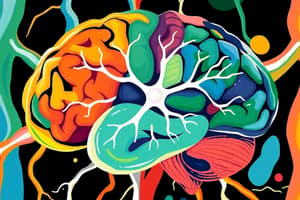
Brain Structure
- The brain is divided into four major parts: the cerebrum, diencephalon, brainstem, and cerebellum.
- The cerebrum is the largest part of the brain and is responsible for higher-level functions like thinking, language, and memory.
- The diencephalon is located below the cerebrum and serves as a relay center for sensory information.
- The brainstem connects the cerebrum and cerebellum to the spinal cord.
- The cerebellum is responsible for coordinating movement and balance.
Neuron Structure
- Neurons are the basic functional units of the nervous system.
- The primary structure of a neuron includes the dendrites, initial segment, axon, and nerve endings.
- Dendrites receive signals from other neurons and transmit them to the cell body.
- The initial segment is where action potentials are generated.
- The axon transmits signals to other neurons or target cells where action potentials arrive at nerve endings.
- Nerve endings (presynaptic terminals) release neurotransmitters that communicate with other neurons.
- Many axons are covered by myelinated sheaths, formed by Schwann cells (PNS) or oligodendrocytes (CNS).
Neuron Excitation & Conduction
- Neurons generate two types of potentials: local (non-propagated) and action potentials.
- Local potentials are graded responses, whereas action potentials are all-or-none responses.
- Action potentials are generated by the opening and closing of voltage-gated ion channels, typically Na+ and K+ channels.
- Ion conductance depends on permeability and resistance of the membrane.
Action Potential Process
- Resting state: Neuron is at rest with no ion movement across the membrane.
- Depolarizing stimulus: Stimulus causes Na+ channels to open, allowing Na+ to enter the cell, leading to threshold potential.
- Rapid depolarization: More Na+ channels open, causing a rapid rise in membrane potential (upstroke).
- Na+ channels inactivate: Membrane potential moves towards +60 mV (Na+ equilibrium potential), but Na+ channels inactivate.
- Repolarization: Voltage-gated K+ channels open, allowing K+ to exit the cell, causing repolarization.
- After-hyperpolarization: Slow closure of K+ channels causes a brief period of hyperpolarization.
- Return to resting state: Membrane potential returns to its resting level after K+ channels close.
Action Potential Properties
- Threshold intensity: The minimum stimulus strength needed to trigger an action potential.
- Strength-duration relationship: Stronger stimuli have shorter durations to reach threshold, whereas weaker stimuli have longer durations.
- Refractory periods: Absolute refractory period (no response during depolarization) and relative refractory period (needs a stronger stimulus to elicit a response).
- All-or-none principle: An action potential will only occur if the threshold level is reached. If subthreshold, no action potential occurs. The size remains constant once threshold is reached.
- Conduction: Action potential propagates as positive charges flow into the region of depolarization, triggering local responses.
Myelinated Axons and Saltatory Conduction
- In myelinated axons, current flows only at the nodes of Ranvier, the gaps between myelin, due to the insulating effect of myelin.
- Depolarization jumps from one node to the next, called saltatory conduction, making signal transmission significantly faster.
Ion Concentration Effects on Excitability
- Decreased Na+ outside: Reduces AP strength but minimal impact on resting potential.
- Increased K+ outside: Lowers AP threshold, making neurons more excitable.
- Decreased K+ outside: Hyperpolarizes the membrane, reducing AP likelihood.
- Decreased Ca2+: Increases membrane excitability.
- Increased Ca2+: Decreases membrane excitability.
Glial Cells - Supporting Role
- Glial cells are non-neuronal cells in the CNS and PNS that support neuron function.
- Types of glial cells: oligodendrocytes, astrocytes, ependymal cells, microglia (CNS); Schwann cells and satellite cells (PNS).
- Functions: surround and hold neurons, supply nutrients and oxygen, insulate neurons from each other, destroy pathogens and remove dead neurons.
Blood-Brain Barrier (BBB)
- BBB is a specialized barrier between the blood and CNS, regulating what substances can enter the brain.
- it protects the brain from harmful substances in the blood.
Neurotrophins - Growth and Development
- Neurotrophins are a family of proteins that regulate the growth, survival, and function of neurons.
- They play essential roles in the development and maintenance of the nervous system.
Axonal Injury and Regeneration
- Peripheral nerve damage can often be repaired. The axon degenerates beyond the injury, but the distal stump survives.
- Axons regenerate from the proximal stump, guided by Schwann cells and neurotrophins from the distal stump.
- CNS axonal injury is more difficult to repair, as CNS neurons lack the growth-promoting chemicals and myelin inhibits axonal growth.
- CNS damage treatments focus on rehabilitation rather than repairing the damaged nerves.
Studying That Suits You
Use AI to generate personalized quizzes and flashcards to suit your learning preferences.