Podcast
Questions and Answers
Welche Aussage trifft auf chemische Synapsen im zentralen Nervensystem (ZNS) am ehesten zu?
Welche Aussage trifft auf chemische Synapsen im zentralen Nervensystem (ZNS) am ehesten zu?
- Sie leiten Signale schneller weiter als elektrische Synapsen.
- Die Mehrheit der Synapsen im ZNS ist chemisch. (correct)
- Sie sind seltener als elektrische Synapsen.
- Sie nutzen immer gap junctions zur Signalübertragung.
Was ist die unmittelbare Folge einer Depolarisation der synaptischen Endigung nach dem Eintreffen eines Aktionspotentials?
Was ist die unmittelbare Folge einer Depolarisation der synaptischen Endigung nach dem Eintreffen eines Aktionspotentials?
- Postsynamptische Strukturen werden aktiviert.
- Transmitter werden durch den synaptischen Spalt transportiert.
- Die Exozytose von vesikelgelagerten Transmittern wird gehemmt.
- Es kommt zum Einstrom von Kalzium-Ionen (Ca2+). (correct)
Welche Aussage beschreibt am besten, wie Neurotransmitter an postsynaptische Strukturen binden?
Welche Aussage beschreibt am besten, wie Neurotransmitter an postsynaptische Strukturen binden?
- Mittels aktiver Ionenkanäle
- Nach dem Prinzip von Angebot und Nachfrage
- Durch Diffusion und osmotischen Druck
- Nach dem Schlüssel-Schloss-Prinzip (correct)
Was ist ein wesentlicher Unterschied zwischen ionotropen und metabotropen Rezeptoren?
Was ist ein wesentlicher Unterschied zwischen ionotropen und metabotropen Rezeptoren?
Welche Aussage ist im Kontext der wichtigsten Transmittersysteme im menschlichen ZNS korrekt?
Welche Aussage ist im Kontext der wichtigsten Transmittersysteme im menschlichen ZNS korrekt?
Welche der folgenden Strukturen gehört nicht zum Zentralnervensystem (ZNS)?
Welche der folgenden Strukturen gehört nicht zum Zentralnervensystem (ZNS)?
Was ist die wichtigste Funktion der Zerebrospinalflüssigkeit?
Was ist die wichtigste Funktion der Zerebrospinalflüssigkeit?
Was versteht man unter dem Begriff 'kaudal' in Bezug auf die anatomische Lagebeschreibung?
Was versteht man unter dem Begriff 'kaudal' in Bezug auf die anatomische Lagebeschreibung?
Welche der genannten Richtungsbezeichnungen ist keine Lagebeschreibung am Gehirn?
Welche der genannten Richtungsbezeichnungen ist keine Lagebeschreibung am Gehirn?
In welcher Ebene erfolgt der Horizontalschnitt (Transversalschnitt) des Gehirns?
In welcher Ebene erfolgt der Horizontalschnitt (Transversalschnitt) des Gehirns?
Was bedeutet der Begriff 'ipsilateral'?
Was bedeutet der Begriff 'ipsilateral'?
Welche Aussage beschreibt die Funktion efferenter Nervenfasern am genauesten?
Welche Aussage beschreibt die Funktion efferenter Nervenfasern am genauesten?
Wie unterscheidet sich das somatische Nervensystem vom vegetativen Nervensystem?
Wie unterscheidet sich das somatische Nervensystem vom vegetativen Nervensystem?
Welche Aussage über gemischte Nerven trifft am ehesten zu?
Welche Aussage über gemischte Nerven trifft am ehesten zu?
Was sind Ganglien?
Was sind Ganglien?
Was ist die Hauptursache für die Symptome der Amyotrophen Lateralsklerose (ALS)?
Was ist die Hauptursache für die Symptome der Amyotrophen Lateralsklerose (ALS)?
Was sind die Hauptmerkmale der Multiplen Sklerose (MS)?
Was sind die Hauptmerkmale der Multiplen Sklerose (MS)?
Was versteht man unter einem Dermatom?
Was versteht man unter einem Dermatom?
Was ist die primäre Funktion von Motoneuronen im Rückenmark?
Was ist die primäre Funktion von Motoneuronen im Rückenmark?
Welche Aussage beschreibt am besten die Lage des Rückenmarks?
Welche Aussage beschreibt am besten die Lage des Rückenmarks?
Wie sind die Vorder- und Hinterhörner der grauen Substanz im Rückenmark hauptsächlich charakterisiert?
Wie sind die Vorder- und Hinterhörner der grauen Substanz im Rückenmark hauptsächlich charakterisiert?
Was ist die Cauda equina?
Was ist die Cauda equina?
Wo haben die aufsteigenden Nervenfasern im Hinterstrang des Rückenmarks ihr Ursprung?
Wo haben die aufsteigenden Nervenfasern im Hinterstrang des Rückenmarks ihr Ursprung?
Welche Aussage beschreibt am besten die Funktion des Rückenmarks?
Welche Aussage beschreibt am besten die Funktion des Rückenmarks?
Wie unterscheidet sich ein Eigenreflex von einem Fremdreflex?
Wie unterscheidet sich ein Eigenreflex von einem Fremdreflex?
Was ist ein typisches Merkmal von Fremdreflexen?
Was ist ein typisches Merkmal von Fremdreflexen?
Welchen ungefähren Anteil am Körpergewicht macht das menschliche Gehirn aus?
Welchen ungefähren Anteil am Körpergewicht macht das menschliche Gehirn aus?
Welche der folgenden Strukturen gehört nicht zu den Hauptabschnitten des menschlichen Gehirns?
Welche der folgenden Strukturen gehört nicht zu den Hauptabschnitten des menschlichen Gehirns?
Welche Hirnstruktur verbindet die beiden Hemisphären miteinander?
Welche Hirnstruktur verbindet die beiden Hemisphären miteinander?
Welche Funktion hat die zerebrospinale Flüssigkeit (Liquor) nicht?
Welche Funktion hat die zerebrospinale Flüssigkeit (Liquor) nicht?
Welche der folgenden Strukturen enthält nicht eine Hirnventrikel?
Welche der folgenden Strukturen enthält nicht eine Hirnventrikel?
Welche Reihenfolge der Hirnhäute ist korrekt, von außen nach innen (vom Schädel zum Gehirn)?
Welche Reihenfolge der Hirnhäute ist korrekt, von außen nach innen (vom Schädel zum Gehirn)?
Woher bezieht das Gehirn hauptsächlich seinen arteriellen Zufluss?
Woher bezieht das Gehirn hauptsächlich seinen arteriellen Zufluss?
Welche Symptome sind typisch für einen Verschluss der Arteria carotis interna?
Welche Symptome sind typisch für einen Verschluss der Arteria carotis interna?
Welche Aussage beschreibt am besten die Organisation von grauer und weißer Substanz im Gehirn?
Welche Aussage beschreibt am besten die Organisation von grauer und weißer Substanz im Gehirn?
Welche Struktur dient im Rückenmark hauptsächlich der Informationsübertragung zwischen Nervenzellen?
Welche Struktur dient im Rückenmark hauptsächlich der Informationsübertragung zwischen Nervenzellen?
Was ist Liquor cerebrospinalis?
Was ist Liquor cerebrospinalis?
Was ist die Funktion des Vorderstrangs und des Hinterstrangs im Rückenmark?
Was ist die Funktion des Vorderstrangs und des Hinterstrangs im Rückenmark?
Flashcards
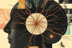
Synapsenarten
Synapsenarten
Chemische und elektrische
Was passiert nach AP?
Was passiert nach AP?
Depolarisierung der synaptischen Endigung
Was folgt auf Depolarisierung?
Was folgt auf Depolarisierung?
Einstrom von Ca2+ und Exozytose von vesikelgelagerten Transmittern
Arten von Rezeptoren
Arten von Rezeptoren
Signup and view all the flashcards
Häufigste exitatorische Transmitter
Häufigste exitatorische Transmitter
Signup and view all the flashcards
Häufigste inhibitorische Transmitter
Häufigste inhibitorische Transmitter
Signup and view all the flashcards
Bestandteile von ZNS und PNS?
Bestandteile von ZNS und PNS?
Signup and view all the flashcards
Somatisches Nervensystem
Somatisches Nervensystem
Signup and view all the flashcards
Vegetatives Nervensystem
Vegetatives Nervensystem
Signup and view all the flashcards
Funktion des PNS
Funktion des PNS
Signup and view all the flashcards
Gemischte Nerven
Gemischte Nerven
Signup and view all the flashcards
Ansammlungen von Nervenzellkörpern
Ansammlungen von Nervenzellkörpern
Signup and view all the flashcards
Was gehört zum ZNS?
Was gehört zum ZNS?
Signup and view all the flashcards
Somatisches NS Funktion
Somatisches NS Funktion
Signup and view all the flashcards
Vegetatives NS Funktion
Vegetatives NS Funktion
Signup and view all the flashcards
Efferente Fasern
Efferente Fasern
Signup and view all the flashcards
Afferente Fasern
Afferente Fasern
Signup and view all the flashcards
ALS Definition
ALS Definition
Signup and view all the flashcards
MS Definition
MS Definition
Signup and view all the flashcards
Funktion Rückenmark
Funktion Rückenmark
Signup and view all the flashcards
Aufbau der Wirbelsäule
Aufbau der Wirbelsäule
Signup and view all the flashcards
Rückenmarkssegmente
Rückenmarkssegmente
Signup and view all the flashcards
Wo beginnt/endet das Rückenmark?
Wo beginnt/endet das Rückenmark?
Signup and view all the flashcards
Ursprung Spinalnerven
Ursprung Spinalnerven
Signup and view all the flashcards
Graue Substanz Rückenmark
Graue Substanz Rückenmark
Signup and view all the flashcards
Aufgaben des Rückenmarks
Aufgaben des Rückenmarks
Signup and view all the flashcards
Eigenreflex
Eigenreflex
Signup and view all the flashcards
Fremdreflex
Fremdreflex
Signup and view all the flashcards
Was ist das Rückenmark?
Was ist das Rückenmark?
Signup and view all the flashcards
Nervenfasern Spinalnerv
Nervenfasern Spinalnerv
Signup and view all the flashcards
Motoneurone
Motoneurone
Signup and view all the flashcards
Sensible Neurone
Sensible Neurone
Signup and view all the flashcards
Interneurone
Interneurone
Signup and view all the flashcards
Das Rückenmark
Das Rückenmark
Signup and view all the flashcards
Aufsteigende Bahnen
Aufsteigende Bahnen
Signup and view all the flashcards
Absteigende Bahnen
Absteigende Bahnen
Signup and view all the flashcards
Volumen und Gewicht Gehirn
Volumen und Gewicht Gehirn
Signup and view all the flashcards
Abschnitte Gehirn
Abschnitte Gehirn
Signup and view all the flashcards
Inhalt graue Substanz
Inhalt graue Substanz
Signup and view all the flashcards
Inhalt weiße Substanz
Inhalt weiße Substanz
Signup and view all the flashcards
Study Notes
- The presentation provides a summary of information coding and the structure and function of the nervous system for engineering students at TU Berlin, instructed by Prof. Dr. Klaus Gramann from the department of Biological Psychology and Neuroergonomics.
Information Coding Summary
- Chemical and electrical synapses exist.
- Most synapses in the central nervous system (ZNS) are chemical.
- An action potential (AP) leads to depolarization of the synaptic terminal.
- Depolarization results in an influx of Calcium, which leads to exocytosis of vesicle-bound transmitters.
- Transmitters travel through the synaptic cleft and attach to postsynaptic structures based on the "lock and key" principle.
- Transmitters can attach to two types of receptors: ionotropic and metabotropic.
- Ionotropic receptors have their own channels, leading to fast postsynaptic potential changes.
- Metabotrope receptors trigger secondary processes that lead to potential changes.
- Major transmitter systems in the human central nervous system include Glutamate & Aspartate, GABA & Glycin, Acetylcholine (Ach), Catecholamines (Dopamine, Noradrenaline, Adrenaline), and Serotonin.
Nervous System Overview
- Key questions addressed: The structure of the spinal cord, the process and types of reflexes, functions of cerebrospinal fluid, and the different sections of the human brain.
Orientation and Position Terminology:
- Cranial (superior): Towards the head or top.
- Dorsal (posterior): Towards the back.
- Ventral (anterior): Towards the front.
- Caudal (inferior): Towards the tail or bottom.
- Medial: Towards the midline.
- Lateral: Away from the midline.
- Rostral (anterior): Toward the front (brain).
- Sagittal Section: Divides body into left and right.
- Frontal Section (Coronal): Divides body into front and back.
- Horizontal Section (Transversal): Divides body into top and bottom.
- Anterior: Toward the front, toward the forehead (brain), toward the tip of the foot (spinal cord).
- Caudal: Toward the back, toward the back of the head (brain), toward the ball of the foot (spinal cord).
- Contralateral: On the opposite side.
- Dorsal: Toward the top (brain), toward the back (spinal cord).
- Ipsilateral: On the same side.
- Lateral: To the side.
- Medial: To the middle.
- Posterior: Toward the back.
- Ventral: Downwards (brain), towards the belly (spinal cord).
Anatomical Organization
- The nervous system branches out from the brain and spinal cord.
- Nerve fibers transport information from the central nervous system to the periphery (efferent) and from the periphery to the central nervous system (afferent).
- The central nervous system includes the brain and spinal cord.
- The peripheral nervous system consists of nerve fibers, fiber bundles, and peripheral nerve cell bodies.
- The nervous system is divided into somatic and vegetative systems.
- The Somatic nervous system controls voluntary movement and awareness (efferent = voluntary control; afferent = perceiving occurrences in the body's periphery)
- The vegetative nervous system controls involuntary functions of inner organs (also known as the visceral nervous system).
- The peripheral nervous system serves the central nervous system as a signal transmission system.
- Information is conveyed from peripherals.
- Commands are forwarded to the end organs (muscles, glands, etc.).
- Most nerves contain both sensory and motor fibers and, therefore, are referred to as "mixed nerves".
- Accumulations of nerve cell bodies outside the brain are called ganglia; inside the brain, they are called nuclei.
- Ganglia and nuclei facilitate information transfer (switching).
- Nerves originating in the brain are called cranial nerves.
- Nerves originating in the spinal cord are called spinal nerves.
Nervous System Pathologies:
- Amyotrophic Lateral Sclerosis (ALS or Lou Gehrig's Disease): characterized by progressive degeneration of motor neurons, leading to muscle weakness (paresis), muscle atrophy (amyotrophy), and is incurable.
- Multiple Sclerosis (MS): characterized by progressive demyelination of central nervous system neurons, accompanied by the destruction of oligodendrocytes.
- Nnerve impulses are interrupted, resulting in impaired perception and motor skills and classified as a slow progressive autoimmune disease.
Nervous System Classification:
- Central Nervous System (ZNS): Brain and spinal cord. * Peripheral Nervous System (PNS): all nerves outside the central nervous system.
- Somatic Nervous System: regulates voluntary motor control.
- Sensory with the exception of inner organs.
- Vegetative Nervous System: regulates the function of the inner organs.
- Sensory input from inner organs.
- Efferent Fibers: Carry information away from the area.
- Afferent Fibers: Carry information toward the area.
Spinal Cord Details
- The spinal cord (Medulla spinalis) innervates extremities and torso.
- It is 40-45 cm long and located in the vertebral column.
- It is surrounded by cerebrospinal fluid (Liquor cerebrospinalis).
- The specific weight of nervous tissue is slightly higher than water so the brain and spinal cord float in fluid, providing mechanical protection from concussions.
- It is composed of 7 cervical vertebrae, 12 thoracic vertebrae, 5 lumbar vertebrae, 5 fused sacral vertebrae, and 4-5 fused coccygeal vertebrae.
- Sections of the spinal cord include 8 cervical segments (C1-C8), 12 thoracic segments (T1-T12), 5 lumbar segments (L1-L5), 5 sacral segments (S1-S5), and 1 coccygeal segment.
- The spinal cord begins at the lower end of the Medulla oblongata and ends at the height of the first or second Lumbar vertebrae.
- Nerve fiber bundles extend further within the vertebral column (Cauda equina).
- Spinal nerves run from the spinal cord to the periphery and exit between vertebral bones.
- Thin bundles unite to form roots (Radix, pl. Radices).
- There is one root on the ventral and one on the dorsal side.
- The thickening before the confluence of the dorsal root threads = spinal ganglion.
- Bilateral front and back bulge of gray matter known known as ventral horn and dorsal horn.
- The Ventral horn contains mostly motor cell groups.
- The Dorsal horn contains mostly sensory cell groups.
- The surrounding white matter in the dorsal column contains ascending nerve fibers.
- The front strand contains descending nerve fibers.
- It has disc-shaped segments from the neck to the sacrum
- The Dorsal root ganglion contains somatic sensory neurons.
- A single spinal nerve has a lateral horn, also known as autonomic motor neurons for visceral sensory functions.
- Descending pathways (anterior column) have branches in different segments in the anterior horn substance and end at the motor neuron.
- Ascending fibers (posterior column) originate in the spinal ganglion or in cell bodies of the posterior horn and end in the brain.
- The main part of the white matter are long fibers from and to the brain and longer fibers located more laterally.
- Important Components: Dorsal root and Ventral root.
- Dermatome: Each spinal nerve innervates a specific area of the body surface.
- Overlapping ensures that each point of the body surface is innervated by a minimum of two spinal nerves.
Spinal Cord Neurons:
- Motor neurons: axons conduct motor impulses to muscles.
- Sensory neurons: receive information from the periphery and transmit it to the brain.
- Interneurons: axons connect to other neurons in the spinal cord.
Spinal Cord Function:
- Transports information (conduction activity.
- Processes information directly (reflex activity).
- Ascending pathways end in the Medulla Oblongata, as well as Formatio Reticularis, Thalamus, Kleinhirn
- Descending paths (front strand) extend from brain to different segments.
Characteristics of Axons:
- Axons can be over a meter in length.
Spinal Reflex Activity Description
- Afferent information turns and goes in one or more Synapsen muscle or gland activity which can be triggered.
- Reflex arches
- Reflex activity depends on switching character
Type of Spinal Reflex
- Exteroceptive
- Interoceptive
Reflexes Examples
- Exteroceptive
- Interoceptive
General Information About Eigenereflex
- Occurs directly
- Very fast reaction time (ca. 10 ms) without fatigue.
- Patellar tendon
- Monosynaptic Connection
General Information About Fremdreflex
-
Occurs indirectly
-
Longer reaction time (ca. 50-150 ms) with fatigue.
-
Polysynaptic Connection
-
The spinal cord is the extension of the brainstem downwards.
-
It runs in the vertebral canal and is washed around by liquor cerebrospinalis.
-
Gray matter consists of ventral horn (motor) and dorsal horn (sensory).
-
White matter of the anterior column primarily transports information from the brain to the spinal cord.
-
White matter of the posterior column primarily transports sensory information from the spinal cord to the brain.
-
Motor nerve fibers extend through the anterior root.
-
Sensory nerve fibers extend through the posterior root to form the spinal nerve on each side.
-
31 spinal nerves arise segmentally.
-
They innervate assigned body areas.
-
Information allows fast, reflex-like reactions to occur.
-
Monosynaptic and polysynaptic reflexes are available.
The Human Brain
- Volume: 1200-1500 cc.
- Weight: 1.35–1.4 kg.
Sections of The Brain
- Grouped from Morphological and Developmental Features
- Medulla Oblongata = extended Mark
- Pons = Bridge
- Cerebellum = Small Brain
- Mesencephalon = Mid-Brain
- Diencephalon = Between Brain
- Telencephalon = Large Brain (End-Brain)
Brain Organization
- The gray matter in the brain harbors motor neurons and cell bodies of interneurons, dendrites, and unmyelinated axons.
- The gray matter forms the cortex, which covers most of the adult brain and discrete internal clusters = cerebral core areas.
- The white matter consists of myelinated axons and lies beneath the gray matter.
- Meninges (cerebral membranes) are connective tissue that separates the soft tissue of the brain from the bone tissue of the skull, encloses and protects blood vessels for brain supply, contains and circulates cerebral spinal fluid, and forms some of the veins that return blood from the brain and contains three layers include dura mater, arachnoid mater, and pia mater.
- The central nervous system is embedded in the Liquor cerebrospinalis.
- The Liquor cerebrospinalis is a fluid system with a buffer function that fills the inner cavities of the brain.
- Ventricles are connected and connected to the central flow of the spinal cord.
- There are 4 ventricles in the brain.
- 2 lateral ventricles, one in each hemisphere, separated through the septum pellucidum.
- 1 third ventricle is within the Diencephalons.
- 1 fourth ventricle located between the pons and cerebellum.
- There are 4 Ventricles including third ventricle, fourth Ventricle, lateral aperture and Cerebrum.
- Cerebrospinal fluid (CSF) functions as a clear, colorless fluid similar to blood plasma, circulating in the ventricles and the cavity of the Aeachcnoida.
- The CSF has three major functions.
- Buoyancy: the brain floats in CSF.
- Protection: CSF creates protection for fast movement.
- Protection: CSF forms a liquid protective buffer for rapid movements.
- Stable Environment: Carries nutrients and removes metabolic end-products from the brain.
- The main amount of arterial inflow is from the inside Halschlagader. arteria carotis interna (bilateral) arteria carotis communis (bilateral).
- Complications From Occlusion Of Carotis Interna Including, halbseitige Lähmung v.a. im Bereich der Arme und des Gesichts, Vorhübergehende Sehbeschwerden, and Mögliche Sprachstörungen bei linkslateralem Verschluss.
- Complications for Occlusion of the Basilararterie include Bei beidseitigem Verschluss mögliches Koma, ,Drop attacks' – plötzliches Stürzen, Hörbeeinträchtigung, and Schluckstörungen.
Studying That Suits You
Use AI to generate personalized quizzes and flashcards to suit your learning preferences.