Podcast
Questions and Answers
Which type of neuron conducts impulses from sensory receptors to the central nervous system?
Which type of neuron conducts impulses from sensory receptors to the central nervous system?
- Sensory neuron (correct)
- Interneuron
- Glial cell
- Motor neuron
Dendrites are responsible for transmitting signals away from the cell body.
Dendrites are responsible for transmitting signals away from the cell body.
False (B)
What is the function of the cell body in a neuron?
What is the function of the cell body in a neuron?
The cell body integrates information from dendrites and controls the neuron's activities.
The ______ is a fatty covering around the axon that speeds up signal transmission.
The ______ is a fatty covering around the axon that speeds up signal transmission.
Match the following neuron structures with their corresponding functions:
Match the following neuron structures with their corresponding functions:
Which of the following is NOT a type of neuroglia cell?
Which of the following is NOT a type of neuroglia cell?
Schwann cells are responsible for producing myelin in the central nervous system (CNS).
Schwann cells are responsible for producing myelin in the central nervous system (CNS).
What is the function of the myelin sheath gaps (nodes of Ranvier) along the axon?
What is the function of the myelin sheath gaps (nodes of Ranvier) along the axon?
The ______ neuron integrates information from sensory neurons and transmits it to motor neurons.
The ______ neuron integrates information from sensory neurons and transmits it to motor neurons.
Which of the following is NOT a function of neuroglia cells?
Which of the following is NOT a function of neuroglia cells?
Astrocytes are responsible for providing structural support and helping to maintain the blood-brain barrier.
Astrocytes are responsible for providing structural support and helping to maintain the blood-brain barrier.
What is the function of the synaptic terminals?
What is the function of the synaptic terminals?
The ______ neuron conducts impulses from the central nervous system to an effector.
The ______ neuron conducts impulses from the central nervous system to an effector.
Which of the following is responsible for producing myelin in the peripheral nervous system?
Which of the following is responsible for producing myelin in the peripheral nervous system?
Microglia are responsible for removing debris and pathogens from the nervous system.
Microglia are responsible for removing debris and pathogens from the nervous system.
What type of neuron has a single dendrite and one axon?
What type of neuron has a single dendrite and one axon?
The primary function of Schwann cells is to produce myelin in the CNS.
The primary function of Schwann cells is to produce myelin in the CNS.
What is the function of the astrocytes?
What is the function of the astrocytes?
The ______ is the outermost and toughest layer of the meninges.
The ______ is the outermost and toughest layer of the meninges.
Which of these is NOT a function of the cerebrospinal fluid (CSF)?
Which of these is NOT a function of the cerebrospinal fluid (CSF)?
Match the following structures with their correct descriptions:
Match the following structures with their correct descriptions:
The anterior horn of the gray matter contains sensory neurons.
The anterior horn of the gray matter contains sensory neurons.
What is the function of the epidural space?
What is the function of the epidural space?
The ______ is a strand of connective tissue that anchors the spinal cord to the coccyx.
The ______ is a strand of connective tissue that anchors the spinal cord to the coccyx.
Which of these is located between the dura mater and the arachnoid mater?
Which of these is located between the dura mater and the arachnoid mater?
The posterior root ganglia contain clusters of neuron cell bodies.
The posterior root ganglia contain clusters of neuron cell bodies.
What is the primary function of the white matter in the spinal cord?
What is the primary function of the white matter in the spinal cord?
The ______ is a shallow groove that divides the spinal cord into left and right halves, located along the posterior midline.
The ______ is a shallow groove that divides the spinal cord into left and right halves, located along the posterior midline.
What is the main function of the satellite cells in the PNS?
What is the main function of the satellite cells in the PNS?
Microglia are responsible for producing myelin in the CNS.
Microglia are responsible for producing myelin in the CNS.
What is the difference between the anterior and posterior roots of spinal nerves?
What is the difference between the anterior and posterior roots of spinal nerves?
The ______ is a space filled with CSF, located between the arachnoid mater and pia mater.
The ______ is a space filled with CSF, located between the arachnoid mater and pia mater.
Which of the following is NOT a function of the pia mater?
Which of the following is NOT a function of the pia mater?
What should the subject do after collecting saliva with a swab dipped in glucose?
What should the subject do after collecting saliva with a swab dipped in glucose?
The pupil diameter should increase when a penlight is flashed into the eye.
The pupil diameter should increase when a penlight is flashed into the eye.
What should the tester monitor during the Diving Reflex Procedure?
What should the tester monitor during the Diving Reflex Procedure?
The subject's mouth should be rinsed with ____ before collecting saliva again.
The subject's mouth should be rinsed with ____ before collecting saliva again.
Match the following steps to their corresponding procedures:
Match the following steps to their corresponding procedures:
During the biceps reflex procedure, where should the tester place their thumb?
During the biceps reflex procedure, where should the tester place their thumb?
The triceps reflex procedure requires the subject to rest their arm completely.
The triceps reflex procedure requires the subject to rest their arm completely.
What should be recorded after conducting the plantar flexion reflex?
What should be recorded after conducting the plantar flexion reflex?
The tester must use the ______ end of the pencil to perform the plantar flexion reflex.
The tester must use the ______ end of the pencil to perform the plantar flexion reflex.
In the salivary reflex demonstration, what should be done first with the cotton swab?
In the salivary reflex demonstration, what should be done first with the cotton swab?
The results from the reflex tests should be recorded in Table 11.1.
The results from the reflex tests should be recorded in Table 11.1.
What is the purpose of rinsing the mouth with water before the salivary reflex test?
What is the purpose of rinsing the mouth with water before the salivary reflex test?
Match the reflex test with its description:
Match the reflex test with its description:
What is one of the functions of the autonomic nervous system mentioned?
What is one of the functions of the autonomic nervous system mentioned?
The iris dilates the pupil when exposed to dim light.
The iris dilates the pupil when exposed to dim light.
What effect does the autonomic nervous system have on heart rate?
What effect does the autonomic nervous system have on heart rate?
The ______ reflects the involuntary response of muscles to stimuli.
The ______ reflects the involuntary response of muscles to stimuli.
Match the reflex types with their descriptions:
Match the reflex types with their descriptions:
What is the normal response for the Achilles reflex?
What is the normal response for the Achilles reflex?
The patellar reflex results in a slight flexion of the lower leg at the knee.
The patellar reflex results in a slight flexion of the lower leg at the knee.
What muscle is primarily responsible for the biceps reflex?
What muscle is primarily responsible for the biceps reflex?
The normal plantar flexion response in adults involves the toes ______.
The normal plantar flexion response in adults involves the toes ______.
Match the reflex to its effectors:
Match the reflex to its effectors:
Which option describes the normal response for the triceps reflex?
Which option describes the normal response for the triceps reflex?
The Babinski sign indicates abnormal plantar flexion in adults.
The Babinski sign indicates abnormal plantar flexion in adults.
At what age is the Babinski sign considered normal?
At what age is the Babinski sign considered normal?
The ______ reflex involves slight extension of the lower leg at the knee.
The ______ reflex involves slight extension of the lower leg at the knee.
What occurs during an abnormal plantar flexion response in adults?
What occurs during an abnormal plantar flexion response in adults?
Flashcards
Saliva Measurement
Saliva Measurement
Collect saliva for three minutes; measure volume without bubbles.
Pupillary Light Reflex
Pupillary Light Reflex
Response of pupil size to light; indicates nervous system function.
Diving Reflex
Diving Reflex
Physiological response when face is submerged in cold water; affects heart rate.
Pulse Oximeter
Pulse Oximeter
Signup and view all the flashcards
Volume of Saliva
Volume of Saliva
Signup and view all the flashcards
Central Nervous System (CNS)
Central Nervous System (CNS)
Signup and view all the flashcards
Peripheral Nervous System (PNS)
Peripheral Nervous System (PNS)
Signup and view all the flashcards
Sensory Neurons
Sensory Neurons
Signup and view all the flashcards
Interneurons
Interneurons
Signup and view all the flashcards
Motor Neurons
Motor Neurons
Signup and view all the flashcards
Dendrites
Dendrites
Signup and view all the flashcards
Cell Body
Cell Body
Signup and view all the flashcards
Axon
Axon
Signup and view all the flashcards
Axon Terminals
Axon Terminals
Signup and view all the flashcards
Schwann Cells
Schwann Cells
Signup and view all the flashcards
Myelin Sheath
Myelin Sheath
Signup and view all the flashcards
Nodes of Ranvier
Nodes of Ranvier
Signup and view all the flashcards
Reflex Arc
Reflex Arc
Signup and view all the flashcards
Neurotransmitters
Neurotransmitters
Signup and view all the flashcards
Neuroglial Cells
Neuroglial Cells
Signup and view all the flashcards
Biceps Reflex
Biceps Reflex
Signup and view all the flashcards
Procedure for Biceps Reflex
Procedure for Biceps Reflex
Signup and view all the flashcards
Triceps Reflex
Triceps Reflex
Signup and view all the flashcards
Procedure for Triceps Reflex
Procedure for Triceps Reflex
Signup and view all the flashcards
Plantar Flexion Reflex
Plantar Flexion Reflex
Signup and view all the flashcards
Procedure for Plantar Flexion Reflex
Procedure for Plantar Flexion Reflex
Signup and view all the flashcards
Salivary Reflex
Salivary Reflex
Signup and view all the flashcards
Procedure for Salivary Reflex
Procedure for Salivary Reflex
Signup and view all the flashcards
Autonomic Nervous System
Autonomic Nervous System
Signup and view all the flashcards
Salivary Glands
Salivary Glands
Signup and view all the flashcards
Pupil Constriction
Pupil Constriction
Signup and view all the flashcards
Deep Tendon Reflexes
Deep Tendon Reflexes
Signup and view all the flashcards
Achilles Reflex
Achilles Reflex
Signup and view all the flashcards
Patellar Reflex
Patellar Reflex
Signup and view all the flashcards
Plantar Flexion
Plantar Flexion
Signup and view all the flashcards
Babinski Sign
Babinski Sign
Signup and view all the flashcards
Gastrocnemius
Gastrocnemius
Signup and view all the flashcards
Vastus Medialis
Vastus Medialis
Signup and view all the flashcards
Biceps Brachii
Biceps Brachii
Signup and view all the flashcards
Triceps Brachii
Triceps Brachii
Signup and view all the flashcards
Multipolar neurons
Multipolar neurons
Signup and view all the flashcards
Bipolar neurons
Bipolar neurons
Signup and view all the flashcards
Unipolar neurons
Unipolar neurons
Signup and view all the flashcards
Astrocytes
Astrocytes
Signup and view all the flashcards
Oligodendrocytes
Oligodendrocytes
Signup and view all the flashcards
Microglia
Microglia
Signup and view all the flashcards
Ependymal cells
Ependymal cells
Signup and view all the flashcards
Satellite cells
Satellite cells
Signup and view all the flashcards
Dura mater
Dura mater
Signup and view all the flashcards
Arachnoid mater
Arachnoid mater
Signup and view all the flashcards
Pia mater
Pia mater
Signup and view all the flashcards
Epidural space
Epidural space
Signup and view all the flashcards
Central canal
Central canal
Signup and view all the flashcards
Anterior median fissure
Anterior median fissure
Signup and view all the flashcards
Study Notes
Nervous System I: Nervous Tissue and the Spinal Cord
- Learning Objectives: Understand the structure and function of the central and peripheral nervous systems, neurons, neuroglial cells, neuron structures, spinal cord structures, reflex arcs, and demonstrate various reflexes.
- Nervous System Overview: The nervous system is a complex network regulating body processes. It receives sensory input, integrates it, and initiates responses to maintain homeostasis (stable internal environment).
- Nervous System Divisions: The human nervous system has two major divisions: Central Nervous System (CNS) and Peripheral Nervous System (PNS). The CNS includes the brain and spinal cord. The PNS consists of nerves, ganglia, and receptors.
- Nervous Tissue Structure: Nervous tissue is composed of two types of cells: neurons (nerve cells) and neuroglia (supporting cells).
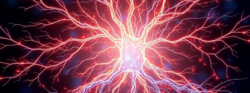
- Neurons: Highly specialized cells for impulse conduction. Three types by function: sensory (afferent), interneurons (association), and motor (efferent). Sensory neurons carry impulses from receptors to the CNS; interneurons integrate sensory information and send signals to motor neurons; and motor neurons transmit impulses from the CNS to effectors (muscles or glands).
- Neuroglia: Support, protect, and nourish neurons. They assist in signal transmission and maintain homeostasis. Different types of neuroglia cells vary in function and location (e.g., oligodendrocytes are in the CNS, Schwann cells are in the PNS).
- Spinal Cord Structure: Protected by the bony vertebral column and extends from the medulla oblongata to the L2 vertebrae. Consists of white matter (bundles of axons carrying sensory/motor information) and gray matter (cell bodies and axons of interneurons).
- Spinal Meninges: Layers of connective tissue surrounding the spinal cord to protect and cushion it. Consists of dura mater (outermost), arachnoid mater, and pia mater (innermost). Spaces between meninges are filled with interstitial fluid and cerebrospinal fluid.
- Reflexes: Rapid, automatic responses to stimuli. The purpose of reflexes is to maintain homeostasis. The reflex arc is the pathway an impulse takes to produce a reflex action, with five basic components: sensory receptor, sensory neuron, integrating center, motor neuron, and effector.
- Reflex Types: Somatic reflexes (skeletal muscle contraction) and autonomic reflexes (smooth/cardiac muscle or glands).
- Reflex Testing Procedures: Various procedures for testing reflexes, including Achilles tendon, patellar, biceps, triceps, plantar, salivary, pupillary, and diving reflexes.
Neuroglia
- Neuroglia (glial cells) perform various functions supporting and protecting neurons.
- Different types of neuroglia have different functions depending on their location (e.g. oligodendrocytes in the CNS and Schwann cells in the PNS).
Spinal Cord Histology
- Observe the cross-section of the spinal cord in vertebra, identify structures like the anterior and posterior gray horns, central canal, and meninges.
- Types of vertebra can be determined by observing the structural features.
Spinal Nerves and Plexuses
- Spinal nerves emerge from the spinal cord, passing through intervertebral foramina to exit the spinal column.
- Nerves are bundles of axons from multiple neurons. Posterior roots contain sensory axons and anterior roots contain motor axons forming mixed nerves when joined.
- Beyond the thoracic spinal nerves, anterior rami form plexuses (networks of motor nerves).
Reflexes
- Reflexes are rapid responses to stimuli, involving a reflex arc - sensory receptor; sensory neuron; integrating center; motor neuron; effector.
- Reflexes are classified as somatic (skeletal muscle contraction) or autonomic (smooth/cardiac muscle or glands).
Studying That Suits You
Use AI to generate personalized quizzes and flashcards to suit your learning preferences.


