Podcast
Questions and Answers
What are the two main subdivisions of the nervous system?
What are the two main subdivisions of the nervous system?
- Voluntary and Involuntary
- Autonomic and Somatic
- Sympathetic and Parasympathetic
- Central and Peripheral (correct)
The brain is located outside the skull.
The brain is located outside the skull.
False (B)
What are the three basic functions of the nervous system?
What are the three basic functions of the nervous system?
Sensory function, integration, and motor function
The spinal cord is encircled by the bones of the ______.
The spinal cord is encircled by the bones of the ______.
Match the following components of the nervous system with their description:
Match the following components of the nervous system with their description:
Which of the following is NOT a component of the nervous system?
Which of the following is NOT a component of the nervous system?
Neuroglia are responsible for transmitting nerve impulses.
Neuroglia are responsible for transmitting nerve impulses.
What is the role of neurotransmitters in the nervous system?
What is the role of neurotransmitters in the nervous system?
What is the primary function of sensory neurons?
What is the primary function of sensory neurons?
The axon carries impulses towards the cell body of a neuron.
The axon carries impulses towards the cell body of a neuron.
What are the two main types of tissue in the nervous system?
What are the two main types of tissue in the nervous system?
The region between myelin sheaths on an axon is called the ______.
The region between myelin sheaths on an axon is called the ______.
Match the following types of neurons with their functions:
Match the following types of neurons with their functions:
What does the cell body of a neuron contain that is similar to other cell types?
What does the cell body of a neuron contain that is similar to other cell types?
Neurons undergo mitosis regularly to replicate.
Neurons undergo mitosis regularly to replicate.
What supports neurons in the nervous system?
What supports neurons in the nervous system?
The swelling at the end of an axon where communication occurs is called the ______.
The swelling at the end of an axon where communication occurs is called the ______.
Which of the following is primarily the functional unit of the nervous system?
Which of the following is primarily the functional unit of the nervous system?
Which type of neuron has one main dendrite and one axon?
Which type of neuron has one main dendrite and one axon?
What type of nerve is involved in voluntary and reflex skeletal muscle contraction?
What type of nerve is involved in voluntary and reflex skeletal muscle contraction?
Motor neurons transmit impulses from sensory receptors to the central nervous system.
Motor neurons transmit impulses from sensory receptors to the central nervous system.
Irritability in neurons refers to their ability to transmit impulses to other neurons.
Irritability in neurons refers to their ability to transmit impulses to other neurons.
What is the primary function of astrocytes in the central nervous system?
What is the primary function of astrocytes in the central nervous system?
What ion is considered the major positive ion in extracellular fluid?
What ion is considered the major positive ion in extracellular fluid?
The condition where a neuron is unable to conduct a nerve impulse is known as the _____ period.
The condition where a neuron is unable to conduct a nerve impulse is known as the _____ period.
The _____ serves to speed the transmission of impulses along myelinated axons.
The _____ serves to speed the transmission of impulses along myelinated axons.
Match the neurotransmitter with its primary function:
Match the neurotransmitter with its primary function:
Match the type of neuron with its function:
Match the type of neuron with its function:
Which of the following cells is responsible for forming myelin around axons in the peripheral nervous system?
Which of the following cells is responsible for forming myelin around axons in the peripheral nervous system?
Which gates open during depolarization?
Which gates open during depolarization?
Neurotransmitters are stored in the dendrites of neurons until released.
Neurotransmitters are stored in the dendrites of neurons until released.
Grey matter is primarily composed of myelinated axons.
Grey matter is primarily composed of myelinated axons.
What happens to neurotransmitters immediately after they stimulate the postsynaptic neuron?
What happens to neurotransmitters immediately after they stimulate the postsynaptic neuron?
What role do microglia play in the central nervous system?
What role do microglia play in the central nervous system?
The _____ cells support neurons in PNS ganglia.
The _____ cells support neurons in PNS ganglia.
During repolarization, _____ ions leave the neuron.
During repolarization, _____ ions leave the neuron.
Match the type of glial cell with its function:
Match the type of glial cell with its function:
Match the type of neuron with its description:
Match the type of neuron with its description:
Which type of neuron is mainly located in the brain and spinal cord?
Which type of neuron is mainly located in the brain and spinal cord?
Which ion is higher inside a neuron at resting potential?
Which ion is higher inside a neuron at resting potential?
Autonomic afferent nerves originate from skin and respond to external stimuli.
Autonomic afferent nerves originate from skin and respond to external stimuli.
The synaptic cleft is the space where neurotransmitters are stored before release.
The synaptic cleft is the space where neurotransmitters are stored before release.
What is the myelin sheath and its function?
What is the myelin sheath and its function?
What type of ion channels open in response to membrane depolarization?
What type of ion channels open in response to membrane depolarization?
A bundle of nerve fibers in the CNS is called a _____ .
A bundle of nerve fibers in the CNS is called a _____ .
An impulse is initiated by stimulation of _____ nerve endings.
An impulse is initiated by stimulation of _____ nerve endings.
What ensures smooth communication and homeostasis within the nervous system?
What ensures smooth communication and homeostasis within the nervous system?
Flashcards
Nervous System Organization
Nervous System Organization
The nervous system is organized into two main divisions: the central nervous system (CNS) and the peripheral nervous system (PNS). The CNS includes the brain and spinal cord, while the PNS includes nerves, ganglia, and sensory receptors.
Central Nervous System (CNS)
Central Nervous System (CNS)
The central nervous system (CNS) is the control center of the body, consisting of the brain and spinal cord.
Peripheral Nervous System (PNS)
Peripheral Nervous System (PNS)
The peripheral nervous system (PNS) consists of nerves, ganglia, and sensory receptors that connect the CNS to the rest of the body.
Brain Location
Brain Location
Signup and view all the flashcards
Spinal Cord Location
Spinal Cord Location
Signup and view all the flashcards
Nervous System Functions
Nervous System Functions
Signup and view all the flashcards
Sensory Function
Sensory Function
Signup and view all the flashcards
Neuron Structure
Neuron Structure
Signup and view all the flashcards
Neuron function
Neuron function
Signup and view all the flashcards
Dendrites function
Dendrites function
Signup and view all the flashcards
Axon function
Axon function
Signup and view all the flashcards
Axon terminal function
Axon terminal function
Signup and view all the flashcards
Sensory neuron
Sensory neuron
Signup and view all the flashcards
Interneuron function
Interneuron function
Signup and view all the flashcards
Motor neuron function
Motor neuron function
Signup and view all the flashcards
Association Neuron
Association Neuron
Signup and view all the flashcards
Motor Neuron
Motor Neuron
Signup and view all the flashcards
Multipolar Neuron
Multipolar Neuron
Signup and view all the flashcards
Bipolar Neuron
Bipolar Neuron
Signup and view all the flashcards
Unipolar Neuron
Unipolar Neuron
Signup and view all the flashcards
Afferent Neuron
Afferent Neuron
Signup and view all the flashcards
Efferent Neuron
Efferent Neuron
Signup and view all the flashcards
Interneuron
Interneuron
Signup and view all the flashcards
Neuroglia
Neuroglia
Signup and view all the flashcards
Astrocytes
Astrocytes
Signup and view all the flashcards
Microglia
Microglia
Signup and view all the flashcards
Oligodendrocytes
Oligodendrocytes
Signup and view all the flashcards
Ependymal Cells
Ependymal Cells
Signup and view all the flashcards
Schwann Cells
Schwann Cells
Signup and view all the flashcards
Somatic Nerve
Somatic Nerve
Signup and view all the flashcards
Autonomic Nerve
Autonomic Nerve
Signup and view all the flashcards
Sympathetic Nervous System
Sympathetic Nervous System
Signup and view all the flashcards
Parasympathetic Nervous System
Parasympathetic Nervous System
Signup and view all the flashcards
Mixed Nerve
Mixed Nerve
Signup and view all the flashcards
Sensory Receptor
Sensory Receptor
Signup and view all the flashcards
Nerve Impulse
Nerve Impulse
Signup and view all the flashcards
Neuron Irritability
Neuron Irritability
Signup and view all the flashcards
Neuron Conductivity
Neuron Conductivity
Signup and view all the flashcards
Synapse
Synapse
Signup and view all the flashcards
Neurotransmitter
Neurotransmitter
Signup and view all the flashcards
Resting Membrane Potential
Resting Membrane Potential
Signup and view all the flashcards
Depolarization
Depolarization
Signup and view all the flashcards
Repolarization
Repolarization
Signup and view all the flashcards
Refractory Period
Refractory Period
Signup and view all the flashcards
Study Notes
Nervous System Overview
- The nervous system is the major control, regulatory, and communication system of the body
- It's the center of mental activities like thoughts, learning, and memory.
- It works together with the endocrine system to regulate and maintain homeostasis
- The nervous system is made up of: brain, cranial nerves & their branches, the spinal cord, spinal nerves & their branches, ganglia, enteric plexuses, and sensory receptors.
Organization of the Nervous System
- The nervous system is organized into two main subdivisions: central nervous system (CNS) and peripheral nervous system (PNS)
- CNS consists of the brain and the spinal cord
- The brain is located in the skull and contains 100 billion neurons
- The spinal cord is surrounded by the vertebral column and contains 100 million neurons connected to the brain.
- The brain and spinal cord are continuous at the foramen magnum
- PNS consists of cranial nerves & branches, spinal nerves & branches, ganglia, enteric plexuses in the small intestine, and sensory receptors in the skin
Peripheral Nervous System (PNS)
- Cranial nerves and their branches
- Spinal nerves and their branches
- Ganglia
- Enteric plexuses in the small intestines
- Sensory receptors in the skin
Function of the Nervous System
- Sensory Function: sensory receptors detect internal or external stimuli. Cranial and spinal nerves carry this sensory information to the brain & spinal cord.
- Integrative Function: the nervous system integrates/processes information via analyzing, storing, and making decisions for appropriate responses.
- Motor Function: carry out the processed response to effectors, (muscle & glands). Stimulations to the effector lead to muscle contraction and gland secretions.
Nervous Tissue
- Two main types of tissue: neurons and neuroglia/glial cells
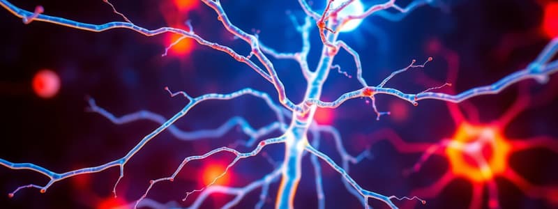
- Neurons are the functional units of the nervous system. They are nerve cells that conduct nerve impulses. They consist of a cell body, one axon, and many dendrites
- Require oxygen and glucose for survival
- Do not undergo mitosis
- Supported by connective tissue—neuroglia
- Neuron cell bodies form the grey matter of the nervous system, found in the periphery of the brain and center of the spinal cord. Groups of cell bodies are called nuclei in the CNS and ganglia in the PNS
- Axons and dendrites form the white matter of the nervous system. Axons and dendrites are extensions of cell bodies, referred to as nerves or nerve fiber outside the brain & spinal cord.
Structure of Neurons (continued)
- Dendrites are shorter and branching extensions. Their function is to receive and carry incoming impulses towards the cell body from another neuron.
- Axons function in carrying impulses.
Structure of Neurons (continued)
- Axons are extensions of cell bodies that carry impulses away from the cell body to another neuron; typically found deep in the brain and nerves.
- Bundles of axons in the CNS are called tracts
- Axons are typically covered in a myelin sheath. Myelin is a white, fatty substance that speeds up the transmission of nerve impulses.
- The unmyelinated region between the myelin is called the nodes of Ranvier.
Synapse and Neurotransmitters
A synapse is the point where the nerve impulse passes from one neuron to another or effector cell.
- Neurotransmitters are chemical substances which release at the synaptic cleft.
Synapse continued
- Synaptic knobs (or terminal buttons): small swellings at the end of axons in nerve fibers; are close to dendrites and cell body of postsynaptic neurons
- Synaptic vesicles: small sacs that contain neurotransmitters in synaptic knobs
Synapse continued
Neurotransmitters are chemical substances which allow transmission of signals from one neuron to the next across synapses; released into the synaptic cleft.
Types of Neurons (Function)
- Sensory neurons: carry messages from the receptors to the Central Nervous System (CNS)
- Interneurons/Association neuron: located in the CNS, receives signals from many neurons, carry out integrative functions (make decisions on responses)
- Motor neurons: carry messages from the CNS to effectors (muscles & glands)
Types of Neurons (Structure)
- Multipolar neuron: have several dendrites & 1 axon; located in the brain and spinal cord
- Bipolar neuron: have 1 main dendrite & 1 axon; found in the eye, ear, & nose
- Unipolar neuron: have 1 axon extension and the cell body is on one side of the axon.
Neuroglia
- Also known as glia, these cells make up about ½ of the volume of the CNS. They do not conduct nerve impulses.
- Their functions include:
- supplying nutrients and oxygen
- surrounding neurons and holding them in place
- destroying pathogens and removing dead neurons
- maintaining homeostasis in the interstitial fluids.
Types of Neuroglia
- Oligodendrocytes: produce and maintain myelin sheath around the axons of CNS neurons.
- Astrocytes: cover the blood vessels in the CNS and form the blood-brain barrier, protecting the brain from potentially harmful substances; Actively involved in the formation & circulation of cerebrospinal fluid (CSF).
- Ependymal cells: actively involved in formation and circulation of cerebrospinal fluid (CSF)
- Microglia: Protect CNS cells from disease by engulfing invading microbes and migrate to areas of injured nerve tissue; clear away debris of dead cells.
Type of Neuroglia (PNS)
- Schwann cells: form the myelin sheath around axons in the PNS and play a role in nerve fiber regeneration
- Satellite cells: support neurons in PNS ganglia and regulate materials exchange between neurons and interstitial fluid
Myelination
- Myelin sheath: white, fatty substance surrounding the axon, covering axons.
- Myelinated axon: axon with myelin
- Unmyelinated axon: axon without myelin
- Nodes of Ranvier: unmyelinated regions between myelin sheath. Function: speed up transmission of nerve impulses.
White and Grey Matter
- White matter: primarily myelinated axons; whitish color due to myelin
- Grey matter: contains neuronal cell bodies, dendrites, unmyelinated axons & axon terminals, neuroglia; appear grey in color
Nerves
- Nerves consist of neurons bundled together. Bundles of nerve fibers in the CNS are referred to as tracts
- Nerve coverings: Endoneurium- covers individual axons or nerve fibers ; Perineurium- encases fascicles (bundles of nerve fibers); Epineurium- encases the entire nerve
Types of Nerves
- Sensory (afferent) nerve: carries information from the body to the spinal cord, then the brain
- Sensory receptors: specialized endings that respond to various stimuli (inside or outside of the body)
- Somatic sense: sensory information from skin; feels pain, touch, heat, and cold
- Proprioceptors sense: information from muscles and joints used for posture and balance.
- Special senses: vision, hearing, balance, smell and taste
- Autonomic afferent nerve: Originates in internal organs, glands, and tissues. E.g. baroreceptors control blood pressure, chemoreceptors control respiration
- Motor (efferent) nerve: originates in brain, spinal cord, or autonomic ganglia. Transmits impulses to effector organs (muscle & glands)
- Two types of motor nerves: somatic nerves involved in voluntary and reflex skeletal muscle contractions, and autonomic nerves (sympathetic and parasympathetic) are involved in cardiac & smooth muscle contractions, and glandular secretions
- Mixed nerves: consist of both sensory and motor fibers
Action Potentials (The Nerve Impulse)
- An impulse is initiated by stimulation of sensory nerve endings (sensory receptors).
- Action potential is due to movement of ions across the nerve cell membrane.
- Two major characteristics of neurons: irritability (ability to initiate nerve impulses in response to stimuli from inside or outside the body) and conductivity (ability to transmit impulses).
Action Potentials Con't
- Ions move along axon membrane, reach synapse, neurotransmitters release, allowing communication between neurons and other cells.
- When a neuron reaches a certain stimulation level, an electrical impulse is generated.
- The principle ions involved are sodium (major positive ion in extracellular fluid) and potassium (major positive ion in intracellular fluid).
- Membrane potential and presence of voltage-gated channels for Na+ and K+ are essential for action potential generation.
- During action potential, voltage-gated Na+ and K+ channels open in sequence.
Generation of Action Potentials
- Resting membrane potential: a neuron not conducting an impulse
- Depolarization: a very rapid process when a nerve cell is stimulated, Na+ channels open, causing inflow of Na+ ions from the extracellular fluid into the neuron that changes the neuronal membrane to be positive.
- Repolarization: occurs at the end of the depolarization phase; K+ gates open; K+ ions leave the neuron; K+ gates close slowly
- Refractory Period: condition where a neuron is recovering; Na channels are closed, and K+ channels are open
- Following action potential, the neuron temporarily cannot conduct another impulse until the Na+ gates have closed.
Action Potentials continued
- An action potential travels down the nerve axon to the terminal region.
- At the terminal, the action potential causes neurotransmitter molecules to be released into the synapse. These molecules stimulate receptors in the next cell and transmit the action potential.
- Local anesthetics and certain neurotoxins prevent opening of voltage-gated Na+ channels preventing nerve impulses from passing.
Transmission of Impulse in Synaptic Cleft
- Once the impulse reaches the presynaptic neuron, synaptic vesicles in the synaptic knobs will release neurotransmitters into the synaptic cleft (exocytosis).
- Neurotransmitter binds to specific receptors on the postsynaptic neuron or effector organ (e.g., muscle).
- Neurotransmitters initiate an electrical response in the postsynaptic neuron, to either excite or inhibit.
- The action is short-lived because postsynaptic receptors are inactivated by enzymes or taken back into the synaptic knob.
Neurotransmitters
- There are about 100 types of neurotransmitters.
- Acetylcholine: most common in CNS & PNS; involved in arousal, dreaming, regulating mood.
- Norepinephrine (noradrenaline): involved in arousal (awakening from deep sleep), dreaming, and regulating mood.
- Dopamine: regulates skeletal muscle tone.
- Serotonin: chemical that allows transmission of signals from one neuron to the next across synapses.
- Histamine: chemical involved in CNS & PNS, other roles.
Studying That Suits You
Use AI to generate personalized quizzes and flashcards to suit your learning preferences.


