Podcast
Questions and Answers
What is the primary function of the thalamus in the brain?
What is the primary function of the thalamus in the brain?
- Controls voluntary muscle movement
- Regulates pituitary hormone secretion
- Integrates sensory inputs (correct)
- Maintains body temperature
Which of the following is true about the sympathetic nervous system?
Which of the following is true about the sympathetic nervous system?
- It stimulates digestion and gut activity.
- It promotes relaxation and conservation of energy.
- It operates only during sleep.
- It is responsible for 'fight or flight' responses. (correct)
What type of neuron carries impulses from the CNS to effectors?
What type of neuron carries impulses from the CNS to effectors?
- Efferent neuron (correct)
- Afferent neuron
- Sensory neuron
- Interneuron
Which division of the peripheral nervous system is responsible for voluntary control of skeletal muscles?
Which division of the peripheral nervous system is responsible for voluntary control of skeletal muscles?
Which of the following actions is NOT associated with the parasympathetic nervous system?
Which of the following actions is NOT associated with the parasympathetic nervous system?
The balance between sympathetic and parasympathetic nervous systems results in what?
The balance between sympathetic and parasympathetic nervous systems results in what?
The autonomic nervous system consists of which two major divisions?
The autonomic nervous system consists of which two major divisions?
What is a function of the autonomic nervous system?
What is a function of the autonomic nervous system?
What is the resting membrane potential of a typical resting cell?
What is the resting membrane potential of a typical resting cell?
What is primarily responsible for maintaining the concentration gradient of Na+ and K+ across the cell membrane?
What is primarily responsible for maintaining the concentration gradient of Na+ and K+ across the cell membrane?
During depolarization, which ion primarily floods into the cell?
During depolarization, which ion primarily floods into the cell?
What is the typical peak value of the action potential after depolarization?
What is the typical peak value of the action potential after depolarization?
What occurs during repolarization of an action potential?
What occurs during repolarization of an action potential?
What is the role of the synaptic cleft in a chemical synapse?
What is the role of the synaptic cleft in a chemical synapse?
Which type of cell is involved in transmitting signals away from a synapse?
Which type of cell is involved in transmitting signals away from a synapse?
What is the overshoot of the resting membrane potential called?
What is the overshoot of the resting membrane potential called?
What type of synapse directly passes action potentials between neurons?
What type of synapse directly passes action potentials between neurons?
What happens to the resting membrane potential after an action potential?
What happens to the resting membrane potential after an action potential?
What is the primary function of acetylcholinesterase (AChE) at the synapse?
What is the primary function of acetylcholinesterase (AChE) at the synapse?
Which type of acetylcholine receptor directly opens membrane channels upon binding with acetylcholine?
Which type of acetylcholine receptor directly opens membrane channels upon binding with acetylcholine?
What effect can binding of acetylcholine to muscarinic receptors have?
What effect can binding of acetylcholine to muscarinic receptors have?
Which class of neurotransmitters does acetylcholine belong to?
Which class of neurotransmitters does acetylcholine belong to?
What is the result of spatial summation of excitatory postsynaptic potentials (EPSPs)?
What is the result of spatial summation of excitatory postsynaptic potentials (EPSPs)?
How are acetate and choline reused after acetylcholine is inactivated?
How are acetate and choline reused after acetylcholine is inactivated?
Which of the following is NOT a characteristic of muscarinic ACh receptors?
Which of the following is NOT a characteristic of muscarinic ACh receptors?
What role do cholinergic fibers play in the nervous system?
What role do cholinergic fibers play in the nervous system?
What triggers the release of neurotransmitters from presynaptic neurons?
What triggers the release of neurotransmitters from presynaptic neurons?
What occurs at the postsynaptic membrane after neurotransmitter binding?
What occurs at the postsynaptic membrane after neurotransmitter binding?
What is the result of a strong enough excitatory postsynaptic potential (EPSP)?
What is the result of a strong enough excitatory postsynaptic potential (EPSP)?
What is an inhibitory postsynaptic potential (IPSP)?
What is an inhibitory postsynaptic potential (IPSP)?
How can the strength of stimuli affect depolarization in postsynaptic neurons?
How can the strength of stimuli affect depolarization in postsynaptic neurons?
What happens when multiple presynaptic neurons release EPSPs simultaneously?
What happens when multiple presynaptic neurons release EPSPs simultaneously?
What is responsible for the removal of neurotransmitters from the synapse?
What is responsible for the removal of neurotransmitters from the synapse?
What occurs when neurotransmitters are released into the synaptic cleft?
What occurs when neurotransmitters are released into the synaptic cleft?
What is the main function of the nervous system?
What is the main function of the nervous system?
Which of the following structures is primarily responsible for receiving stimuli?
Which of the following structures is primarily responsible for receiving stimuli?
What type of neuron carries signals from sensory receptors to the central nervous system?
What type of neuron carries signals from sensory receptors to the central nervous system?
Which type of neuroglia is responsible for forming myelin sheaths in the peripheral nervous system (PNS)?
Which type of neuroglia is responsible for forming myelin sheaths in the peripheral nervous system (PNS)?
Which part of the nervous system consists of the brain and spinal cord?
Which part of the nervous system consists of the brain and spinal cord?
What is the primary role of interneurons in the nervous system?
What is the primary role of interneurons in the nervous system?
Which centers are controlled by the medulla oblongata?
Which centers are controlled by the medulla oblongata?
Which part of the brain is responsible for balance and posture?
Which part of the brain is responsible for balance and posture?
The forebrain includes which of the following structures?
The forebrain includes which of the following structures?
What function do astrocytes serve in the central nervous system?
What function do astrocytes serve in the central nervous system?
Which lobe of the brain is associated with decision-making and planning?
Which lobe of the brain is associated with decision-making and planning?
What is the primary role of the spinal cord?
What is the primary role of the spinal cord?
Which structure serves as a protective barrier for the central nervous system?
Which structure serves as a protective barrier for the central nervous system?
Which neuroglial cell type is involved in defending the nervous system?
Which neuroglial cell type is involved in defending the nervous system?
Which division of the peripheral nervous system is responsible for carrying impulses from receptors to the central nervous system?
Which division of the peripheral nervous system is responsible for carrying impulses from receptors to the central nervous system?
What is the primary role of the hypothalamus in the brain?
What is the primary role of the hypothalamus in the brain?
In what situation is the sympathetic nervous system primarily activated?
In what situation is the sympathetic nervous system primarily activated?
What distinguishes the function of the somatic nervous system from the autonomic nervous system?
What distinguishes the function of the somatic nervous system from the autonomic nervous system?
Which of the following is an action associated with the sympathetic nervous system?
Which of the following is an action associated with the sympathetic nervous system?
How do the sympathetic and parasympathetic systems generally work together?
How do the sympathetic and parasympathetic systems generally work together?
Which type of sensory neuron transmits information from internal organs to the central nervous system?
Which type of sensory neuron transmits information from internal organs to the central nervous system?
What is the impact of the autonomic nervous system on cardiac muscles?
What is the impact of the autonomic nervous system on cardiac muscles?
What enzyme is responsible for the breakdown of acetylcholine in the synaptic cleft?
What enzyme is responsible for the breakdown of acetylcholine in the synaptic cleft?
Which receptors directly open membrane channels when acetylcholine binds to them?
Which receptors directly open membrane channels when acetylcholine binds to them?
What occurs when acetylcholine binds to muscarinic receptors?
What occurs when acetylcholine binds to muscarinic receptors?
What is a characteristic of nicotinic acetylcholine receptors?
What is a characteristic of nicotinic acetylcholine receptors?
What is the role of acetylcholinesterase in neurotransmission?
What is the role of acetylcholinesterase in neurotransmission?
Which class of neurotransmitters does include serotonin and norepinephrine?
Which class of neurotransmitters does include serotonin and norepinephrine?
What is the primary effect of excitatory postsynaptic potentials (EPSPs)?
What is the primary effect of excitatory postsynaptic potentials (EPSPs)?
What happens to acetate and choline after acetylcholine is inactivated?
What happens to acetate and choline after acetylcholine is inactivated?
What is the primary ion responsible for the depolarization phase of an action potential?
What is the primary ion responsible for the depolarization phase of an action potential?
What mechanism is involved in restoring the resting membrane potential during repolarization?
What mechanism is involved in restoring the resting membrane potential during repolarization?
What is the typical resting membrane potential (RMP) of a neuron?
What is the typical resting membrane potential (RMP) of a neuron?
Which structure conducts signals away from a synapse?
Which structure conducts signals away from a synapse?
What type of synapse uses neurotransmitters to transmit signals?
What type of synapse uses neurotransmitters to transmit signals?
What event occurs when an excitatory postsynaptic potential (EPSP) is strong enough?
What event occurs when an excitatory postsynaptic potential (EPSP) is strong enough?
What occurs during hyperpolarization of a neuron?
What occurs during hyperpolarization of a neuron?
What action does the Na+/K+ ATPase pump primarily accomplish?
What action does the Na+/K+ ATPase pump primarily accomplish?
What best describes the 'all-or-none response' of action potentials?
What best describes the 'all-or-none response' of action potentials?
What is the function of gap junctions in electrical synapses?
What is the function of gap junctions in electrical synapses?
What role do dendrites play in a neuron?
What role do dendrites play in a neuron?
Which type of neuron conducts signals from sensory receptors to the central nervous system?
Which type of neuron conducts signals from sensory receptors to the central nervous system?
What is the main function of astrocytes in the central nervous system?
What is the main function of astrocytes in the central nervous system?
Which part of the nervous system is responsible for involuntary reflex actions?
Which part of the nervous system is responsible for involuntary reflex actions?
What is the primary role of the myelin sheath?
What is the primary role of the myelin sheath?
What part of the nervous system contains cranial and spinal nerves?
What part of the nervous system contains cranial and spinal nerves?
Which structure is not part of the brainstem?
Which structure is not part of the brainstem?
What is one of the functions performed by the medulla oblongata?
What is one of the functions performed by the medulla oblongata?
Which part of the cerebrum is responsible for high-level functions such as language and memory?
Which part of the cerebrum is responsible for high-level functions such as language and memory?
What is the role of microglia in the nervous system?
What is the role of microglia in the nervous system?
What type of neuron connects afferent and efferent neurons within the central nervous system?
What type of neuron connects afferent and efferent neurons within the central nervous system?
Which lobe of the brain is primarily involved in planning and decision-making?
Which lobe of the brain is primarily involved in planning and decision-making?
What function do Schwann cells serve in the peripheral nervous system?
What function do Schwann cells serve in the peripheral nervous system?
What is the primary function of the hypothalamus?
What is the primary function of the hypothalamus?
What is the primary role of calcium ions (Ca2+) during neurotransmitter release?
What is the primary role of calcium ions (Ca2+) during neurotransmitter release?
Which potential is characterized by the inward diffusion of Na+ and results in depolarization?
Which potential is characterized by the inward diffusion of Na+ and results in depolarization?
What occurs when EPSPs produced by multiple presynaptic neurons coincide?
What occurs when EPSPs produced by multiple presynaptic neurons coincide?
What is the consequence of the postsynaptic membrane not reaching the threshold potential?
What is the consequence of the postsynaptic membrane not reaching the threshold potential?
Which process describes the periods of depolarization and hyperpolarization based on neurotransmitter activity on the postsynaptic membrane?
Which process describes the periods of depolarization and hyperpolarization based on neurotransmitter activity on the postsynaptic membrane?
What triggers the removal of neurotransmitters from the synapse?
What triggers the removal of neurotransmitters from the synapse?
How does the strength of a stimulus affect postsynaptic depolarization?
How does the strength of a stimulus affect postsynaptic depolarization?
What defines an inhibitory postsynaptic potential (IPSP)?
What defines an inhibitory postsynaptic potential (IPSP)?
Flashcards are hidden until you start studying
Study Notes
Function of Nervous System
- The nervous system detects internal and external changes.
- It interprets these changes and responds appropriately.
- The nervous system is also responsible for learning, memory, and intelligence.
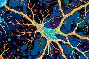
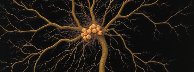
Neuron
- The neuron is the basic unit of the nervous system.
- It consists of a cell body, dendrites, and an axon.
- Dendrites receive stimuli.
- Axons conduct nerve impulses away from the cell body.
- Axons are covered in a myelin sheath, which speeds up signal transmission.
- Myelin sheaths are interrupted by nodes of Ranvier, which help to facilitate signal transmission.
Types of Neurons
- Afferent (sensory) neurons transmit signals from sensory receptors to the CNS.
- Efferent (motor) neurons transmit signals from the CNS to effector organs (muscles and glands).
- Interneurons connect afferent and efferent neurons within the CNS.
Neuroglia
- Neuroglia support and assist neuron function.
- They physically support neurons, form myelin sheaths, and regulate the extracellular fluid composition.
Types of Neuroglia
- Schwann cells form myelin sheaths for axons in the PNS.
- Oligodendrocytes form myelin sheaths for axons in the CNS.
- Astrocytes help to form the blood-brain barrier.
- Microglia function as phagocytic cells for defense.
Nervous System Organization
- The nervous system consists of the central nervous system (CNS) and the peripheral nervous system (PNS).
- The CNS includes the brain and the spinal cord.
- The PNS includes nerves that extend from the CNS to the muscles, glands, and sensory organs.
Spinal Cord and Reflexes
- The spinal cord transmits nerve impulses to and from the brain.
- It also has reflex centers that control involuntary actions.
Brain
- The brain is responsible for integrating information and higher-level functions.
- It consists of the forebrain, brainstem, and cerebellum.
Brainstem
- The brainstem controls involuntary movements.
- It also integrates cardiovascular and respiratory activity.
- The medulla oblongata is the lowest part of the brainstem; it contains the following centers:
- Cardiac center to control heart rate and strength.
- Respiratory center to control rate and depth of breathing.
- Vasomotor center to control blood vessel diameter.
- Autonomic reflex center to induce vomiting, coughing, and sneezing.
Cerebellum
- The cerebellum is involved in maintaining posture, balance, and skeletal muscle function.
- It also plays a role in some forms of learning.
Forebrain
- The forebrain consists of the cerebrum and the diencephalon.
Cerebrum
- The cerebrum has two hemispheres.
- The cerebral cortex is the outermost layer of the cerebrum, and it is responsible for higher brain functions, including memory, learning, language, planning, and decision-making.
- The cerebrum has four lobes: frontal, parietal, occipital, and temporal.
Diencephalon
- The thalamus integrates sensory inputs.
- The hypothalamus regulates pituitary hormone secretion and controls homeostatic functions, such as water balance and body temperature.
Peripheral Nervous System
- The peripheral nervous system carries impulses from sensory receptors to the CNS and from the CNS to effector organs.
- It is divided into the sensory (afferent) and motor (efferent) divisions.
Sensory (Afferent) Division
- The sensory (afferent) division carries impulses from the receptors to the CNS.
- It consists of somatic and visceral components.
- Somatic: stimuli originates from the skin and skeletal muscles.
- Visceral: stimuli originates from internal organs.
Motor (Efferent) Division
- The motor (efferent) division carries impulses from the CNS to effectors.
- It is divided into the somatic and autonomic nervous systems.
- Somatic: voluntary control of skeletal muscles.
- Autonomic: involuntary control of cardiac muscle, smooth muscle, and glands.
- The autonomic nervous system consists of the sympathetic and parasympathetic nervous systems.
Autonomic Nervous System
- The autonomic nervous system is part of the PNS.
- It consists of sympathetic and parasympathetic nerves.
- These two divisions act antagonistically.
- The balance between the two divisions regulates organ function.
Sympathetic Nervous System
- The sympathetic nervous system is responsible for the "fight-or-flight" response.
- It prepares the body to deal with stress or strong emotions.
- Its functions include:
- Increasing heart rate.
- Dilating bronchi.
- Dilating arteries to muscles.
- Constricting arteries to the gut.
- Converting glycogen to glucose.
- Increasing epinephrine secretion.
Parasympathetic Nervous System
- The parasympathetic nervous system promotes conservation and relaxation.
- Its effects oppose the effects of the sympathetic system.
- It is associated with the body's quiet state and well-being.
Neuron Function
- Neurons transmit electrochemical signals.
- This is accomplished through the generation and propagation of action potentials.
Resting Membrane Potential (RMP)
- The RMP is a negative charge difference across the cell membrane.
- This is maintained by the Na+/K+ ATPase pump.
- The resting membrane is more permeable to potassium (K+) than sodium (Na+).
- The RMP is approximately -70 mV.
Na+/K+ ATPase Pump
- The Na+/K+ ATPase pump maintains and establishes the concentration gradient of Na+ and K+ across the cell membrane.
- It pumps three Na+ ions out of the cell for every two K+ ions pumped in.
Action Potential (AP)
- Action potentials are the electrochemical signals that travel along neurons.
- They involve a rapid, localized change in the RMP that results in a brief reversal of the membrane potential.
- An AP is characterized by a rapid depolarization phase, followed by a repolarization phase and a brief hyperpolarization phase.
- AP's are all-or-none responses.
- They can travel long distances without diminishing in strength.
AP Phases
- Depolarization: The cell membrane becomes more permeable to Na+ ions, which flow into the cell due to their concentration gradient. This causes the RMP to move toward a positive value (+40 mV).
- Repolarization: The cell membrane becomes less permeable to Na+ and more permeable to K+ ions, which move out of the cell due to their concentration gradient. This causes the RMP to return to its resting value (-70 mV).
- Hyperpolarization: The membrane potential briefly becomes more negative than the RMP. This is caused by the K+ channels remaining open for a brief period after the repolarization phase.
Synapse
- A synapse is the junction between a neuron and another neuron, muscle, or gland cell.
- It is where neurotransmitters are released.
- Presynaptic neuron: The neuron that transmits signals toward the synapse.
- Postsynaptic neuron: The neuron that receives signals from the synapse.
- Synaptic knob: The terminal of the presynaptic neuron where neurotransmitters are stored.
- Synaptic cleft: The narrow gap between the pre- and postsynaptic neurons.
Types of Synapses
- Electrical Synapse: Direct transfer of action potential through gap junctions between neurons.
- Chemical Synapse: Transmission of signals by release of neurotransmitters from the presynaptic neuron to the postsynaptic neuron.
Neurotransmitter Release at a Chemical Synapse
- An action potential arrives at the presynaptic terminal.
- Voltage-gated Ca2+ channels open and Ca2+ flows into the presynaptic terminal.
- Ca2+ triggers the release of neurotransmitters from synaptic vesicles via exocytosis.
- Neurotransmitters diffuse across the synaptic cleft and bind to receptors on the postsynaptic membrane.
- Binding of the neurotransmitter to its receptor activates ion channels, causing a postsynaptic potential.
- After its action, neurotransmitters are removed from the synapse by enzymatic degradation or reuptake into the presynaptic terminal.
Synaptic Potentials
- Synaptic potentials are postsynaptic depolarizations or hyperpolarizations caused by neurotransmitters.
- Excitatory postsynaptic potential (EPSP): A depolarization of the postsynaptic membrane that brings the membrane potential closer to the threshold for firing an action potential.
- Inhibitory postsynaptic potential (IPSP): A hyperpolarization of the postsynaptic membrane that makes the membrane potential more negative and less likely to fire an action potential.
Summation of Synaptic Potentials
- Spatial summation: Multiple presynaptic neurons release neurotransmitters at the same time, and their EPSP's summate to reach the threshold for firing an action potential.
- Temporal summation: A single presynaptic neuron releases multiple neurotransmitters in rapid succession, and their EPSP's summate to reach the threshold for firing an action potential.
Neurotransmitters
- Acetylcholine (ACh): Important neurotransmitter at neuromuscular junctions.
- Biogenic Amines: Includes norepinephrine, epinephrine, and serotonin.
- Amino Acids: Includes glutamic acid and gamma-aminobutyric acid (GABA).
- Neuropeptides: Includes endorphins and substance P.
Acetylcholine (ACh)
- ACh is a common neurotransmitter at neuromuscular junctions.
- Cholinergic fibers release ACh.
- ACh is rapidly destroyed by the enzyme acetylcholinesterase (AChE) to terminate its effect at the synapse.
Types of ACh Receptors
- Nicotinic ACh receptors: Found at neuromuscular junctions. They are ionotropic, meaning they are directly linked to ion channels that open upon binding of ACh. This leads to depolarization and EPSPs.
- Muscarinic ACh receptors: Present in the heart, smooth muscle, and glands. They are metabotropic, meaning they indirectly activate second messenger systems. This can lead to either EPSPs or IPSPs.
Important Notes
- The brain is comprised of more glial cells than neurons.
- The nervous system is a vast and complex network that plays a vital role in regulating and coordinating the body's activities.
- There are a variety of diseases that affect the nervous system, including Alzheimer's disease, Parkinson's disease, multiple sclerosis, and stroke.
Function of Nervous System
- Senses changes in the body and external environment.
- Interprets changes.
- Responds to changes by initiating responses.
- Assimilates experiences for memory, learning, and intelligence.
Neurons
- Basic unit of the nervous system.
- Composed of:
- Cell body: Contains cytoplasm and a nucleus.
- Dendrites: Receive stimuli.
- Axon: Conducts nerve impulses away from the cell body.
- Wrapped with myelin sheath.
- Has nodes of Ranvier: Gaps between segments of myelin.
Types of Neurons
- Afferent (sensory) neurons: Conduct signals from sensory receptors to the central nervous system (CNS).
- Efferent (motor) neurons: Conduct signals from the CNS to effector organs (muscles and glands).
- Interneurons: Located within the CNS, connect afferent and efferent neurons.
Neuroglia
- Supporting cells in the nervous system that assist neurons.
- Functions:
- Physically support neurons.
- Form myelin sheaths.
- Regulate extracellular fluid composition.
Types of Neuroglia
- Schwann cells: Form myelin sheaths around peripheral nervous system (PNS) axons.
- Oligodendrocytes: Form myelin sheaths around CNS axons.
- Astrocytes: Help form the blood-brain barrier.
- Microglia: Phagocytic cells for defense.
Nervous System
- Central nervous system (CNS): Brain and spinal cord.
- Located inside the skull and vertebral column.
- Responsible for integration.
- Peripheral nervous system (PNS): Nerves extending from the CNS to muscles, glands, and sense organs.
- Includes cranial nerves and spinal nerves.
Spinal Cord, Spinal Nerves and Spinal Reflex
- Spinal cord:
- Carries sensory nerve fibers through the dorsal root.
- Carries motor nerve fibers through the ventral root.
- Functions of the spinal cord:
- Controls involuntary reflex actions.
- Provides a pathway for transmitting nerve impulses to and from the brain for voluntary actions.
Brain
- Divided into:
- Forebrain: Cerebrum and diencephalon.
- Brainstem: Midbrain, pons, and medulla oblongata.
- Cerebellum.
Brainstem
- Midbrain, pons, and medulla oblongata.
- Control involuntary movements.
- Integrating centers for cardiovascular and respiratory activity.
Medulla Oblongata
- Where the spinal cord arises.
- Functions:
- Cardiac center: Controls heart rate and force.
- Respiratory center: Controls breathing rate and depth.
- Vasomotor center: Controls blood vessel diameter.
- Autonomic reflex center: Induces vomiting, coughing, and sneezing.
Cerebellum
- Controls posture, balance, and skeletal muscle functions.
- Involved in some forms of learning.
Forebrain
- Cerebrum: Right and left cerebral hemispheres.
- Diencephalon: Thalamus and hypothalamus.
Cerebrum
- Right and left cerebral hemispheres.
- Cerebral cortex:
- Four lobes: frontal, parietal, occipital, and temporal.
Cerebrum Functions
- Highest sensory and motor center.
- Higher-level brain functions: memory, learning, language, planning, decision-making.
Motor and Sensory Areas of the Cerebral Cortex
- Specific areas are dedicated to sensory and motor functions.
Diencephalon
- Thalamus: Integrating centers for sensory inputs.
- Hypothalamus:
- Regulates pituitary hormone secretion.
- Controls homeostatic regulation (e.g., water balance, body temperature).
Peripheral Nervous System (PNS)
- Sensory (afferent) division: Carries impulses from receptors to the CNS.
- Somatic: Stimuli come from the skin and skeletal muscles.
- Visceral: Stimuli come from internal organs.
- Motor (efferent) division: Carries impulses from the CNS to effectors.
- Somatic: Voluntary control of skeletal muscles.
- Autonomic: Involuntary control of cardiac and smooth muscles and glands.
- Sympathetic nervous system.
- Parasympathetic nervous system.
Autonomic Nervous System (ANS)
- Part of the PNS.
- Consists of sympathetic and parasympathetic nerves.
- Acts antagonistically to each other.
- Maintains coordinated regulation of organs and glands.
Sympathetic Nervous System
- "Fight or flight" response.
- Activated during stress or strong emotions.
- Functions:
- Increases heart rate.
- Dilates bronchi.
- Dilates arteries to muscles.
- Constricts arteries to the gut.
- Converts glycogen to glucose.
- Increases epinephrine secretion.
Parasympathetic Nervous System
- "Conservation" response.
- Effects opposite to the sympathetic system.
- Concerned with body preservation in a quiet state.
Antagonistic Characters
- Sympathetic and parasympathetic systems have opposing effects.
The Autonomic Nervous System
- Sympathetic: Red
- Parasympathetic: Blue
How Does a Neuron Function?
- Neurons transmit electrochemical signals called action potentials.
Resting Membrane Potential (RMP)
- A difference in electrical charge across a neuron's cell membrane.
- Established and maintained by the Na+/K+ ATPase pump.
- K+ is higher inside the cell, Na+ is higher outside the cell.
- Resting membrane is more permeable to K+ than Na+.
- RMP is approximately -70 mV.
Na+/K+ ATPase Pump
- Maintains and establishes the concentration gradient of Na+ and K+ across the cell membrane.
Action Potential (AP)
- The transmission of electrochemical signals along the cell membrane.
- Produced in excitable cells (e.g., neurons, muscle cells, sensory cells).
- Dramatic change in RMP to produce electrical signals:
- Involves localized inversion of RMP to +40mV followed by a return to -70mV.
- Long-distance transmission without decrease in signal strength.
- All-or-none response: action potentials occur maximally or not at all.
Action Potential Stages
- Depolarization: Inversion of RMP (-70mV to +40mV) due to Na+ influx.
- Repolarization: Restoration of RMP (+40mV to -70mV) due to K+ efflux.
- Hyperpolarization: Overshoot of RMP to -90mV.
Synapse
- The functional connection between a neuron and another neuron, muscle, or gland cell.
- Presynaptic neuron: Conducts signal toward the synapse.
- Postsynaptic neuron: Conducts signal away from the synapse.
- Synaptic knob: Terminal of the presynaptic neuron.
- Synaptic cleft: The narrow gap between pre- and postsynaptic neurons.
Types of Synapses
- Electrical synapse: Directly passes the action potential through gap junctions.
- Chemical synapse: Transmits signals by releasing neurotransmitters from presynaptic neuron endings to postsynaptic neurons.
Mechanism of Neurotransmitter Release in a Chemical Synapse
- AP arrives at the presynaptic terminal.
- Opening of Ca2+ channels and influx of Ca2+.
- Release of neurotransmitter (e.g., acetylcholine) into the synaptic cleft.
- Diffusion of neurotransmitter across the synaptic clef to the postsynaptic membrane.
- Binding of neurotransmitter to receptors on the postsynaptic membrane.
- Activation of ion channels on the postsynaptic membrane and generation of a postsynaptic potential.
- Generation of a postsynaptic AP.
- Removal of neurotransmitter from the synapse.
Synaptic Potentials
- Depolarization or hyperpolarization of the postsynaptic neuron after neurotransmitter binding.
- Excitatory postsynaptic potential (EPSP): Depolarization of the postsynaptic membrane.
- Inhibitory postsynaptic potential (IPSP): Hyperpolarization of the postsynaptic membrane.
Neurotransmitters
- Classes:
- Acetylcholine (ACh).
- Biogenic amines (e.g., norepinephrine, epinephrine, serotonin).
- Amino acids (e.g., glutamic acid, gamma-aminobutyric acid (GABA)).
- Neuropeptides.
Acetylcholine (ACh)
- Common neurotransmitter at neuromuscular junctions in the PNS.
- Cholinergic fibers: Nerve fibers that release ACh.
- Acetylcholinesterase: Enzyme that rapidly destroys ACh for synaptic cleft clearance.
- Two types of ACh receptors (cholinergic receptors):
- Nicotinic ACh receptors.
- Muscarinic ACh receptors.
Nicotinic ACh Receptors
- Enclose membrane channels and open when ACh binds.
- Cause depolarization (EPSP).
Muscarinic ACh Receptors
- Open ion channels indirectly via G-proteins.
- Can cause EPSP or IPSP (hyperpolarization).
Acetylcholinesterase (AChE)
- Inactivates ACh after it acts at the synapse.
Studying That Suits You
Use AI to generate personalized quizzes and flashcards to suit your learning preferences.