Podcast
Questions and Answers
Which type of cells line the fluid-filled ventricles of the brain and central canal of the spinal cord?
Which type of cells line the fluid-filled ventricles of the brain and central canal of the spinal cord?
- Ependymal cells (correct)
- Astrocytes
- Microglia
- Oligodendrocytes
What is a primary function of astrocytes in the brain?
What is a primary function of astrocytes in the brain?
- Insulating axons
- Producing myelin
- Regulating extracellular fluid ionic concentration (correct)
- Generating action potentials
What feature distinguishes fibrous astrocytes from protoplasmic astrocytes?
What feature distinguishes fibrous astrocytes from protoplasmic astrocytes?
- Fibrous astrocytes have shorter processes.
- Fibrous astrocytes are abundant in gray matter.
- Protoplasmic astrocytes are more numerous. (correct)
- Protoplasmic astrocytes have long radiating processes.
What role do the cilia at the apical ends of ependymal cells serve?
What role do the cilia at the apical ends of ependymal cells serve?
Which cell type has the unique marker GFAP (glial fibrillary acid protein)?
Which cell type has the unique marker GFAP (glial fibrillary acid protein)?
Where within a neuron is the Golgi apparatus located?
Where within a neuron is the Golgi apparatus located?
What initiates the action potential in a neuron?
What initiates the action potential in a neuron?
What occurs when sodium channels open during action potential?
What occurs when sodium channels open during action potential?
At what membrane potential is a neuron typically considered to be at rest?
At what membrane potential is a neuron typically considered to be at rest?
What is the primary role of dendrites in a neuron?
What is the primary role of dendrites in a neuron?
What occurs during the repolarization phase of an action potential?
What occurs during the repolarization phase of an action potential?
What effect do local anesthetics have on action potentials?
What effect do local anesthetics have on action potentials?
What is the result of depolarizing a neuron to +30 mV?
What is the result of depolarizing a neuron to +30 mV?
What type of neurons receive stimuli from receptors?
What type of neurons receive stimuli from receptors?
What is the primary function of autonomic motor nerves?
What is the primary function of autonomic motor nerves?
Which of the following best describes the morphology of the cell body in neurons?
Which of the following best describes the morphology of the cell body in neurons?
Which treatment is typically used to address the loss of dopamine-producing neurons?
Which treatment is typically used to address the loss of dopamine-producing neurons?
What are interneurons primarily responsible for?
What are interneurons primarily responsible for?
What is the primary characteristic of anaxonic neurons?
What is the primary characteristic of anaxonic neurons?
What type of staining is used to visualize the nucleus of a typical neuron?
What type of staining is used to visualize the nucleus of a typical neuron?
What term describes the trophic center of a neuron?
What term describes the trophic center of a neuron?
Where are the cell bodies of the preganglionic sympathetic nerves located?
Where are the cell bodies of the preganglionic sympathetic nerves located?
What neurotransmitter is commonly associated with preganglionic sympathetic nerves?
What neurotransmitter is commonly associated with preganglionic sympathetic nerves?
Where are the second neurons of the parasympathetic nervous system primarily located?
Where are the second neurons of the parasympathetic nervous system primarily located?
What condition describes the changes in a neuron when it undergoes chromatolysis?
What condition describes the changes in a neuron when it undergoes chromatolysis?
Which cells are known to proliferate at injured sites in the nervous system?
Which cells are known to proliferate at injured sites in the nervous system?
What is the role of astrocytes in the central nervous system?
What is the role of astrocytes in the central nervous system?
Which cell type originates from neural progenitor cells?
Which cell type originates from neural progenitor cells?
What distinguishes Schwann cells from oligodendrocytes?
What distinguishes Schwann cells from oligodendrocytes?
What feature characterizes the cerebral cortex?
What feature characterizes the cerebral cortex?
What is the primary function of microglia in the CNS?
What is the primary function of microglia in the CNS?
How do astrocytes communicate with one another?
How do astrocytes communicate with one another?
Which staining technique visualizes nuclei in tissue samples?
Which staining technique visualizes nuclei in tissue samples?
What happens to microglial cells when activated by damage or microorganisms?
What happens to microglial cells when activated by damage or microorganisms?
Flashcards are hidden until you start studying
Study Notes
Nervous System Cells
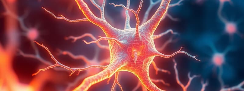
- Neurons
- Specialized cells that transmit nerve impulses
- Types:
- Anaxonic
- No apparent axon or dendrites
- Found in brain and retina
- Multipolar
- Multiple dendrites
- Single axon
- Most common type of neuron
- Bipolar
- One dendrite and one axon
- Found in sensory organs
- Anaxonic
- Function: Receive, process, and transmit information
- Difficult to classify under a microscope
Neuron Structure
- Cell Body (Perikaryon or Soma)
- Contains the nucleus
- Trophic center: produces most of the cytoplasm
- Has a large, euchromatic nucleus with a prominent nucleolus
- Cytoplasm:
- Many free ribosomes
- Highly developed RER
- Basophilic
- Chromatophilic or Nissl substance/bodies
- Golgi apparatus is only in the cell body
- Mitochondria is throughout the cell (abundant in axon terminals)
- Actin and intermediate filaments
- Neurofilaments: Neurofibrils when stained with silver stains and viewed under a light microscope
- Lipofuscin: pigment inclusion made up of residual bodies from lysosomal digestion.
Neuron Structure (cont)
- Dendrites
- Site of signal reception and processing in neurons
- Short, small processes
- Covered by synapses
- Thin as they branch
- Dendrite spine synapses on dendrites in the CNS
- Axon
- Responsible for transmitting information
- True axon: single, long process extending from the cell body
- Contains action potential
- Initiated at the axon hillock
- Propagated along the axon as a "wave" of membrane depolarization
- Produced by voltage-gated Na+ and K+ channels
- Resting potential: -65 mV
- Na is increased outside the cell
- K is increased inside the cell
- Depolarization (+30 mV): influx of Na+
- Myelin: multiple compacted layers of cell membrane
- Formed from moving out of the cytoplasm during wrapping of the axon
- Insulates axons and facilitates rapid transmission of nerve impulses
Nervous System Cells (cont)
- Glial Cells - Supportive cells of the nervous system:
- Astrocytes
- "astro" = star
- Most numerous glial cells in the brain
- Functions:
- Cover synapses
- Regulates ECF ionic concentration (buffers K+ levels)
- Guide and support movement of neurons during CNS development
- Form the glial limiting membrane that lines meninges at the external CNS
- Form astrocytic scar that fills tissue defects after CNS injury
- Large number of long, radiating, branching processes
- Proximal regions: glial fibrillary acid protein (GFAP)
- Unique marker
- Distal region: + GFAP
- Types:
- Fibrous astrocytes: abundant in white matter
- Protoplasmic astrocytes: shorter processes in gray matter
- Oligodendrocytes (myelin-forming cells of CNS)
- Form myelin sheaths for axons
- Can myelinate multiple axons
- Functions:
- Insulate axons and speed up nerve impulse transmission
- Support and organize axons
- Schwann Cells
- "neurolemmocytes"
- Functions:
- Provide myelin sheaths around one axon only
- Trophic interactions
- Microglia
- Small cells with actively mobile processes that are evenly distributed
- Migrate throughout gray and white matter
- Functions:
- Removes damaged or effete synapses
- Major mechanism of immune defense in the CNS
- Secretes cytokines
- Originate from monocytes (macrophage, antigen-presenting cell)
- Ependymal cells
- Line the fluid-filled ventricles of the brain and the central canal of the spinal cord
- Functions:
- Apical ends have cilia that move CSF
- Long microvilli for absorption
- Joined apically by apical junctional complexes
- Elongated basal ends that extend to adjacent neuropil
- Astrocytes
CNS Structure
- Major structures:
- Cerebrum:
- Cerebral nuclei: localized darker areas containing a large number of aggregated cell bodies
- Cerebral cortex:
- Has 6 layers
- Contains pyramidal neurons: most conspicuous
- Cerebrum:
- Different types of neurons and their functions:
- Sensory:
- Afferent
- Receive stimuli from receptors
- Motor:
- Efferent
- Send impulses to effector organs
- Somatic motor: voluntary control of skeletal muscles
- Autonomic motor: involuntary control of glands, heart, and smooth muscles
- Sensory:
- Interneurons:
- Form complex functional networks (circuits) in the CNS
- Can be either multipolar or anaxonic
Autonomic Nervous System
- Divisions:
- Sympathetic (fight-or-flight response)
- Preganglionic sympathetic nerve cell bodies are in the thoracic and lumbar segments of the spinal cord
- Sympathetic 2nd neurons are in small ganglia of the vertebral column
- Parasympathetic (rest-and-digest response)
- Preganglionic parasympathetic nerves are in the medulla, midbrain, and sacral portion of the spinal cord
- Parasympathetic 2nd neurons are in small ganglia near the effector organ
- Sympathetic (fight-or-flight response)
- Intramural ganglia: in the walls of the GI tract
- Two circuits:
- Preganglionic fiber (in CNS): Its axon synapses with a postganglionic peripheral ganglion system
- Postganglionic fiber (in peripheral ganglion system)
- Acetylcholine: neurotransmitter in preganglionic fibers of the autonomic nervous system
Neural Plasticity & Regeneration
- Controlled by neurotrophins:
- Promote anabolic events of axon regeneration
- Neuronal stem cells:
- Located in the ependyma
- Astrocytes:
- Can proliferate at injured sites
- Chromatolysis:
- Cell body swells
- Nissl substance diminishes
- Nucleus migrates to a peripheral position in the perikaryon
- New Schwann cells align, guiding regrowing axons
Local Anesthetics
- Low molecular weight compounds
- Bind to voltage-gated Na+ channels
- Interfere with Na+ influx
- Inhibits action potential:
- By interfering with the influx of Na+
- Prevents depolarization of the membrane
- Resting potential (-65 mV)
- Na+ channel opens: Na+ influx
- Depolarization (+30 mV)
- Na+ channel closes
- K+ Channel opens: K+ influx
- Membrane returns to resting potential
Studying That Suits You
Use AI to generate personalized quizzes and flashcards to suit your learning preferences.


