Podcast
Questions and Answers
What is a key function of myosin-II in muscle tissue?
What is a key function of myosin-II in muscle tissue?
- It is involved in nutrient absorption.
- It helps in DNA replication.
- It facilitates muscle contraction. (correct)
- It serves as a structural component of the cell membrane.
Which structural feature is unique to myosin-II among motor proteins?
Which structural feature is unique to myosin-II among motor proteins?
- It contains no light chains.
- It has a single globular head.
- It forms a coiled-coil structure with identical heavy chains. (correct)
- It does not utilize ATP for movement.
What is the primary role of titin in the sarcomere?
What is the primary role of titin in the sarcomere?
- Facilitates the binding of myosin heads to actin filaments
- Provides elasticity and stability during muscle relaxation (correct)
- Regulates the length of the thick filaments
- Promotes the attachment of actin to the Z disc
What role do light chains play in the myosin-II structure?
What role do light chains play in the myosin-II structure?
How do the thick and thin filaments interact during muscle contraction according to the sliding filament model?
How do the thick and thin filaments interact during muscle contraction according to the sliding filament model?
How do bacteria utilize the actin cytoskeleton to invade host tissues?
How do bacteria utilize the actin cytoskeleton to invade host tissues?
What is the function of capping proteins such as capZ and tropomodulin in a sarcomere?
What is the function of capping proteins such as capZ and tropomodulin in a sarcomere?
Which component of myosin-II is crucial for its motor function?
Which component of myosin-II is crucial for its motor function?
What defines the actin-based motor proteins as a family?
What defines the actin-based motor proteins as a family?
What is the primary structural arrangement of the myofibrils in skeletal muscle?
What is the primary structural arrangement of the myofibrils in skeletal muscle?
Which of the following describes the role of nebulin in the sarcomere?
Which of the following describes the role of nebulin in the sarcomere?
In the context of muscle contraction, which property of myosin-II is crucial for its function?
In the context of muscle contraction, which property of myosin-II is crucial for its function?
What is the primary consequence of the phosphorylation of myosin light chains by myosin light chain kinase (MLCK)?
What is the primary consequence of the phosphorylation of myosin light chains by myosin light chain kinase (MLCK)?
Which of the following acts as an upstream regulator in the signaling cascade that activates myosin light chain kinase?
Which of the following acts as an upstream regulator in the signaling cascade that activates myosin light chain kinase?
Which characteristic differentiates myosin II in non-muscle cells from that in skeletal muscle cells?
Which characteristic differentiates myosin II in non-muscle cells from that in skeletal muscle cells?
How does ATP hydrolysis influence the myosin head domain?
How does ATP hydrolysis influence the myosin head domain?
What is a significant function of the small bipolar filaments formed by myosin II in non-muscle cells?
What is a significant function of the small bipolar filaments formed by myosin II in non-muscle cells?
Which property of myosin V distinguishes it from myosin II?
Which property of myosin V distinguishes it from myosin II?
What role does alpha actinin play in non-muscle cells concerning myosin?
What role does alpha actinin play in non-muscle cells concerning myosin?
What is the effect of activating the signaling cascade that stimulates Rho GTPase?
What is the effect of activating the signaling cascade that stimulates Rho GTPase?
What distinguishes the structural organization of myosin II in non-muscle cells?
What distinguishes the structural organization of myosin II in non-muscle cells?
Which of the following describes a function of myosin I?
Which of the following describes a function of myosin I?
What is the primary function of the myosin head domain during muscle contraction?
What is the primary function of the myosin head domain during muscle contraction?
What occurs in the absence of ATP in muscle fibers?
What occurs in the absence of ATP in muscle fibers?
How does the hydrolysis of ATP affect the conformation of the myosin head domain?
How does the hydrolysis of ATP affect the conformation of the myosin head domain?
What structural feature of myosin filaments allows them to interact with actin filaments?
What structural feature of myosin filaments allows them to interact with actin filaments?
What is rigor mortis and how is it related to muscle energy?
What is rigor mortis and how is it related to muscle energy?
What happens to myosin's interaction with actin after the hydrolysis of ATP?
What happens to myosin's interaction with actin after the hydrolysis of ATP?
What method can be used to visualize myosin and actin interactions in vitro?
What method can be used to visualize myosin and actin interactions in vitro?
What is the consequence of the conformational change in the myosin head after releasing inorganic phosphate?
What is the consequence of the conformational change in the myosin head after releasing inorganic phosphate?
What causes the myosin head to move towards the plus end of the actin filament?
What causes the myosin head to move towards the plus end of the actin filament?
What is the primary role of the C-terminal domain of myosin proteins?
What is the primary role of the C-terminal domain of myosin proteins?
How does myosin VI differ from other myosin isoforms regarding its movement?
How does myosin VI differ from other myosin isoforms regarding its movement?
What type of organelles do myosin V primarily transport?
What type of organelles do myosin V primarily transport?
Which type of myosin is specialized in promoting the dynamics of actin filaments near the plasma membrane?
Which type of myosin is specialized in promoting the dynamics of actin filaments near the plasma membrane?
What is a characteristic feature of skeletal muscle cells (muscle fibers)?
What is a characteristic feature of skeletal muscle cells (muscle fibers)?
What is the diameter of a typical adult human skeletal muscle cell?
What is the diameter of a typical adult human skeletal muscle cell?
Which statement accurately describes the mechanism of myosin II in muscle contraction?
Which statement accurately describes the mechanism of myosin II in muscle contraction?
In which direction do most myosin motor proteins move cargo along actin filaments?
In which direction do most myosin motor proteins move cargo along actin filaments?
Which types of motor proteins are responsible for movement along microtubules?
Which types of motor proteins are responsible for movement along microtubules?
What is the primary function of myosin motor proteins in eukaryotic cells?
What is the primary function of myosin motor proteins in eukaryotic cells?
Flashcards are hidden until you start studying
Study Notes
Myosin and Muscle Contraction
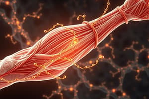
- Myosin is a major motor protein essential for muscle contraction, particularly myosin-II.
- Myosin-II forms a dimer consisting of two identical heavy chains with a globular ATPase head and a long coiled-coil tail.
- The head domain of myosin can hydrolyze ATP, enabling conformational changes necessary for its motor function.
- Myosin filaments, or thick filaments, consist of many myosin-II dimers arranged antiparallel, projecting heads that interact with actin.
Mechanism of Muscle Contraction
- The contraction cycle begins when ATP binds to the myosin head, inducing its release from actin.
- ATP is then hydrolyzed, which repositions the myosin head towards the plus end of the actin filament.
- The release of inorganic phosphate promotes a strong interaction between myosin and actin, triggering the power stroke where myosin pulls the actin filament.
- The continuous cycling of ATP binding and hydrolysis facilitates sliding of actin filaments relative to myosin, resulting in muscle contraction.
Non-Muscle Functions of Myosin
- Myosin-II isoforms also function in non-muscle cells, participating in cellular contractility and movement.
- In non-muscle cells, myosin-II is not organized into sarcomeres but forms small bipolar filaments regulated by phosphorylation from myosin light-chain kinase (MLCK).
- MLCK is activated by signaling pathways, influencing myosin activity and enabling contractile properties in non-muscle cells.
Diverse Myosin Isoforms
- Myosin-I operates as a monomer, facilitating movement near the cell membrane and supporting cellular processes like vesicle trafficking.
- Myosin-V transports organelles, such as mitochondria, along actin filaments, functioning as a cargo carrier with specific cargo recognition.
- Myosin-V consists of two head domains that allow it to move towards the plus end of actin for organelle distribution.
- Myosin-VI is unique, moving toward the minus end of actin filaments due to a distinct insertion in its head domain.
Key Attributes of Skeletal Muscle Cells
- Skeletal muscle fibers, or cells, are multinucleated structures resulting from the fusion of precursor cells (myoblasts).
- Muscle fibers typically have a diameter of 50 μm and can be several centimeters long.
- Myofibrils within muscle fibers, the contractile units, are cylindrical and aligned in parallel, playing a critical role in muscle contraction.
Visualization and Experimental Approaches
- Techniques like electron microscopy and atomic force microscopy are utilized to observe myosin and actin interactions during movement.
- Fluorescent labeling allows visualization of actin and myosin dynamics in vitro, aiding understanding of muscle contraction mechanisms.
- The study of these motor proteins identifies potential therapeutic targets for muscular disorders and provides insights into cell physiology.### Sarcomeres in Skeletal Muscle
- Sarcomeres are the basic contractile units of myofibrils, approximately 2.2 μm long, contributing to the striated muscle appearance.
- Each sarcomere consists of two Z discs, a dark band (formed by superimposed thick and thin filaments), and a light band at the periphery.
- Thick filaments (myosin) are in the middle, while thin filaments (actin) are attached to the Z discs on both sides.
- Contraction occurs as the myosin heads interact with actin, causing actin filaments to slide toward the center of the sarcomere, effectively reducing the Z disc distance.
Sliding Filament Model
- Thick filaments are surrounded by thin filaments in a hexagonal arrangement, crucial for muscle contraction.
- Myosin head domains bind transiently to actin filaments, allowing for multiple simultaneous interactions that facilitate contraction.
- The process relies on brief interactions, differing from vesicular transport, where motor proteins remain attached for longer durations.
Capping Proteins
- Proteins such as CapZ and tropomodulin cap the ends of actin filaments, preventing their elongation and maintaining stability.
- Alpha actinin is present at the Z disc, maintaining proper spacing between actin filaments.
- Titin anchors thick filaments to Z discs, functioning like a spring during muscle relaxation.
- Nebulin acts as a molecular ruler, ensuring proper length of actin filaments.
Accessory Proteins
- Accessory proteins are crucial for maintaining the structure and function of the sarcomere.
- Alpha actinin contributes to the distance between actin filaments at the Z disc, while capping proteins regulate filament dynamics.
- The assembly of actin filaments is aided by proteins such as formin, ensuring proper structure formation during muscle development.
Signaling Events in Muscle Contraction
- Tropomyosin stabilizes actin filaments and blocks myosin binding sites when muscles are relaxed.
- Upon stimulation, calcium ions released from the sarcoplasmic reticulum bind to troponin C, inducing conformational changes that expose actin binding sites.
- Troponin I inhibits myosin binding when calcium is absent, while calcium binding removes this inhibition.
Calcium Ion Release
- T tubules, invaginations of the plasma membrane, are in contact with the sarcoplasmic reticulum (SR) and contain voltage-gated calcium channels.
- Action potential from nerve cells causes depolarization, opening calcium channels in both T tubules and the SR, releasing calcium into the cytosol.
- Muscle relaxation requires the removal of calcium ions from the cytosol back to the sarcoplasmic reticulum via calcium pumps, which consume ATP.
Energy Requirements for Muscle Contraction
- Muscle contraction and relaxation require significant energy, primarily for myosin head movement and calcium pumping back into the SR.
- ATP is vital for both myosin activity and calcium ion regulation, highlighting the energy-intensive nature of muscle function.
Studying That Suits You
Use AI to generate personalized quizzes and flashcards to suit your learning preferences.