Podcast
Questions and Answers
Which ion's release from the sarcoplasmic reticulum is crucial for exposing the myosin-binding sites on actin?
Which ion's release from the sarcoplasmic reticulum is crucial for exposing the myosin-binding sites on actin?
- Sodium (Na+)
- Potassium (K+)
- Chloride (Cl-)
- Calcium (Ca2+) (correct)
What prevents the binding of myosin to actin in muscle's noncontractile state?
What prevents the binding of myosin to actin in muscle's noncontractile state?
- Calcium ions being absent
- Troponin binding to actin
- Tropomyosin covering the binding sites (correct)
- Myosin being phosphorylated
What is the primary function of acetylcholine in the neuromuscular junction?
What is the primary function of acetylcholine in the neuromuscular junction?
- To bind to calcium ions and release them
- To trigger the opening of ion channels in the sarcolemma (correct)
- To initiate muscle fiber contraction directly
- To activate the actin-myosin crossbridge cycle
Which structure is primarily responsible for storing calcium ions in muscle cells?
Which structure is primarily responsible for storing calcium ions in muscle cells?
During muscle contraction, which event occurs immediately after calcium binds to troponin?
During muscle contraction, which event occurs immediately after calcium binds to troponin?
What initiates the power stroke during the actin-myosin crossbridge cycle?
What initiates the power stroke during the actin-myosin crossbridge cycle?
During the actin-myosin crossbridge cycle, which step occurs immediately after the binding of ATP to myosin?
During the actin-myosin crossbridge cycle, which step occurs immediately after the binding of ATP to myosin?
Which component directly causes the exposure of the myosin-binding site on actin?
Which component directly causes the exposure of the myosin-binding site on actin?
What is the role of the autonomic nervous system in muscle function?
What is the role of the autonomic nervous system in muscle function?
What is the result of ATP hydrolysis in the actin-myosin crossbridge cycle?
What is the result of ATP hydrolysis in the actin-myosin crossbridge cycle?
Match each type of bone with its primary characteristic or function:
Match each type of bone with its primary characteristic or function:
Match the following components of long bones with their descriptions:
Match the following components of long bones with their descriptions:
Match each bone type with an example or characteristic:
Match each bone type with an example or characteristic:
Match the type of bone with its role in the body:
Match the type of bone with its role in the body:
Match the characteristic with the relevant type of marrow:
Match the characteristic with the relevant type of marrow:
What is the primary role of the metaphysis in bone structure?
What is the primary role of the metaphysis in bone structure?
What are the primary components that make up the osteon structure?
What are the primary components that make up the osteon structure?
How do Volkmann's canals contribute to bone health?
How do Volkmann's canals contribute to bone health?
What is the significance of lacunae in the bone structure?
What is the significance of lacunae in the bone structure?
What is the primary function of cancellous bone?
What is the primary function of cancellous bone?
What occurs when blood calcium levels are too high?
What occurs when blood calcium levels are too high?
Which type of ossification is responsible for the formation of long bones?
Which type of ossification is responsible for the formation of long bones?
What type of joint allows for full mobility and flexibility?
What type of joint allows for full mobility and flexibility?
Which characteristic defines ligaments within the skeletal system?
Which characteristic defines ligaments within the skeletal system?
What is the role of osteoblasts during the process of ossification?
What is the role of osteoblasts during the process of ossification?
What primary role do melanocytes serve in the epidermis?
What primary role do melanocytes serve in the epidermis?
Which layer of the skin is primarily responsible for providing strength and elasticity?
Which layer of the skin is primarily responsible for providing strength and elasticity?
Which sensory receptor is specifically responsive to light touch?
Which sensory receptor is specifically responsive to light touch?
What is the primary function of the hypodermis?
What is the primary function of the hypodermis?
Which layer of the epidermis is the site of proliferation for keratinocytes?
Which layer of the epidermis is the site of proliferation for keratinocytes?
Match the type of gland with its primary function:
Match the type of gland with its primary function:
Match the physiological process with its description:
Match the physiological process with its description:
Match the gland type with its location:
Match the gland type with its location:
Match the skin function with the relevant mechanism:
Match the skin function with the relevant mechanism:
Match the type of sweat gland with its characteristic:
Match the type of sweat gland with its characteristic:
What is the primary function of bile in the digestive system?
What is the primary function of bile in the digestive system?
Which of the following describes the role of albumin in the bloodstream?
Which of the following describes the role of albumin in the bloodstream?
What is the main purpose of the loop of Henle in the nephron?
What is the main purpose of the loop of Henle in the nephron?
Which hormone is responsible for regulating sodium and potassium levels in the body?
Which hormone is responsible for regulating sodium and potassium levels in the body?
What is the function of the proximal convoluted tubule in the nephron?
What is the function of the proximal convoluted tubule in the nephron?
In the context of renal function, what does the term 'filtration' refer to?
In the context of renal function, what does the term 'filtration' refer to?
What is the process of osmoregulation primarily responsible for?
What is the process of osmoregulation primarily responsible for?
Which aspect characterizes the descending loop of Henle?
Which aspect characterizes the descending loop of Henle?
Which structure in the nephron is involved in collecting filtered fluid?
Which structure in the nephron is involved in collecting filtered fluid?
What role does the exocrine pancreas play in the digestive system?
What role does the exocrine pancreas play in the digestive system?
What is the primary role of salivary amylase in digestion?
What is the primary role of salivary amylase in digestion?
What occurs in the stomach after food is mixed with gastric juices?
What occurs in the stomach after food is mixed with gastric juices?
Which cells in the stomach lining are responsible for producing hydrochloric acid?
Which cells in the stomach lining are responsible for producing hydrochloric acid?
What type of digestion is primarily accomplished through mastication?
What type of digestion is primarily accomplished through mastication?
Which function do microvilli serve in the small intestine?
Which function do microvilli serve in the small intestine?
Which hormone stimulates the secretion of gastric acid in the stomach?
Which hormone stimulates the secretion of gastric acid in the stomach?
What roles do lacteals play in the digestive system?
What roles do lacteals play in the digestive system?
What is the function of pepsin in the stomach?
What is the function of pepsin in the stomach?
Which process propels food through the digestive tract after it leaves the esophagus?
Which process propels food through the digestive tract after it leaves the esophagus?
What is the main role of gut flora in the gastrointestinal tract?
What is the main role of gut flora in the gastrointestinal tract?
Flashcards are hidden until you start studying
Study Notes
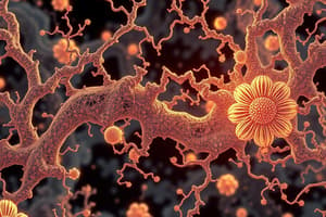
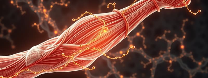
Muscle Contraction Overview
- Muscle fibers contain thin (actin) and thick (myosin) filaments, which are essential for skeletal muscle contractions.
- The actin-myosin crossbridge cycle is responsible for muscle contraction.
Neuromuscular Junction
- Signal transmission begins at the motor neurons in the somatic nervous system.
- The interaction occurs at the neuromuscular junction's motor endplate, where motor neurons connect with muscle fibers via a chemical synapse.
Role of Acetylcholine
- Activation of motor neurons leads to the release of acetylcholine into the synaptic cleft.
- Acetylcholine binds to the sarcolemma (muscle cell膜), initiating ion channel opening and depolarization.
Action Potential and Calcium Release
- The depolarization results in the propagation of an action potential through T-tubules.
- This action triggers the release of Ca2+ ions from the sarcoplasmic reticulum, essential for muscle contraction.
Interaction of Proteins
- In a resting state, tropomyosin and troponin regulate the interaction between actin and myosin.
- Tropomyosin covers the myosin-binding sites on actin, preventing contraction.
Calcium's Role in Muscle Contraction
- The released Ca2+ ions bind to troponin, causing a structural change in the protein complex.
- This structural shift in tropomyosin exposes the myosin-binding sites on actin, allowing for the formation of actin-myosin crossbridges.
Actin-Myosin Crossbridge Cycle Steps
- Resting State: Myosin head is in a high-energy state with ADP and inorganic phosphate (Pi) bound.
- Ca2+ Activation: Calcium ions bind to troponin, triggering a conformational change that exposes binding sites on actin for myosin.
- Power Stroke Mechanism: The myosin head attaches to actin, releasing ADP and Pi, which generates the power stroke, sliding the myosin along the actin filament.
- ATP Binding: New ATP molecule binds to myosin, causing it to detach from actin, completing the cycle.
- ATP Hydrolysis: ATP is hydrolyzed to ADP and Pi, resetting the myosin head to the cocked position, enabling the cycle to repeat as long as Ca2+ is present.
Muscle Function
- Muscle movement, both voluntary and involuntary, is regulated by the nervous system.
- Shivering: An involuntary response for thermoregulation controlled by the autonomic nervous system.
- Autonomic Nervous System: Divided into sympathetic and parasympathetic divisions, influencing muscle behavior.
- Sympathetic Response: Increases heart rate, dilates blood vessels, and slows down the digestive tract.
- Parasympathetic Response: Opposes sympathetic actions and primarily conserves energy, regulating various body functions.
Types of Bones
- Short bones: Equal width and length; primarily located in the wrist; enhance mobility.
- Flat bones: Serve as protective structures for vital organs; the skull exemplifies flat bone composition.
- Sesamoid bones: Found embedded within tendons; improve muscle functionality; the kneecap (patella) is a prominent example.
- Irregular bones: Have complex shapes; fulfill specific functions; the pelvis is categorized as an irregular bone.
- Long bones: Characterized by a dense outer cortical layer and an inner spongy structure; include key components such as:
- Epiphysis: Forms joints and contains red bone marrow, crucial for blood cell production.
- Diaphysis: Contains the medullary cavity, housing red and yellow bone marrow; yellow marrow serves as fat storage.
- Medullary Cavity: Central cavity within long bones; holds marrow.
- Metaphysis: Region where diaphysis meets epiphysis, involved in bone growth.
- Epiphyseal plate: Cartilaginous zone that allows for lengthening of bones during growth.
Metaphysis
- Located between the medullary cavity and the epiphyseal plates.
- Plays a crucial role in bone growth and development.
- Contains the epiphyseal plate, or "growth plate," made of hyaline cartilage.
- Stimulates lengthening of the diaphysis through growth and ossification.
- In mature bones, the epiphyseal plate transitions to become the epiphyseal line.
Bone Microstructures
Cortical Bone
- Hard outer layer of long bones, providing strength and support.
- Comprised of osteons (Haversian systems), which are cylindrical structures.
- Osteons are organized around Haversian canals that contain blood vessels for nutrient supply.
Lamellae
- Concentric layers forming the osteons, contributing to structural integrity.
- Contain lacunae, which are small cavities housing osteocytes, specialized bone cells.
- Canaliculi are minute channels connecting lacunae, allowing nutrient exchange among osteocytes.
Volkmann's Canals
- Connect Haversian canals to the periosteum, the outer layer of bone.
- Facilitate the distribution of nutrients from blood vessels to bone tissues.
Cancellous Bone
- Inner layer of bone characterized by a spongy network of trabeculae.
- Plays a critical role in absorbing red bone marrow, essential for blood cell production.
- Supports the structural framework of the cancellous bone, enhancing overall durability.
Conclusion
- The interplay among cortical bone, lamellae, Volkmann's canals, and cancellous bone is essential for maintaining bone strength and functionality.
- These structures collectively contribute to the skeletal system's durability and overall function.
Blood Calcium Regulation
- High blood calcium levels trigger the release of calcitonin from the thyroid, reducing calcium concentrations.
- Stimulates osteoblasts to deposit calcium in bones, restoring homeostasis.
- Low blood calcium levels lead to the secretion of parathyroid hormone (PTH) from the parathyroid, increasing calcium levels.
- Osteoclasts are activated to demineralize bone, releasing calcium into the bloodstream, resetting calcium levels.
Ossification Types
- Intramembranous ossification occurs within fibrous membranes, crucial for flat bone development like the skull.
- Osteoblasts secrete osteoid that mineralizes to form cortical bone.
- Endochondral ossification utilizes a cartilage model for the development of long bones, shaping their structures.
- Cartilage model calcification initiates ossification centers, and osteoblasts replace cartilage with bone tissue.
- Key examples include humerus, femur, and tibia which mature through this process.
Connective Tissues and Joint Structures
- Tendons connect muscles to bones, facilitating movement and stabilization.
- Ligaments link bones together, maintaining joint integrity.
- Cartilage is a flexible, resilient connective tissue, lacking blood vessels and nerves, crucial for the skeletal system.
- Hyaline cartilage promotes smooth joint movement, predominantly in early development, eventually turning into bone.
Joint Types
- Synarthroses allow no movement, linked by dense connective tissue.
- Amphiarthroses permit limited movement through cartilage connections.
- Diarthroses (synovial joints) are designed for full mobility, encased in a joint capsule.
Bone Membranes
- Periosteum includes a vascularized outer fibrous layer and a cambium layer that connects it to the cortical bone.
- Endosteum resides between cortical and cancellous bone, playing a key role in bone regeneration and maintenance.
Skin Structure
- Skin is the body's largest organ, derived from the ectoderm and composed of three layers: epidermis, dermis, and hypodermis.
Epidermis
-
The epidermis is the outermost layer, further divided into five strata:
- Stratum corneum: Contains flattened keratinocytes for pathogen protection.
- Stratum lucidum: Present in hairless areas such as palms.
- Stratum granulosum: Contains dead keratinocytes.
- Stratum spinosum: Houses Langerhans cells and live keratinocytes, participating in immune functions.
- Stratum basale: Site of keratinocyte proliferation, containing melanocytes and stem cells.
-
Key Cell Types in the Epidermis:
- Keratinocytes: Produce keratin for protection.
- Melanocytes: Generate melanin for UV protection.
- Langerhans cells: Immune system macrophages aiding in response activation.
Dermis
-
The dermis supports various skin structures and is rich in collagen and elastic fibers, allowing for strength and elasticity.
-
Composed of two regions:
- Papillary region: Upper 20%, connects to the epidermis via papillae, crucial for nourishment and temperature regulation; houses Meissner's corpuscles for light touch sensitivity.
- Reticular region: Denser and houses oil glands, sweat ducts, fat, and hair follicles, making up most of the dermis. Responsible for skin strength, elasticity, and forming stretch marks when torn.
-
Sensory Receptors in the Dermis:
- Tactile Corpuscles (Meissner's Corpuscles): Sensitive to light touch.
- Bulbous Corpuscles (Ruffini Endings): Detect skin stretching.
- Pacinian Corpuscles: Respond to vibration and pressure.
-
Dermis serves as a site for tattooing, where ink is trapped in dermal layers and becomes integrated into the skin during healing.
Hypodermis
- The hypodermis is the innermost layer, containing connective tissue that anchors skin to muscles and bones.
- Primarily composed of adipose tissue for thermal insulation and shock absorption, providing protection and energy storage.
Glands Overview
- The integumentary system contains various glands crucial for maintaining bodily equilibrium.
- Glands are classified into sudoriferous (sweat) glands and sebaceous glands.
Eccrine Glands
- Eccrine glands are found extensively across the body surface.
- They play a key role in regulating body temperature by secreting a watery fluid.
- The secretion helps cool the body, especially during high temperatures.
Apocrine Glands
- Apocrine glands are localized in areas such as the armpits and groin.
- These glands release their secretions into hair follicles.
- They are responsible for producing substances like earwax (via ceruminous glands) and milk (via mammary glands), serving functions beyond mere temperature regulation.
Sebaceous Glands
- Sebaceous glands are distributed throughout the body except on the palms and soles.
- They produce sebum, a mixture of oils and wax that moisturizes and lubricates skin and hair.
- Sebum acts as a natural barrier against external elements, contributing to skin health.
Homeostasis and Skin Function
- The skin is integral in maintaining the body's internal balance, particularly through water impermeability.
- It contains structures essential for body temperature regulation.
- Shivering generates heat through rapid muscle contractions, converting energy to thermal energy.
- Piloerection (goosebumps) raises hair to trap heat, aiding in warmth retention.
- Vasoconstriction narrows blood vessels to conserve heat in cold conditions.
- In hot environments, sweat glands release a mixture of water and ions for cooling.
- Evaporative cooling occurs when sweat evaporates, absorbing heat and reducing body temperature.
- Vasodilation expands blood vessels, allowing heat transfer from the blood to skin surface for effective cooling.
Digestion and Mastication
- Mastication involves chewing food to break it into smaller pieces for easier digestion.
- Mechanical digestion encompasses physical breakdown methods such as chewing, churning in the stomach, and muscular movements along the digestive tract.
Salivary Enzymes
- Salivary amylase initiates starch breakdown into simpler sugars, including maltose.
- Salivary lipase begins fat digestion by breaking down triglycerides into fatty acids and glycerol.
Digestive Structures
- A bolus is a rounded mass of chewed food mixed with saliva, readied for swallowing.
- The pharynx connects the nasal cavity and mouth to the esophagus and trachea, facilitating the passage of food and air.
- The esophagus is a muscular tube that transports food from the mouth to the stomach.
Digestive Processes
- Peristalsis refers to wavelike muscle contractions that push food through the digestive tract.
- The stomach mixes food with gastric juices to digest proteins and regulate food passage to the small intestine.
Cellular Functions in the Stomach
- Parietal cells in the stomach lining produce hydrochloric acid, creating an acidic environment crucial for digestion.
- Chief cells secrete pepsinogen, which converts to pepsin to break down proteins into smaller peptides.
- Gastrin, a hormone produced by the stomach, stimulates gastric acid secretion.
Chyme and Absorption
- Chyme is a semi-fluid mass of partially digested food exiting the stomach into the small intestine.
- Microvilli are tiny projections in the small intestine's lining that enhance surface area for nutrient absorption.
Nutritional Components
- Fat-soluble vitamins (K, A, D, E) dissolve in fats and are stored within the body's fatty tissues.
- Lacteals are lymphatic vessels in the small intestine that absorb fats and fat-soluble vitamins into the lymphatic system.
Gut Flora and Waste Management
- Gut flora consists of beneficial microorganisms in the gastrointestinal tract that assist in digestion and immune functions.
- The rectum stores feces before elimination from the body, regulated by anal sphincters.
Enteric Nervous System
- The enteric nervous system governs the gastrointestinal tract's functionality independently of the central nervous system.
Liver and Related Organs
- The liver produces bile, detoxifies, and stores nutrients, playing a vital metabolic role.
- Bile emulsifies fats, aiding their digestion and absorption.
- Albumin, a protein generated by the liver, transports various substances in the blood.
- The gallbladder stores and concentrates bile for fat digestion.
Pancreas Functions
- The pancreas produces digestive enzymes and hormones like insulin and glucagon, integral for metabolic processes.
- Exocrine glands in the pancreas release enzymes and substances directly onto surfaces or into ducts.
Brush-Border Enzymes
- Brush-border enzymes facilitate nutrient breakdown at the intestinal surface, aiding absorption.
- Disaccharidases split disaccharides into monosaccharides for absorption.
- Peptidases break down proteins into smaller peptides and amino acids.
Nephron and Kidney Functions
- Nephrons are the kidney's functional units, responsible for filtration, secretion, and reabsorption to form urine.
- Bowman’s capsule encases the glomerulus, collecting filtered fluid from blood.
- The glomerulus filters blood to initiate urine formation.
Kidney Segments
- The proximal convoluted tubule reabsorbs water, ions, and nutrients.
- The loop of Henle further reabsorbs water and ions, with descending and ascending limbs having specific permeability properties.
- The countercurrent exchange system in the loop of Henle enhances water reabsorption efficiency.
Hormonal Regulation and Homeostasis
- The distal convoluted tubule fine-tunes electrolyte balance and water reabsorption regulated by hormones.
- The collecting duct facilitates additional water reabsorption, producing concentrated urine.
Kidney Structure
- The medulla houses renal pyramids and loops of Henle, while the cortex contains glomeruli and convoluted tubules.
- The renal capsule is a protective layer around the kidney.
Hormones and Homeostasis
- Aldosterone regulates sodium and potassium levels and is produced by adrenal glands.
- Renin, an enzyme by the kidneys, assists in blood pressure and electrolyte balance regulation.
- Antidiuretic hormone (ADH), produced by the pituitary gland, controls water reabsorption in kidneys.
- Osmoregulation maintains fluid and ion balance critical for proper cell function.
Studying That Suits You
Use AI to generate personalized quizzes and flashcards to suit your learning preferences.