Podcast
Questions and Answers
What are Russell bodies?
What are Russell bodies?
- Macrophages found in bone marrow
- Inclusions containing DNA in the plasma cells
- Globular inclusions of immunoglobulin in the cytoplasm of plasma cells (correct)
- Chemokines produced by T helper cells
Which body part is primarily affected by hypercalcemia in Multiple Myeloma?
Which body part is primarily affected by hypercalcemia in Multiple Myeloma?
- Liver
- Lungs
- Bones (correct)
- Kidneys
What does the presence of Dutcher bodies indicate?
What does the presence of Dutcher bodies indicate?
- Significant bone marrow damage
- High levels of light chains in urine
- Accumulation of immunoglobulin in the cytoplasm
- Accumulation of immunoglobulin in the nucleus of plasma cells (correct)
What condition is often associated with hypercalcemia caused by neoplastic plasma cells?
What condition is often associated with hypercalcemia caused by neoplastic plasma cells?
What is Bence Jones proteinuria a result of?
What is Bence Jones proteinuria a result of?
What is the primary consequence of increased osteoclast activity in Multiple Myeloma?
What is the primary consequence of increased osteoclast activity in Multiple Myeloma?
Elevated free light chains in plasma typically indicates an excess of which component?
Elevated free light chains in plasma typically indicates an excess of which component?
What symptom is commonly associated with severe hypercalcemia?
What symptom is commonly associated with severe hypercalcemia?
Patients with Multiple Myeloma often demonstrate what kind of immunoglobulin abnormality?
Patients with Multiple Myeloma often demonstrate what kind of immunoglobulin abnormality?
What characteristic appearance do plasma cells exhibit when they are neoplastic?
What characteristic appearance do plasma cells exhibit when they are neoplastic?
What is the primary indicator of primary myelofibrosis progression?
What is the primary indicator of primary myelofibrosis progression?
What is a definitive characteristic used to diagnose Reed Sternberg cells in Hodgkin Lymphoma?
What is a definitive characteristic used to diagnose Reed Sternberg cells in Hodgkin Lymphoma?
Which factor significantly contributes to abnormal blood flow and platelet function in patients with primary myelofibrosis?
Which factor significantly contributes to abnormal blood flow and platelet function in patients with primary myelofibrosis?
Which feature is often associated with Nodular Sclerosis variant of Hodgkin Lymphoma?
Which feature is often associated with Nodular Sclerosis variant of Hodgkin Lymphoma?
What are the common symptoms associated with sustained congestion in primary myelofibrosis?
What are the common symptoms associated with sustained congestion in primary myelofibrosis?
What abnormality indicates a diagnosis of Acute Myeloid Leukemia (AML)?
What abnormality indicates a diagnosis of Acute Myeloid Leukemia (AML)?
What is the main consequence of extensive marrow fibrosis in myelofibrosis?
What is the main consequence of extensive marrow fibrosis in myelofibrosis?
Which condition is a potential first indication of thrombotic episodes in primary myelofibrosis patients?
Which condition is a potential first indication of thrombotic episodes in primary myelofibrosis patients?
Which mutation is commonly associated with therapy-related Acute Myeloid Leukemia?
Which mutation is commonly associated with therapy-related Acute Myeloid Leukemia?
In what form does Myelodysplastic Syndrome (MDS) often present clinically?
In what form does Myelodysplastic Syndrome (MDS) often present clinically?
What role does histamine play in the symptoms of primary myelofibrosis?
What role does histamine play in the symptoms of primary myelofibrosis?
What is a common complication associated with organomegaly in primary myelofibrosis patients?
What is a common complication associated with organomegaly in primary myelofibrosis patients?
Which cells are indicative of high risk transformation in Myelodysplastic Syndromes?
Which cells are indicative of high risk transformation in Myelodysplastic Syndromes?
What is commonly found in the marrow of patients with acute promyelocytic leukemia (t(15,17))?
What is commonly found in the marrow of patients with acute promyelocytic leukemia (t(15,17))?
What is the relationship between TGF-B and primary myelofibrosis?
What is the relationship between TGF-B and primary myelofibrosis?
Which condition is characterized by an increased proliferation and presence of mutated hematopoietic stem cells?
Which condition is characterized by an increased proliferation and presence of mutated hematopoietic stem cells?
Which of the following is NOT a characteristic of primary myelofibrosis?
Which of the following is NOT a characteristic of primary myelofibrosis?
What is a characteristic feature of Lymphocyte-Rich Hodgkin Lymphoma?
What is a characteristic feature of Lymphocyte-Rich Hodgkin Lymphoma?
What does the term 'extramedullary hematopoiesis' refer to in primary myelofibrosis?
What does the term 'extramedullary hematopoiesis' refer to in primary myelofibrosis?
What is the clinical consequence of an increased number of immature myeloid blasts in Acute Myeloid Leukemia?
What is the clinical consequence of an increased number of immature myeloid blasts in Acute Myeloid Leukemia?
Flashcards are hidden until you start studying
Study Notes
Mycosis Fungoides/Sezary Syndrome
- Tumor involving CD4+ T helper cells primarily affecting the skin
- Features cutaneous lesions that progress through: inflammatory premycotic phase, plaque phase, and tumor phase
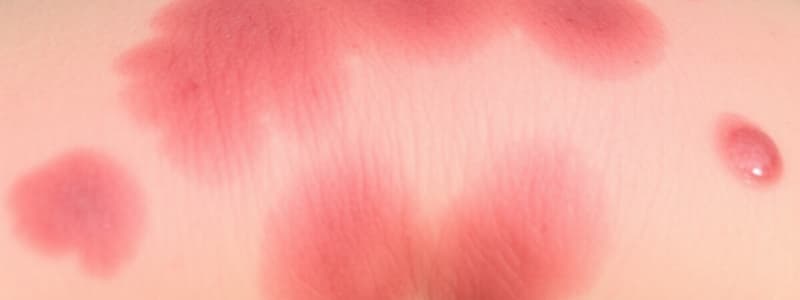
- Neoplastic T cells infiltrate the epidermis and upper dermis, leading to significant abnormalities in the skin
- Characteristic of a persistent rash that does not resolve
- Sezary syndrome manifests as generalized exfoliative erythroderma
- Indolent course with a median survival of approximately 10 years
- Histological evidence includes infolding of nuclear membrane and cerebriform appearance of the cells
Plasma Cell Neoplasms
- Consist of B cell proliferations generating neoplastic plasma cells
- Plasma cells secrete monoclonal immunoglobulin (Ig) or fragments, functioning as tumor markers
- Some tumors may exclusively produce light chains or rarely only heavy chains
- M component refers to monoclonal Ig present in the blood
Multiple Myeloma
- Characterized by IGH locus translocations leading to neoplastic plasma cells
- Neoplastic plasma cells produce CCL3, a chemokine that enhances osteoclast formation and bone destruction
- Diagnosis requires detection of clonal plasma cells in bone marrow and presence of CRAB symptoms: hypercalcemia, renal dysfunction, anemia, and bone lesions
- Predominantly affects men, particularly those aged 65-70, and African Americans
- Elevated free light chain levels indicate a skewed balance favoring one light chain
- Bence Jones proteins, small free light chains, can be excreted in urine
Clinical Features and Diagnosis
- Lytic bone lesions typically occur in the axial skeleton and include punched-out defects leading to pathologic fractures and chronic pain
- Hypercalcemia can cause neurologic issues such as confusion and lethargy
- The immune system becomes impaired due to decreased production of normal immunoglobulins, leading to recurrent infections
- Laboratory findings include elevated Ig levels in blood and/or light chains in urine, alongside the identification of monoclonal Igs in electrophoresis
- Common monoclonal Ig identified is IgG, followed by IgA, leading to hyperviscosity syndromes
Smoldering Myeloma vs. MGUS
- Smoldering myeloma is characterized by serum M protein >3g/dL or ≥10% clonal plasma cells in the marrow, with a 75% chance of progression to multiple myeloma
- MGUS (Monoclonal Gammopathy of Undetermined Significance) typically presents in asymptomatic patients with serum M protein <3g/dL and <10% clonal plasma cells
- No evidence of lytic bone lesions or myeloma-related organ complications in MGUS
Symptomatic Plasma Cell Myeloma
- Exhibits CRAB features indicating organ and tissue impairment
- Neoplastic giant cells, known as Reed-Sternberg cells, are a hallmark of this condition
- Differential diagnoses may include infectious mononucleosis and large cell non-Hodgkin lymphoma
- Staging is critically important for prognosis, influencing treatment decisions and outcomes.### Hodgkin Lymphoma
- Characterized by the presence of Reed-Sternberg cells, which are large cells that may appear as single multilobular nuclei or multiple nuclei resembling owl eyes.
- Lacunar cells are a form of Reed-Sternberg cells seen in nodular sclerosis, featuring delicate, folded, multilobate nuclei and pale cytoplasm.
- L&H cells indicative of lymphohistiocytic variants have polypoid nuclei, inconspicuous nucleoli, and abundant cytoplasm.
- Progression often begins in lymph nodes, later affecting the spleen, liver, bone marrow, and other tissues.
- Subtypes include:
- Nodular sclerosis: CD15+, CD30+, EBV-; frequent lacunar cells
- Mixed cellularity: CD15+, CD30+, 70% EBV+; frequent mononuclear and Reed-Sternberg cells
- Lymphocyte-rich: CD15+, CD30+, 40% EBV+; frequent mononuclear Reed-Sternberg cells
- Lymphocyte-depleted: CD15+, CD30+, most EBV+; frequent Reed-Sternberg cells
- Nodular lymphocyte-predominant: CD15-, CD30-, EB-; frequent L&H cells
Myeloid Neoplasms
Acute Myeloid Leukemia (AML)
- Caused by acquired oncogenic mutations that hinder myeloid differentiation, leading to an accumulation of immature myeloid forms (blasts).
- Replacement of bone marrow with blasts results in marrow failure, causing anemia, thrombocytopenia, and neutropenia.
- WHO classification includes subtypes with specific genetic aberrations such as inv(16) and t(15,17).
- Auer rods, distinctive granules in myeloblasts, may be noted, particularly in acute promyelocytic leukemia (APL).
- Common clinical features include rapid onset of symptoms like anemia, infections, and bleeding due to dysfunctional hematopoiesis.
Myelodysplastic Syndrome (MDS)
- Group of clonal stem cell disorders characterized by maturation defects in myeloid progenitors and ineffective hematopoiesis.
- Presentation of cytopenias and high risk of transformation to AML.
- Associated chromosomal abnormalities include monosomy 5 and 7, as well as deletions of 5q, 7q, and 20q.
- Morphological findings reveal dysplastic features in erythroid, granulocytic, and megakaryocytic lineages, such as ring sideroblasts and pseudo-Pelger-Huet cells.
Myeloproliferative Neoplasms
- Characterized by increased production of one or more types of blood cells due to mutated, activated tyrosine kinases.
- Common features include extramedullary hematopoiesis and potential transformation to acute leukemia or spent phase with marrow fibrosis and cytopenias.
Chronic Myeloid Leukemia (CML)
- Driven by the BCR-ABL fusion gene from the Philadelphia chromosome (t(9,22)), leading to increased granulocytic precursors and hypercellular marrow.
- Clinical hallmarks include leukocytosis, splenomegaly, and the presence of sea-blue histiocytes.
- Progression can lead to an accelerated phase, characterized by anemia and thrombocytopenia, ultimately leading to a blast crisis.
Additional Conditions
Polycythemia Vera
- Triggered by activating mutations in JAK2, leading to increased production of red blood cells, granulocytes, and platelets.
- Patients exhibit low serum EPO levels, hyperuricemia, and symptoms related to increased red cell mass.
- Rare transformation to AML occurs in approximately 1% of cases, with notable features including marked reticulin fiber increase in marrow.### Congestion and Organomegaly
- Congestion leads to mild organomegaly, particularly in the spleen and liver due to extramedullary hematopoiesis.
- Peripheral blood shows an increased number of basophils and abnormally large platelets.
Progression and Spent Phase
- Progression into the spent phase is characterized by extensive marrow fibrosis that displaces hematopoietic cells.
- Organomegaly results from extramedullary hematopoiesis, predominantly in the spleen and liver.
Symptoms and Complications
- Symptoms include headaches, dizziness, hypertension (HTN), gastrointestinal issues, and thromboses in portal and mesenteric veins.
- Hepatic thrombosis can lead to Budd-Chiari syndrome, a complication of blood flow stagnation and deoxygenation.
Role of Basophils
- Basophils release histamine, which causes intense pruritus and can lead to peptic ulceration.
Blood Flow and Hemostasis
- Abnormal blood flow and impaired platelet function increase risks for bleeding and thrombotic events.
- Initial signs of thrombosis can manifest as deep vein thrombosis (DVT), myocardial infarction (MI), or stroke.
Platelet Characteristics
- Platelets are notably giant and defective in aggregation, contributing to hemostatic issues.
Primary Myelofibrosis
- Activating mutations in JAK2, CALR, or MPL are central to the pathology of primary myelofibrosis.
- Transforming growth factor-beta (TGF-B) promotes collagen deposition and angiogenesis in the marrow.
Hallmarks of Primary Myelofibrosis
- Development of obliterative marrow fibrosis replaces hematopoietic tissue, leading to cytopenia and reliance on extramedullary hematopoiesis.
- Extensive collagen deposition by non-neoplastic fibroblasts impairs normal marrow function and hematopoiesis.
Studying That Suits You
Use AI to generate personalized quizzes and flashcards to suit your learning preferences.


