Podcast
Questions and Answers
What differentiates a 'simple' fracture from a 'compound' fracture?
What differentiates a 'simple' fracture from a 'compound' fracture?
- The involvement of multiple bone fragments.
- The exposure of bone through the skin. (correct)
- The location of the fracture on the bone.
- Whether the fracture line is straight or angled.
Which of the following best describes a 'comminuted' fracture?
Which of the following best describes a 'comminuted' fracture?
- The fracture extends completely through both cortices of the bone.
- The fracture is still in alignment.
- The fracture results in multiple bone fragments. (correct)
- The fracture only goes through one cortex of the bone.
A vet observes a fracture running diagonally along the diaphysis. How should this fracture be classified?
A vet observes a fracture running diagonally along the diaphysis. How should this fracture be classified?
- Spiral.
- Transverse.
- Oblique. (correct)
- Segmental.
What is the key characteristic of a 'spiral' fracture?
What is the key characteristic of a 'spiral' fracture?
What is the primary characteristic of an avulsion fracture?
What is the primary characteristic of an avulsion fracture?
Salter-Harris fractures are unique because they involve which part of the bone?
Salter-Harris fractures are unique because they involve which part of the bone?
What is the primary cause of bucked shins in young racehorses?
What is the primary cause of bucked shins in young racehorses?
Which of the following is the most accurate description of periostitis?
Which of the following is the most accurate description of periostitis?
What is the typical treatment approach for distal splint bone fractures?
What is the typical treatment approach for distal splint bone fractures?
Why are proximal splint bone fractures typically considered more serious than distal fractures?
Why are proximal splint bone fractures typically considered more serious than distal fractures?
What anatomical location is most commonly affected by cannon bone fractures?
What anatomical location is most commonly affected by cannon bone fractures?
What is the primary goal when surgically repairing cannon bone fractures?
What is the primary goal when surgically repairing cannon bone fractures?
Which statement best describes a "chip fracture" of the carpal bone?
Which statement best describes a "chip fracture" of the carpal bone?
In proximal sesamoid fractures, what dictates whether a fragment can be removed or needs to be reattached?
In proximal sesamoid fractures, what dictates whether a fragment can be removed or needs to be reattached?
What is the purpose of hyaluronic acid in synovial fluid?
What is the purpose of hyaluronic acid in synovial fluid?
Why is subchondral bone more adaptable to changes in force compared to trabecular bone?
Why is subchondral bone more adaptable to changes in force compared to trabecular bone?
What is the relationship between Degenerative Joint Disease and Osteoarthritis?
What is the relationship between Degenerative Joint Disease and Osteoarthritis?
What is the role of inflammation in the development of non-infectious osteoarthritis?
What is the role of inflammation in the development of non-infectious osteoarthritis?
What are the symptoms of osteoarthritis?
What are the symptoms of osteoarthritis?
What is the primary characteristic of vertebral spondylosis?
What is the primary characteristic of vertebral spondylosis?
In "kissing spine," which part of the vertebrae is primarily affected?
In "kissing spine," which part of the vertebrae is primarily affected?
What is the underlying cause of "kissing spine"?
What is the underlying cause of "kissing spine"?
Infectious osteoarthritis, also know as septic arthritis involves what?
Infectious osteoarthritis, also know as septic arthritis involves what?
How is infectious osteoarthritis treated?
How is infectious osteoarthritis treated?
What pathological process defines subchondral bone disease?
What pathological process defines subchondral bone disease?
How is subchondral bone disease diagnosed?
How is subchondral bone disease diagnosed?
What is disrupted in utero, to cause OCD?
What is disrupted in utero, to cause OCD?
What characterizes physitis?
What characterizes physitis?
Typical signalment for physitis is?
Typical signalment for physitis is?
What factor contributes to angular limb deformities?
What factor contributes to angular limb deformities?
The carpus being in valgus means?
The carpus being in valgus means?
What causes flexural limb deformities?
What causes flexural limb deformities?
What is the aim for treatment for flexural limb deformities?
What is the aim for treatment for flexural limb deformities?
With common digital extensor rupture, what is the best diagnosis?
With common digital extensor rupture, what is the best diagnosis?
Tendons can be injured by?
Tendons can be injured by?
When is the common time to see tendonitis?
When is the common time to see tendonitis?
When performing lidocaine blocks of specific nerves, what is the goal?
When performing lidocaine blocks of specific nerves, what is the goal?
Swelling on the distal plantar aspect of the tarsus is?
Swelling on the distal plantar aspect of the tarsus is?
Tenosynovitis is the inflammation of what?
Tenosynovitis is the inflammation of what?
What anatomical structure is most commonly affected by tenosynovitis in working horses?
What anatomical structure is most commonly affected by tenosynovitis in working horses?
Extensor tendon laceration occurs because?
Extensor tendon laceration occurs because?
Calcanean bursitis affects what anatomical area?
Calcanean bursitis affects what anatomical area?
Rupture of the peroneus tertius is caused by.
Rupture of the peroneus tertius is caused by.
Classic sign of peroneus tertius rupture is.
Classic sign of peroneus tertius rupture is.
Exertional rhabdomyolysis is which type of disease?
Exertional rhabdomyolysis is which type of disease?
Flashcards
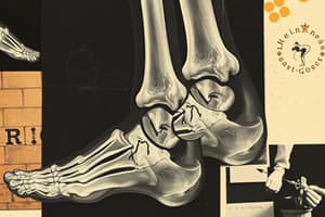
Fracture
Fracture
A break in the continuity of a bone.
Simple Fracture
Simple Fracture
Fracture where all fracture edges remain within the skin.
Compound Fracture
Compound Fracture
Fracture where part of the bone is exposed through a break in the skin.
Incomplete Fracture
Incomplete Fracture
Signup and view all the flashcards
Complete Fracture
Complete Fracture
Signup and view all the flashcards
Comminuted Fracture
Comminuted Fracture
Signup and view all the flashcards
Segmental Fracture
Segmental Fracture
Signup and view all the flashcards
Non-displaced Fracture
Non-displaced Fracture
Signup and view all the flashcards
Displaced Fracture
Displaced Fracture
Signup and view all the flashcards
Transverse Fracture
Transverse Fracture
Signup and view all the flashcards
Oblique Fracture
Oblique Fracture
Signup and view all the flashcards
Spiral Fracture
Spiral Fracture
Signup and view all the flashcards
Avulsion Fracture
Avulsion Fracture
Signup and view all the flashcards
Salter-Harris Fracture
Salter-Harris Fracture
Signup and view all the flashcards
Bucked Shins
Bucked Shins
Signup and view all the flashcards
Distal Splint Bone Fracture
Distal Splint Bone Fracture
Signup and view all the flashcards
Proximal Splint Bone Fracture
Proximal Splint Bone Fracture
Signup and view all the flashcards
Cannon Bone Fracture
Cannon Bone Fracture
Signup and view all the flashcards
Carpal Chip Fracture
Carpal Chip Fracture
Signup and view all the flashcards
Carpal Slab Fracture
Carpal Slab Fracture
Signup and view all the flashcards
Proximal Sesamoid Fracture
Proximal Sesamoid Fracture
Signup and view all the flashcards
Osteoarthritis (OA)
Osteoarthritis (OA)
Signup and view all the flashcards
Synovial Fluid
Synovial Fluid
Signup and view all the flashcards
Synovial membrane
Synovial membrane
Signup and view all the flashcards
Articular cartilage
Articular cartilage
Signup and view all the flashcards
Subchondral Bone
Subchondral Bone
Signup and view all the flashcards
Degenerative Joint Disease (DJD)
Degenerative Joint Disease (DJD)
Signup and view all the flashcards
Vertebral Spondylosis
Vertebral Spondylosis
Signup and view all the flashcards
"Kissing Spine"
"Kissing Spine"
Signup and view all the flashcards
Infectious Osteoarthritis
Infectious Osteoarthritis
Signup and view all the flashcards
Subchondral Bone Disease
Subchondral Bone Disease
Signup and view all the flashcards
OCD (Osteochondritis Dissecans)
OCD (Osteochondritis Dissecans)
Signup and view all the flashcards
Physitis
Physitis
Signup and view all the flashcards
Angular Limb Deformities
Angular Limb Deformities
Signup and view all the flashcards
Flexural Limb Deformities
Flexural Limb Deformities
Signup and view all the flashcards
Common Digital Extensor Rupture
Common Digital Extensor Rupture
Signup and view all the flashcards
Tendonitis
Tendonitis
Signup and view all the flashcards
"Bowed Tendon"
"Bowed Tendon"
Signup and view all the flashcards
Suspensory Desmitis
Suspensory Desmitis
Signup and view all the flashcards
Curb
Curb
Signup and view all the flashcards
Tenosynovitis
Tenosynovitis
Signup and view all the flashcards
Study Notes
Musculoskeletal Pathologies Overview
- Familiarize with common musculoskeletal pathologies (fractures, degenerative disease, developmental disease, and soft tissue pathologies).
Fracture Classifications
- Fractures are classified based on their relation to the skin, the pattern of the fracture, and the bone fragments involved.
Classification of Fractures by Skin Relation
- Simple Fracture: All fracture edges remain within the skin.
- Compound Fracture: Part of the bone is exposed through a break in the skin.
Fracture Classifications by Cortex Involvement
- Incomplete Fracture: Only goes through one cortex of the bone.
- Complete Fracture: Goes through both cortices of the bone.
Fracture Classifications by Fragmentation
- Comminuted Fracture: Involves multiple bone fragments.
- Segmental Fracture: Two or more separate fractures are present in the same bone.
Fracture Classifications by Alignment
- Non-displaced Fracture: Fragments are still in alignment.
- Displaced Fracture: Fragments are out of alignment.
Fracture Patterns
- Transverse Fracture: A straight break across, perpendicular to the diaphysis (shaft) of the bone.
- Oblique Fracture: A diagonal break to the bone, can be short or long.
- Spiral Fracture: An oblique fracture that creates a spiral around the long axis, likely caused by a traumatic twisting motion.
Special Fracture Patterns
- Avulsion Fracture: Occurs at the point of soft tissue attachment.
- Salter Harris fractures: Involve the growth plate.
Types of Salter-Harris Fractures
- The different types are differentiated by the direction of the fracture line and whether it passes through or along the growth plate, epiphysis, or metaphysis.
Bucked Shins
- It is a type of dorsal metacarpal disease, characterized by painful periostitis (inflammation of the periosteum) on the dorsal aspect of the MCIII (cannon bone).
- The condition is painful to the touch and may involve a small, incomplete stress fracture.
- Usually results from high-strain cyclic fatigue (racing) on a continuously stressed bone.
- It is most commonly seen in young racing Thoroughbreds (around 2 years old).
- Treatment includes rest, anti-inflammatories, cold therapy, and reduced training.
Distal Splint Bone (MCII/IV) Fracture
- It is also known as "Splints” or "Popping a splint".
- It can occur due to direct trauma or suspensory desmitis.
- Inflammation associated with the fracture causes lameness, which often resolves after a few days of rest.
- Diagnosis is confirmed by radiography, but the suspensory ligament may also be examined with ultrasound.
- Treatment involves removing the fractured tip and any remaining abnormal bone.
Proximal Splint Bone Fracture
- The proximal part of the bone is responsible for bearing some weight and is involved in the carpal joint, requiring surgical intervention.
- Potential surgical interventions include removal of the bone fragment(s) or stabilization of the proximal part of the bone.
Cannon Bone (MCIII or MTIII) Fracture
- Typically is located at the distal articular surface, especially the lateral condyle.
- It is commonly seen in young racehorses due to repetitive strain.
- TBs tend to have this fracture more in MCIII (forelimb), while Standardbreds (SBs) have it equally in MTIII and MCIII.
- A noticeable sign is acute lameness after exercise with effusion in the fetlock joint.
- The preferred treatment is to repair the fracture with screws, ensuring they are flush to prevent abnormal joint wear.
Carpal Bone Fracture
- Usually are "chip fractures”, where the corner of a carpal bone is sheared off.
- It is almost always secondary to trauma from running at high speed.
- It usually occurs on the dorsal surface due to the shape of the carpal bones.
- A "Slab fracture" is a fracture that goes all the way through two articular surfaces ("complete").
- Management either involves removing the fractured piece if it's not very big, or reattaching it with screws if it's large.
Proximal Sesamoid Fracture
- A fracture that is classified based on its location within the bone
- Typically caused by overextension of the leg
- Usually associated with damage to the suspensory ligament
- Acute lameness with heat and swelling is associated with this issue.
- Increased pain when you flex the fetlock - can help to diagnose this issue.
Osteoarthritis Overview
- A review of joint components includes synovial fluid (clear, viscous, lubricates, and HA is the main component), Synovial membrane, and Articular cartilage.
- Surface between bones in contact with one another, that is avascular and aneural.
- Subchondral bone is the bone just deep to the calcified cartilage.
- It is more adaptable to lots of change in force than the adjacent trabecular bone.
Non-Infectious Osteoarthritis
- Is a leading cause of equine lameness, formerly known as Degenerative Joint Disease.
- Inflammation of the connective tissue damages the articular cartilage, due to age, wear, or abnormalities.
- Damaged articular cartilage cannot repair itself, leading to progressive damage/wear.
- Signs include heat, pain, swelling, stiffness, and crepitus in the affected joints.
- Various treatments are NSAIDs, steroids, injections, surgery, supplements, and alternative therapies.
Non-Infectious osteoarthritis
- It specifically affects the spine.
- It is characterized by bony proliferation on the ventral, ventrolateral, or lateral aspects.
- Tends to occur at T11-13.
- Not always painful.
- Can be related to age, injury/trauma, poorly fitting tack, or heredity.
- Treatment choices vary.
"Kissing Spine"
- This condition typically occurs between T10-18, potentially also between L1-6.
- It affects the dorsal part of the spinous processes, causing "Kissing" or overriding lesions.
- Proliferative bone forms in reaction to bone trauma & loss.
- The exact cause is unknown, but is likely due to poor conformation, riding technique, and core muscle engagement.
- It's found in racing TBs (Thoroughbreds), often without pain, but is rare and very painful in SBs (Saddlebreds).
Kissing Spine Grading System:
- Grade 1: narrowing of the interspinal space
- Grade 2: densification of the margins
- Grade 3: bone lysis adjacent to the margins
- Grade 4: severe remodeling
Infectious Osteoarthritis
- It is also referred to as Septic arthritis and is a bacterial infection of the joint causing inflammation.
- It may result from penetrating injuries, contaminated injections, surgery, or systemic infections.
- In foals, it's known as "navel ill,” where bacteria enters through the umbilicus.
Infectious Osteoarthritis: Diagnosing and Treatment
- It entails severe lameness & joint swelling.
- Associated with cloudy/thin synovial fluid where bacteria can be cultured.
- Systemic and local treatment with antibiotics is a must.
- The treatment plan is to flush out the dead & infectious material out of joint, and infuse the joint & nearby vessels with antimicrobials
Subchondral Bone Disease
- The term refers to the degeneration and necrosis of subchondral bone, often in the carpus & stifle.
- It can be caused by cyclic trauma that damages the subchondral bone.
- Can result in loss of innervation & blood supply and the eventual death of tissue.
- Lameness, effusion, and reduced performance are indicators, which often precedes slab fractures of carpal bones in racehorses.
- Diagnosis is confirmed by radiographs that reveal round or oval small lesions on articular surfaces.
- Treatment consists of removing necrotic tissue.
Developmental Orthopedic Conditions
- OCD (Osteochondritis Dissecans) is a common disease, only ~ ½ of horses have clinical signs of lameness.
- Cartilage/bone formation is disrupted in utero causing abnormal and weak cartilage.
- Creates cartilage & bone flaps & loose pieces that cause irritation, inflammation, and a subsequent arthritis
- Aggravated by rapid growth, diet, trauma &/or routine exercise.
Diagnosing OCD
- Typically confirmed by radiographic evidence of the fragment or Osteophytes
- Most common treatment is removal of the pieces.
- Usually seen in young horses capable to heal or be treated to have full careers.
- Shoulder OCD lesions can be hard to treat.
Developmental Orthopedic Condition Physitis
- It entails the swelling & inflammation around growth plate.
- Its affect long bones such as the tiba, radius, MCIII, PI, etc.
- Typically seen in fast-growing heavy foals around 306 months old.
- This is due to excessive load on immature growth plates plus a decreased bone and cartilage
- Joint has "boxy* appearance.
- There is a flaring at the growth plate associated with the swelling.
- Confirm with radiographs.
Physitis Treatment/Diagnosis:
- Reduce excess feeding to assist reducing growth rate
- Keep foal on soft surface with stall rest
- Supplement Calcium and Phosphorus with vitamin D supplementation
- NSAIDS can help reduce pain
Angular Limb Deformities
- Varus & Valgus are two typical angular limb deformities.
- It stems from many factors which is poor conformation, abnormal cartilage development, endochondral ossification, trauma, & hypothyroidisms.
- Typically these occur in the front limbs however they can occur in just one or all limbs.
- These conditions can cause lamess if harsh
Angular Limb Deformities: Diagnosis and Treatment
- To check for other potential abnormalities, a radiograph is taken
- Severe angular limb issues may need a cast/splint for a few weeks
- Goal is to stabilize the joint while encouraging light movement/exercise
- Highly severe cases will require corrective surgery ASAP
Flexural Limb Deformities
- Another name for this condition is, “contracted tendons
- Common in young foals and congenital
- Ca be cause by uterine malposition, fused joints, genetic defect
- This is most common in forelimbs
- Many times effects both legs however one is more severe
- Common in the fetlock and interphalangeal joints
Flexural Limb Deformities: Treatment and Prevention
- Conditions may cause toe abscess and physitis
- Causes a Chronic pain and inability to walk at all (using the affected joint as a knuckling joint)
- Treatment includes: splints/casts, high dosages of oxytetracycline, corrective hoof trimming, and possible surgery to cut flexor tendons
- Ensure that the mare is appropriately fed to increase health of gestational fetus
Common Digital Extensor Rupture
- Rupture is present when the foal is born and shortly after
- Often associated wit carpal or fetlock flexural deformities
- Typically in the forelimb
- Note a soft fluid swelling over the dorsolateral aspect of the carpus where there is an injured digital extensor
- Diagnosis and treatment
- Use ultrasounds to confirm condition
- Rest, heavy bandaging/splinting may prevent further injury
- If unrelated to other limb issues, prognosis is excellent
Soft Tissue Pathology
Tendonitis
- Inflammation of the tendon
- Can be either acute (trauma) or chronic (wear and tear)
- Generally is due to repetitive trauma, typically while racing
- In racehorses the SDF tendon is the most affected
- Barrel races are more likely to have DDF injuries
Tendonitis Diagnosis and Treatment
- If the case is neglected for too long it can become chornic, the tendons fibroses, and becomes overly thickened and hardened.
- Ultrasound is the best method to diagnose the the tissue damage.
- Rest and anti imflammatories is key!
- Treatment is much better if done early
Bowed Tendon
- Term for the visible swelling & “bowed” look of the tendon associated with tendonitis
- Can refer to either the SDF or DDF tendon
- Anywhere along the length of the tendon can occur
Soft Tissue Pathology: Suspensory Desmitis
- Means “inflammation of the ligament”
- Injury can occur in the proximal portion, body, or either branches of their respected tendon
- Localize the pain by blocking certain nerves
- Ultrasound is used to determine exact location and the severity of the desmitis of the tendon’
Soft Tissue Pathology Curb
- General name used to describe tissue swelling and inflammation
- Happens on the distal aspect of the taus
- Can be called tendinitis or tenosynovitis
- +/- Lamness
- Common in other breed and other athletes
- To diagnose use some general ultrasounds on the affected area to check whats going on
Tenosynovitis
- Inflammation of the tendon sheath
- Protective layers of tendons are not longer useful; generally damage
- Happens common in flexor tendor sheaths and working horses
- No lamness if not secondary to infection, can be painful without open wound or laceration.
- Referred to as "Wind puffs"
Extensor Tendon Laceration
-
- Trauma is NOT related to digital extensor tendon rupture*
- occurs because of tendon is unproted by being other than the skin or the limb
- More common in end limb
- When lacerated below it. The horse can not extend distal limb fully
Bursitis
- Its when the calcean and bicepitial busae are the too that are often damage
- Imflmation and damage of the busae can cause tendons
- May be related to associated tindinitis, injury or infection
- Use septic and agressilvy flushed
- Antibiotic therapy is key
###Peroneus Tertius Rupture
- Generally effects to reciprocal apparatus. That responds to tension of the stifle
- Causes severe hyperextension of the lower leg
- Commonly ruptures in the stifle
- Generally needs stall rest along with reduced exercise
Peroneus Tertius Rupture Signs
-
Can move stifle and hock
-
Cannot hold weight normally, with surrounding musles very flacid
-
Definite lameness
-
Charestic dimlping of the calcanea When hock is extended
-
Muscle pathology*
-
Exertional Rhabdomyolysis
-
""Tying up"" is a muscle fiber disease
-
Generally no history of this disease
-
Generally duce to exercise or training for a high physical animal
-
causes anxiety and metabolic imbalsnces
Muscle Injury: Exertional Diagnosis and Treatment
- Diagnosis needs blood rest and possibly electron test to further
- They can gradually return but needs precautions
- Provide elxtrolystes
- Change diet plans
- new training techniques
- Hyperkalemic Periodic Paralysis (HYPP)
- Hyperkalemic Periodic Paralysis (HYPP)*
- Is a mutation in the sodium channel gene which is not genetic
- causes pores to leak with potassium
- Makes mucule overly excitable and contact at a rapidly high pace
- Hyperkalemic Periodic Muscle Diagnosis* -Muscle are diseased with genetics that were passed down with dominant autosomal that can affect the gene mutation -Qhs generally have the Impressive gene This gene has sixed a total of 2250 foals in a life time
Studying That Suits You
Use AI to generate personalized quizzes and flashcards to suit your learning preferences.