Podcast
Questions and Answers
Which part of the superficial pectoral runs cranially?
Which part of the superficial pectoral runs cranially?
- Transverse part
- Deep part
- Descending part (correct)
- Ascending part
What is the primary action of the deep pectoral muscle?
What is the primary action of the deep pectoral muscle?
- Flexes the elbow
- Lifts the scapula
- Adducts and retracts the limb (correct)
- Stabilizes the shoulder joint
What is the nerve supply for the rhomboideus muscle?
What is the nerve supply for the rhomboideus muscle?
- Long thoracic nerve
- Accessory nerve
- Dorsal branches of cervical and thoracic spinal nerves (correct)
- Caudal pectoral nerve
Which action is primarily performed by the triceps brachii?
Which action is primarily performed by the triceps brachii?
Which of the following muscles is located deep to the trapezius?
Which of the following muscles is located deep to the trapezius?
What is the origin of the anconeus muscle?
What is the origin of the anconeus muscle?
What action does the serratus ventralis perform?
What action does the serratus ventralis perform?
Which intrinsic muscle of the forelimb is a shoulder extensor?
Which intrinsic muscle of the forelimb is a shoulder extensor?
Which nerve is responsible for supplying the extensor group of antebrachial muscles?
Which nerve is responsible for supplying the extensor group of antebrachial muscles?
Which of the following flexor muscles has three heads?
Which of the following flexor muscles has three heads?
Which muscle is absent in carnivores but present in horses and goats?
Which muscle is absent in carnivores but present in horses and goats?
What is the primary action of the supinator muscle?
What is the primary action of the supinator muscle?
What area does the cranial part of the deep pectoral lie in relation to the superficial pectoral?
What area does the cranial part of the deep pectoral lie in relation to the superficial pectoral?
Which muscle specifically acts to flex the carpus?
Which muscle specifically acts to flex the carpus?
How does the pronator quadratus function in movement?
How does the pronator quadratus function in movement?
Which muscle can flex the digits as well as the carpus?
Which muscle can flex the digits as well as the carpus?
Which muscle is primarily responsible for advancing and adducting the limb while also flexing the neck laterally?
Which muscle is primarily responsible for advancing and adducting the limb while also flexing the neck laterally?
What is the insertion point of the Cleidobrachialis muscle?
What is the insertion point of the Cleidobrachialis muscle?
Which nerve innervates the Latissimus dorsi muscle?
Which nerve innervates the Latissimus dorsi muscle?
What action is NOT performed by the Trapezius muscle?
What action is NOT performed by the Trapezius muscle?
The Brachiocephalicus is divided into which two parts?
The Brachiocephalicus is divided into which two parts?
What is the origin of the Omotransversarius muscle?
What is the origin of the Omotransversarius muscle?
Which of the following muscles is part of the deep group of extrinsic muscles of the forelimb?
Which of the following muscles is part of the deep group of extrinsic muscles of the forelimb?
What is the primary action of the Latissimus dorsi muscle?
What is the primary action of the Latissimus dorsi muscle?
Which muscles are innervated by the radial nerve?
Which muscles are innervated by the radial nerve?
What is a common clinical sign of median nerve dysfunction in dogs?
What is a common clinical sign of median nerve dysfunction in dogs?
Which statement correctly describes the pathway of the median nerve?
Which statement correctly describes the pathway of the median nerve?
Which muscle is primarily innervated by the thoracodorsal nerve?
Which muscle is primarily innervated by the thoracodorsal nerve?
Which of the following muscles is NOT innervated by the ulnar nerve?
Which of the following muscles is NOT innervated by the ulnar nerve?
What reflex is associated with the lateral thoracic nerve?
What reflex is associated with the lateral thoracic nerve?
How many spinal nerves are involved in the brachial plexus?
How many spinal nerves are involved in the brachial plexus?
Which branch of the pectoral nerve innervates the pectoral muscles?
Which branch of the pectoral nerve innervates the pectoral muscles?
Which muscle is innervated by the suprascapular nerve?
Which muscle is innervated by the suprascapular nerve?
What is a primary action of the musculocutaneous nerve?
What is a primary action of the musculocutaneous nerve?
The brachioradialis muscle primarily functions to:
The brachioradialis muscle primarily functions to:
Which muscle is NOT supplied by the brachial plexus?
Which muscle is NOT supplied by the brachial plexus?
Where does the suprascapular nerve travel?
Where does the suprascapular nerve travel?
The radial nerve is responsible for innervating which area?
The radial nerve is responsible for innervating which area?
What condition may result from injury to the suprascapular nerve?
What condition may result from injury to the suprascapular nerve?
The axillary nerve innervates which of the following muscles?
The axillary nerve innervates which of the following muscles?
Flashcards are hidden until you start studying
Study Notes
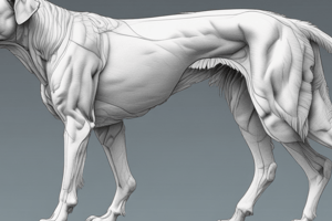
Muscles of the Forelimb
- Muscles of the forelimb can be classified by their location as either extrinsic or intrinsic muscles.
- Extrinsic muscles originate outside of the thoracic limb (e.g., cervical vertebrae), and insert on the thoracic limb (e.g., scapula or humerus).
- Intrinsic muscles both originate and insert on the thoracic limb.
- Extrinsic muscles can be further categorized into superficial and deep groups.
Extrinsic Muscles
-
Superficial Group
-
Brachiocephalicus
- Divided into Cleidobrachialis and Cleidocephalicus
- Cleidobrachialis inserts on the cranial border of the humerus.
- Cleidocephalicus further divides into Pars mastoidea, Pars cervicis, and Pars occipitalis.
- Pars mastoidea inserts on the mastoid process of the skull.
- Pars cervicis inserts on the neck.
- Pars occipitalis inserts on the occipital bone of the skull.
- Action: Lateral and ventral flexion of the head and neck, shoulder extension.
- Innervation: Accessory and Axillary nerves.
-
Omotransversarius: Strap-like muscle adjacent to the Brachiocephalicus.
- Originates on the wing of the atlas (C1).
- Inserts on the distal scapular spine.
- Action: Advances and adducts the limb, laterally flexes the neck.
- Innervation: Accessory nerve.
-
Latissimus dorsi: Broad, flat, triangular muscle caudal to the scapula
- Originates on the thoracolumbar fascia.
- Inserts on the teres major tuberosity.
- Action: Flexes the shoulder; advances the limb.
- Innervation: Thoracodorsal nerve.
-
Trapezius: Broad, flat, triangular muscle caudal to the scapula.
- Divided into Trapezius cervicis and Trapezius thoracis.
- Originates from C2 to T10.
- Inserts on the spine of the scapula.
- Action: Elevates the shoulder, draws the scapula cranially and caudally.
- Innervation: Accessory nerve.
-
Superficial Pectoral: Two parts - descending and transverse.
- Originates on the cranial part of the sternum.
- Inserts on the crest of the greater tubercle.
- Action: Adducts and advances the limb.
- Innervation: Cranial pectoral nerve
-
-
Deep Group
-
Deep Pectoral: Also called Pectoralis Profundus.
- Has cranial and caudal parts.
- Cranial part is deep to the superficial pectoral.
- Caudal part is larger and called the ascending part.
- Originates on the ventral part of the sternum.
- Inserts on the greater and lesser tubercle of the humerus.
- Action: Adducts the limb, retracts the limb, draws the trunk toward the limb.
- Innervation: Caudal pectoral nerve and lateral thoracic nerve.
-
Rhomboideus: Three parts - Cervicis, Thoracis, and Capitis (canines and pigs only).
- Located deep to the Trapezius.
- Originates from C2 to T7 (Capitis from nuchal crest).
- Inserts on the dorsal border of the scapula.
- Action: Elevates the forelimb and draws the scapula against the trunk.
- Innervation: Dorsal branches of cervical and thoracic spinal nerves.
-
Serratus Ventralis: Large, fan-shaped muscle with two parts - cervical and thoracic.
- Originates on the transverse processes of C3-5 and Ribs 1-7 or 8.
- Inserts on the serrated faces of the medial surface of the scapula (facies serrata).
- Action: Protracts and stabilizes the scapula, helps with inspiration.
- Innervation: Long thoracic nerve and ventral branches of cervical spinal nerves.
-
Subclavius: Absent in carnivores, present in horses and goats, smaller in oxen.
-
Intrinsic Muscles
- Intrinsic muscles of the forelimb can be categorized based on their location and action.
Scapular Muscles
-
Scapular muscles act on the shoulder joint.
-
Lateral Muscles:
-
Supraspinatus: Occupies the supraspinous fossa (cranial).
- Acts as the lateral collateral ligament of the shoulder joint for stabilization.
- Action: Extends the shoulder.
-
Infraspinatus: Located in the infraspinous fossa (caudal).
- Action: Externally rotates the shoulder.
-
-
Medial Muscles:
-
Subscapularis: Occupies the subscapular fossa (medial).
- Action: Internally rotates the shoulder.
-
Coracobrachialis: Located between the subscapularis and biceps brachii.
- Action: Adducts the shoulder.
-
-
Caudal Muscles:
-
Teres Major: Located caudal to the scapula, deep to the latissimus dorsi.
- Action: Extends and adducts the shoulder.
-
Teres Minor: Located caudal to the scapula, lateral to the teres major.
- Action: Externally rotates the shoulder.
-
Deltoideus: Located on the caudolateral aspect of the scapula.
- Action: Abducts the shoulder.
-
Antebrachial Muscles
-
Antebrachial muscles act on the carpal and digital joints.
-
Extensor Muscles (Craniolateral)
-
Extensor Carpi Radialis: Extends the carpus, Flexes the elbow.
-
Common Digital Extensor: Extends the carpus and digits.
-
Lateral Digital Extensor: Extends the carpus and lateral digits.
-
Ulnaris Lateralis: Flexes the carpus.
-
-
Flexor Muscles (Caudomedial)
-
Superficial Digital Flexor: Flexes the carpus and digits.
-
Deep Digital Flexor: Flexes the carpus and digits.
-
Flexor Carpi Radialis: Flexes the carpus.
-
Flexor Carpi Ulnaris: Flexes the carpus.
-
-
Pronator Muscles
-
Pronator Teres: Pronates the paw (turn paw downward).
-
Pronator Quadratus: Pronates the paw.
-
-
Supinator Muscles
-
Supinator: Supinates the paw (turn paw upward).
-
Brachioradialis: Supinates the paw.
-
Nerves of the Forelimb
-
Brachial Plexus: Network of nerves formed by the ventral branches of the last three cervical and first two thoracic spinal nerves (C6,7,8 & T1,2).
-
Supplies all muscles of the forelimb except trapezius, omotransversarius, brachiocephalicus, rhomboideus, and the skin over the upper shoulder region.
-
Nerves originating from the brachial plexus:
- Suprascapular Nerve
- Subscapular Nerve
- Musculocutaneous Nerve
- Axillary Nerve
- Lateral Thoracic Nerve
- Radial Nerve
- Median Nerve
- Ulnar Nerve
- Thoracodorsal Nerve
- Pectoral Nerves
- Long Thoracic Nerve
-
Suprascapular Nerve (C6, C7)
- Innervates Supraspinatus and Infraspinatus muscles.
- Muscles actions: Stabilizes and extends the shoulder joint.
- CLINICAL: Susceptible to injury due to over abduction or violent retraction, causing Shoulder Slip/Sweeney.
-
Subscapular Nerve (C6, C7)
- Innervates Subscapularis muscle.
- Muscle action: Adducts the limb and rotates the humerus medially.
- Also, a branch of the Subscapular Nerve, known as the Thoracodorsal Nerve (C8) innervates the Latissimus Dorsi.
-
Musculocutaneous Nerve (C6, C7, C8)
- Innervates Biceps Brachii, Brachialis, and Coracobrachialis muscles.
- Muscles actions: Shoulder extensors and elbow flexors.
- CLINICAL: Dysfunction of musculocutaneous nerve in dogs may cause little change in gait due to redundancy but may make it difficult to offer a paw.
-
Axillary Nerve (C7, C8)
- Innervates Deltoideus, Teres major, Teres minor, and part of the Subscapularis muscles.
- Muscles actions: Flexors of the shoulder joint; Rotates the brachium.
-
Radial Nerve (C7, C8, T1, T2)
- Innervates Extensors of carpus and digits (Extensor carpi radialis, Common digital extensor, Lateral digital extensor), and Triceps Brachii muscle (elbow extension).
- CLINICAL:
- High Radial Nerve Paralysis (more common): Damage occurs proximal to the middle of the arm.
- Low Radial Nerve Paralysis: Damage occurs distal to the triceps brachii innervation site.
-
Median Nerve (C8, T1, T2):
- Innervates most flexors of the carpus and digits (Flexor carpi radialis, Superficial digital flexor, Pronator teres, Pronator quadratus, and the humeral, radial, and ulnar heads of the Deep digital flexor).
- CLINICAL: Dysfunction of median nerve in dogs can cause carpus extension and “raised claws”.
-
Ulnar Nerve (C8, T1, T2)
- Innervates Flexor carpi ulnaris, interosseous muscles, and provides sensory innervation to the caudal forelimb and lateral digits.
- Dorsal branch: Supplies skin on the lateral aspect of the forefoot.
- Palmar branch: Supplies interosseous and other small muscles of the foot, and supplies sensory branches to skin and deep structures.
- Innervates Flexor carpi ulnaris, interosseous muscles, and provides sensory innervation to the caudal forelimb and lateral digits.
-
Lateral Thoracic Nerve (C8, T1):
- Innervates the Cutaneous Trunci muscle and provides sensory innervation to the ventral part of the thorax and abdomen.
- Muscle action: Panniculus reflex
- CLINICAL: Sensory function in the lateral thorax is important for assessing for nerve damage.
-
Long Thoracic Nerve:
- Innervates the Serratus Ventralis muscle.
-
Pectoral Nerves (C6-T1):
- Innervates the superficial and deep pectoral muscles, including the subclavius muscle.
- Muscle action: Adducts the limb.
-
Additional Notes on the Brachial Plexus:
- Number of Spinal Nerves: While mammals have 7 cervical vertebrae, they also possess 8 cervical spinal nerves, with one nerve traveling with each vertebra in the cervical region and the 8th nerve traveling with the first rib.
- Important Note: The specific nerve supply to muscles may vary slightly between different species.
Studying That Suits You
Use AI to generate personalized quizzes and flashcards to suit your learning preferences.


