Podcast
Questions and Answers
What muscle(s) are primarily responsible for the formation of the medial aspect of the nasolabial fold?
What muscle(s) are primarily responsible for the formation of the medial aspect of the nasolabial fold?
- Levator labii superioris alaeque nasi (correct)
- Depressor septi nasi
- Procerus
- Nasalis
What is the functional classification of the trigeminal nerve?
What is the functional classification of the trigeminal nerve?
- GSA, GVE, SVE
- GSA, SVE (correct)
- GSA, GSE
- GVE, SVE
Which muscle's overactivity is often associated with a "gummy smile"?
Which muscle's overactivity is often associated with a "gummy smile"?
- Zygomaticus major
- Levator labii superioris alaeque nasi (correct)
- Levator anguli oris
- Depressor anguli oris
What action does the buccinator muscle perform during chewing?
What action does the buccinator muscle perform during chewing?
Where are the cell bodies of the sensory neurons of the trigeminal nerve located?
Where are the cell bodies of the sensory neurons of the trigeminal nerve located?
Which of the following statements about the trigeminal ganglion is TRUE?
Which of the following statements about the trigeminal ganglion is TRUE?
What is the Modiolus?
What is the Modiolus?
What type of neuron is found in the trigeminal ganglion?
What type of neuron is found in the trigeminal ganglion?
Which of the following muscles are NOT part of the circumorbital and palpebral group?
Which of the following muscles are NOT part of the circumorbital and palpebral group?
Which muscle(s) is/are a common target for Botox injections?
Which muscle(s) is/are a common target for Botox injections?
Which of the following is NOT a division of the trigeminal nerve?
Which of the following is NOT a division of the trigeminal nerve?
Which muscle is NOT directly involved in the action of closing the mouth?
Which muscle is NOT directly involved in the action of closing the mouth?
What type of innervation is provided by the trigeminal nerve to the muscles of mastication?
What type of innervation is provided by the trigeminal nerve to the muscles of mastication?
What is the significance of the danger triangle of the face?
What is the significance of the danger triangle of the face?
What muscle is considered the "muscle of the cheek"?
What muscle is considered the "muscle of the cheek"?
Which muscle is responsible for wrinkling the skin over the bridge of the nose?
Which muscle is responsible for wrinkling the skin over the bridge of the nose?
Which of the following cranial nerves provides motor innervation to the parotid gland?
Which of the following cranial nerves provides motor innervation to the parotid gland?
Which of the following actions is NOT performed by the orbicularis oris muscle?
Which of the following actions is NOT performed by the orbicularis oris muscle?
What branch of the ophthalmic nerve (V1) provides sensory innervation to the medial upper eyelid, medial conjunctiva, and the superior aspect of the nose?
What branch of the ophthalmic nerve (V1) provides sensory innervation to the medial upper eyelid, medial conjunctiva, and the superior aspect of the nose?
Which two branches of the ophthalmic nerve exit the orbit via different pathways?
Which two branches of the ophthalmic nerve exit the orbit via different pathways?
Which nerve is commonly associated with chronic frontal migraines if entrapped or compressed?
Which nerve is commonly associated with chronic frontal migraines if entrapped or compressed?
Which nerve is responsible for providing sensory innervation to the lateral upper eyelid and lateral conjunctiva?
Which nerve is responsible for providing sensory innervation to the lateral upper eyelid and lateral conjunctiva?
What is the name of the opening through which the maxillary nerve (V2) leaves the cranial cavity?
What is the name of the opening through which the maxillary nerve (V2) leaves the cranial cavity?
Which branch of the maxillary nerve (V2) is responsible for providing sensory innervation the skin over the cheek and lateral aspect of the nose?
Which branch of the maxillary nerve (V2) is responsible for providing sensory innervation the skin over the cheek and lateral aspect of the nose?
What is the name of the opening through which the zygomatic nerve exits the orbit?
What is the name of the opening through which the zygomatic nerve exits the orbit?
Which of the following branches of the maxillary nerve (V2) is responsible for providing sensory innervation to the lower eyelid, upper lip, and cheek?
Which of the following branches of the maxillary nerve (V2) is responsible for providing sensory innervation to the lower eyelid, upper lip, and cheek?
Which of the following statements accurately describes the role of the superior cervical ganglion in salivary gland innervation?
Which of the following statements accurately describes the role of the superior cervical ganglion in salivary gland innervation?
Sympathetic innervation of the salivary glands primarily results in:
Sympathetic innervation of the salivary glands primarily results in:
Which of the following anatomical structures is responsible for relaying postganglionic sympathetic fibers to the salivary glands?
Which of the following anatomical structures is responsible for relaying postganglionic sympathetic fibers to the salivary glands?
Which of the following statements accurately describes the origin of preganglionic sympathetic fibers that innervate salivary glands?
Which of the following statements accurately describes the origin of preganglionic sympathetic fibers that innervate salivary glands?
The parasympathetic innervation of salivary glands, unlike sympathetic innervation, primarily results in:
The parasympathetic innervation of salivary glands, unlike sympathetic innervation, primarily results in:
What is the primary function of the SMAS (Superficial Musculo-Aponeurotic System) in relation to the facial nerve?
What is the primary function of the SMAS (Superficial Musculo-Aponeurotic System) in relation to the facial nerve?
Which of these features is NOT directly associated with the buccal fat pad?
Which of these features is NOT directly associated with the buccal fat pad?
Considering the information provided, what is a key distinction between other facial fat and the buccal fat pad, in terms of their behavior with age?
Considering the information provided, what is a key distinction between other facial fat and the buccal fat pad, in terms of their behavior with age?
Identify the correct statement regarding the role of the SMAS in relation to facial expressions and emotions.
Identify the correct statement regarding the role of the SMAS in relation to facial expressions and emotions.
Which of the following statements accurately describes the relationship between the tympanic plexus and the lesser petrosal nerve?
Which of the following statements accurately describes the relationship between the tympanic plexus and the lesser petrosal nerve?
What is the primary function of the parasympathetic innervation of the parotid gland?
What is the primary function of the parasympathetic innervation of the parotid gland?
Which of the following accurately describes the relationship between the lesser petrosal nerve and the otic ganglion?
Which of the following accurately describes the relationship between the lesser petrosal nerve and the otic ganglion?
What is the primary role of the auriculotemporal nerve in parasympathetic innervation of the parotid gland?
What is the primary role of the auriculotemporal nerve in parasympathetic innervation of the parotid gland?
Which of the following statements correctly identifies the pathway of preganglionic parasympathetic fibers from their origin to the otic ganglion?
Which of the following statements correctly identifies the pathway of preganglionic parasympathetic fibers from their origin to the otic ganglion?
What is the significance of the foramen ovale in the context of the glossopharyngeal nerve and parasympathetic innervation?
What is the significance of the foramen ovale in the context of the glossopharyngeal nerve and parasympathetic innervation?
Based on the information provided, which structure is NOT directly involved in the parasympathetic innervation of the parotid gland?
Based on the information provided, which structure is NOT directly involved in the parasympathetic innervation of the parotid gland?
Which of the following best represents the overall flow of parasympathetic signals related to parotid gland function?
Which of the following best represents the overall flow of parasympathetic signals related to parotid gland function?
Flashcards
Buccal Fat Pad
Buccal Fat Pad
A fat structure present in the cheeks that supports suckling in neonates and protects neurovascular structures.
SMAS
SMAS
Superficial Musculo-Aponeurotic System, a network of fibers that supports facial nerve branches and anchors muscles to the skin.
Muscles of Facial Expression
Muscles of Facial Expression
Muscles located within superficial fascia that express emotions and control facial openings.
Glabella
Glabella
Signup and view all the flashcards
Philtrum
Philtrum
Signup and view all the flashcards
Epicranial group
Epicranial group
Signup and view all the flashcards
Occipitofrontalis
Occipitofrontalis
Signup and view all the flashcards
Orbicularis oculi
Orbicularis oculi
Signup and view all the flashcards
Corrugator supercilii
Corrugator supercilii
Signup and view all the flashcards
Procerus
Procerus
Signup and view all the flashcards
Nasalis
Nasalis
Signup and view all the flashcards
Levator labii superioris alaeque nasi
Levator labii superioris alaeque nasi
Signup and view all the flashcards
Zygomaticus major
Zygomaticus major
Signup and view all the flashcards
Buccinator
Buccinator
Signup and view all the flashcards
Modiolus
Modiolus
Signup and view all the flashcards
Danger Triangle
Danger Triangle
Signup and view all the flashcards
Trigeminal Nerve
Trigeminal Nerve
Signup and view all the flashcards
Trigeminal Ganglion
Trigeminal Ganglion
Signup and view all the flashcards
Pseudounipolar Neurons
Pseudounipolar Neurons
Signup and view all the flashcards
Sensory Ganglia
Sensory Ganglia
Signup and view all the flashcards
Motor Ganglia
Motor Ganglia
Signup and view all the flashcards
Ophthalmic Division (V1)
Ophthalmic Division (V1)
Signup and view all the flashcards
Innervation of Facial Muscles
Innervation of Facial Muscles
Signup and view all the flashcards
Ophthalmic Nerve (V1)
Ophthalmic Nerve (V1)
Signup and view all the flashcards
Nasociliary Nerve
Nasociliary Nerve
Signup and view all the flashcards
Frontal Nerve
Frontal Nerve
Signup and view all the flashcards
Lacrimal Nerve
Lacrimal Nerve
Signup and view all the flashcards
Zygomatic Nerve
Zygomatic Nerve
Signup and view all the flashcards
Zygomaticofacial Nerve
Zygomaticofacial Nerve
Signup and view all the flashcards
Infraorbital Nerve
Infraorbital Nerve
Signup and view all the flashcards
Supratrochlear Nerve
Supratrochlear Nerve
Signup and view all the flashcards
Sympathetic innervation
Sympathetic innervation
Signup and view all the flashcards
Inferior salivatory nucleus
Inferior salivatory nucleus
Signup and view all the flashcards
Preganglionic sympathetic neurons
Preganglionic sympathetic neurons
Signup and view all the flashcards
Superior cervical ganglion
Superior cervical ganglion
Signup and view all the flashcards
Postganglionic sympathetic axons
Postganglionic sympathetic axons
Signup and view all the flashcards
Glossopharyngeal Nerve
Glossopharyngeal Nerve
Signup and view all the flashcards
Preganglionic Parasympathetic Fibers
Preganglionic Parasympathetic Fibers
Signup and view all the flashcards
Tympanic Nerve
Tympanic Nerve
Signup and view all the flashcards
Tympanic Plexus
Tympanic Plexus
Signup and view all the flashcards
Lesser Petrosal Nerve
Lesser Petrosal Nerve
Signup and view all the flashcards
Otic Ganglion
Otic Ganglion
Signup and view all the flashcards
Auriculotemporal Nerve
Auriculotemporal Nerve
Signup and view all the flashcards
Study Notes
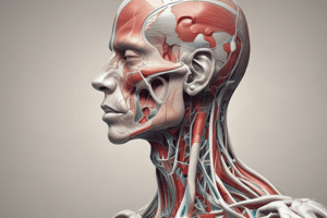
Superficial Face Anatomy
- The superficial face is comprised of facial features, muscles of facial expression, perfusion and drainage patterns, the parotid gland, and its innervation.
- Skin landmarks include the glabella, philtrum, nasolabial folds, mentolabial folds/sulcus, labial commissure, oral fissure, and nasal ala.
Learning Objectives
- List the muscles of facial expression and describe their actions: Understand the significance of these muscles in controlling orbital, nasal, and oral openings, and the role of the SMAS.
- Detail the trigeminal nerve's divisions: Describe the axons, foramina traveled through, branches supplying sensation to the superficial face. Also, note their relationships with other structures.
- Define trigeminal and geniculate ganglia: Understand their specific functions and definitions.
- Describe the pathway of the facial nerve through the skull: Outline its path from the brain, its exit from the skull, and the naming of its branches to the face.
- Describe the external carotid artery branches and internal jugular tributaries: Describe their branching patterns and contributions to facial blood supply and venous drainage.
- Identify the parotid gland and duct: Determine the location of these structures and, importantly, the structures that pass through the gland, along with the autonomic innervation.
Facial Features overview
- Facial features and fascia
- Muscles of facial expression
- Perfusion and drainage patterns
- Parotid gland
- Innervation
Facial Muscles of Expression
- Lie within superficial fascia.
- Originate from bone (or deep fascia) and insert into skin (or other facial muscles).
- Anchored to skin via SMAS.
- Exhibit emotion and control facial openings (sphincters).
- Innervated by branches of the facial nerve.
- Organized into groups: epicranial, circumorbital & palpebral, nasal, and buccolabial.
Muscles of Facial Expression (specific groups)
- Epicranial group: occipitofrontalis & temporoparietalis.
- Circumorbital & palpebral group: orbicularis oculi, corrugator supercilii & levator palpebrae superioris.
- Nasal group: procerus, nasalis & dilator naris anterior.
- Buccalabial group: elevators, evertors, depressors of the lips, orbicularis oris & buccinator.
- Zygomaticus major, zygomaticus minor, risorius, depressor anguli oris and depressor labii inferioris, mentalis.
- Buccinator muscles: chewing/expelling air from cheeks during activities like blowing or chewing, and speaking
- Platysma
Additional Facial Muscles (not facial expression)
- Masseter
- Temporalis
Parotid Gland
- Largest salivary gland.
- Location: anterior to the external ear, superficial to the masseter muscle/ramus of the mandible, extends between zygomatic arch & angle of mandible.
- Structures passing through the parotid gland: facial nerve proper & its branches, auriculotemporal nerve, external carotid artery, superficial temporal vessels, transverse facial vessels, maxillary vessels, and retromandibular vein.
- Salivary gland neoplasms account for 2-6% of head & neck neoplasms. Most (80-90%) are benign. Treatment is typically parotidectomy (total or partial).
Mumps
- Viral infection affecting major salivary glands.
- Initial symptoms similar to the flu, swelling of the glands is the key symptom.
- Prevention via MMR vaccine (~86% effective).
Parotid Duct
- Also called Stenson's duct.
- Travels over the masseter muscle.
- Opens into the oral cavity at the parotid papilla.
Facial Blood Supply and Venous Drainage
- Blood Supply: Predominantly from branches of the external carotid artery (ECA). Includes superior thyroid, ascending pharyngeal, lingual, facial, occipital, posterior auricular, maxillary, and superficial temporal arteries, some contributions from internal carotid artery (ICA).
- Branches of Facial A.: Superior & inferior labial arteries, angular artery.
- Superficial temporal artery and transverse facial artery.
- Venous Drainage: Facial vein continues as angular vein, joining with ophthalmic veins (entering the cavernous sinus). The retromandibular vein merges with the superior and inferior divisions. Retromandibular vein empties into the internal jugular or external jugular regions.
- Danger Triangle: Region on the face that if infected, cavernous sinus infection is possible. Covers area from upper lip to bridge of nose.
Facial Nerve Innervation
- Key concept: provides motor innervation to muscles of facial expression (SVE).
- Arise from pons/geniculate ganglion.
- 6 main branches; Posterior auricular, Temporal, Zygomatic, Buccal, Mandibular, and Cervical.
- Posterior Auricular: does NOT enter parotid gland
- Temporal branches: Frontalis, orbicularis oculi, corrugator supercilii
- Zygomatic branches: Orbicularis oculi, Zygomaticus major & minor
- Buccal branches: Orbicularis oris, buccinator, risorius.
- Mandibular branches: Mentalis, depressor labii inferioris, depressor anguli oris.
- Cervical branches: Platysma.
Trigeminal Nerve
- Largest cranial nerve (CN V) found pons; trigeminal (semilunar) ganglion in middle cranial fossa.
- Sensory components: involved in sensation of face (GSA), & motor in mastication (SVE)
- Three divisions from the ganglion; Ophthalmic V1, Maxillary V2, and Mandibular V3.
- Ophthalmic V1: exits via superior orbital fissure, branches include nasociliary, frontal, and lacrimal.
- Maxillary V2: exits via foramen rotundum, branches include zygomatic nerve (zygomaticofacial and zygomaticotemporal), and infraorbital nerve.
- Mandibular V3: exits via foramen ovale, branches include mental nerve, buccal nerve, and auriculotemporal nerve.
- no direct parasympathetic fibers.
Glossopharyngeal Nerve
- Arise from the medulla exits via jugular foramen.
- Functional components: GSA, GVA, SVE, GVE, SVA.
- GVE originates as fibers from inferior salivary nucleus.
- Innervates, the parotid gland.
- Lesser petrosal nerve: travels through foramen ovale; carries preganglionic parasympathetic fibers to the otic ganglion; postganglionic fibers innervate parotid gland.
- Preganglionic sympathetic nerves from lateral horn of T1-T2 hitchhike on ECA branches to reach the gland.
- Important branches include lesser petrosal, tympanic, lingual and pharyngeal nerves
Studying That Suits You
Use AI to generate personalized quizzes and flashcards to suit your learning preferences.