Podcast
Questions and Answers
Which of the following best describes the relationship between muscle synergists and muscle antagonists?
Which of the following best describes the relationship between muscle synergists and muscle antagonists?
- Synergists oppose movement, while antagonists assist in movement.
- Synergists and antagonists are the same muscles, just activated at different times.
- Synergists and antagonists both contract simultaneously to create rigid movements.
- Synergists assist in the movement of a joint, while antagonists oppose the action of another muscle. (correct)
A patient experiences difficulty initiating voluntary movements after a stroke. Which motor system is MOST likely affected?
A patient experiences difficulty initiating voluntary movements after a stroke. Which motor system is MOST likely affected?
- The pyramidal motor system due to its role in direct voluntary control. (correct)
- The sensory processing system, affecting feedback mechanisms.
- The vestibular system, influencing balance and spatial orientation.
- The extrapyramidal motor system, impacting coordination.
What would be the MOST likely effect of damage to the nigrostriatal pathway?
What would be the MOST likely effect of damage to the nigrostriatal pathway?
- Uncontrolled activation of D2-like neurons within the striatum.
- Increased dopamine release in the basal ganglia, leading to jerky movements.
- Decreased dopamine release in the basal ganglia, potentially leading to Parkinson's-like symptoms. (correct)
- Impaired sensory processing, particularly of tactile stimuli.
Why might a drug that selectively blocks D2 receptors in the basal ganglia affect motor control?
Why might a drug that selectively blocks D2 receptors in the basal ganglia affect motor control?
How does sensory adaptation affect our perception of stimuli?
How does sensory adaptation affect our perception of stimuli?
What distinguishes phasic receptors from tonic receptors?
What distinguishes phasic receptors from tonic receptors?
Damage to the somatosensory cortex would MOST likely result in deficits in which of the following?
Damage to the somatosensory cortex would MOST likely result in deficits in which of the following?
How might increased use of a particular finger (e.g., frequent texting) alter the representation of that finger in the somatosensory cortex?
How might increased use of a particular finger (e.g., frequent texting) alter the representation of that finger in the somatosensory cortex?
Why are Pacinian corpuscles classified as phasic receptors?
Why are Pacinian corpuscles classified as phasic receptors?
If a patient reports a loss of sensation in a specific area of skin, what could be the MOST likely explanation related to dermatomes?
If a patient reports a loss of sensation in a specific area of skin, what could be the MOST likely explanation related to dermatomes?
How is the frequency of a sound wave perceived by the auditory system?
How is the frequency of a sound wave perceived by the auditory system?
What is the function of the ossicles in the middle ear?
What is the function of the ossicles in the middle ear?
How do hair cells in the organ of Corti transduce sound vibrations into electrical signals?
How do hair cells in the organ of Corti transduce sound vibrations into electrical signals?
What is the role of glutamate in auditory signal transmission within the cochlea?
What is the role of glutamate in auditory signal transmission within the cochlea?
How does the auditory system determine the location of a sound?
How does the auditory system determine the location of a sound?
What is the tonotopic organization of the primary auditory cortex (A1)?
What is the tonotopic organization of the primary auditory cortex (A1)?
What is the role of the cupula in the vestibular system?
What is the role of the cupula in the vestibular system?
Which of the following provides information about linear acceleration?
Which of the following provides information about linear acceleration?
How does the vestibular-ocular reflex (VOR) contribute to stable vision during head movements?
How does the vestibular-ocular reflex (VOR) contribute to stable vision during head movements?
Which of the following is NOT a primary input that contributes to the perception of flavor?
Which of the following is NOT a primary input that contributes to the perception of flavor?
What is the function of filiform papillae on the tongue?
What is the function of filiform papillae on the tongue?
How does salt dissolution lead to the perception of salty taste?
How does salt dissolution lead to the perception of salty taste?
How do T1R receptors contribute to taste perception?
How do T1R receptors contribute to taste perception?
What is the role of ATP in the gustatory pathway?
What is the role of ATP in the gustatory pathway?
How do olfactory receptor proteins initiate the sense of smell?
How do olfactory receptor proteins initiate the sense of smell?
What is the function of glomeruli in the olfactory bulb?
What is the function of glomeruli in the olfactory bulb?
How does the vomeronasal organ (VNO) contribute to chemosensation?
How does the vomeronasal organ (VNO) contribute to chemosensation?
What determines the brightness of a light source?
What determines the brightness of a light source?
How do photoreceptors convert light into electrical signals?
How do photoreceptors convert light into electrical signals?
What distinguishes rods from cones in the retina?
What distinguishes rods from cones in the retina?
How do on-center bipolar cells respond to glutamate released by photoreceptors?
How do on-center bipolar cells respond to glutamate released by photoreceptors?
What is the significance of the optic disc in the retina?
What is the significance of the optic disc in the retina?
How does the eye adapt to changes in light levels?
How does the eye adapt to changes in light levels?
How do the three types of cones (S, M, L) contribute to color vision?
How do the three types of cones (S, M, L) contribute to color vision?
What occurs at the optic chiasm?
What occurs at the optic chiasm?
What is the function of the suprachiasmatic nucleus (SCN) in the visual pathway?
What is the function of the suprachiasmatic nucleus (SCN) in the visual pathway?
How does the hierarchical model describe visual processing?
How does the hierarchical model describe visual processing?
What characterizes simple cells in the primary visual cortex (V1)?
What characterizes simple cells in the primary visual cortex (V1)?
Damage to the dorsal stream of the visual cortex would MOST likely result in deficits in which of the following?
Damage to the dorsal stream of the visual cortex would MOST likely result in deficits in which of the following?
What is akinetopsia, and what brain area is MOST associated with it?
What is akinetopsia, and what brain area is MOST associated with it?
According to the Law of Specific Nerve Energies, how is sensory information coded and perceived?
According to the Law of Specific Nerve Energies, how is sensory information coded and perceived?
Flashcards
Skeletal Muscles
Skeletal Muscles
Muscles attached to bones, responsible for voluntary movements.
Tendons
Tendons
Connective tissues attaching muscles to bones.
Muscle Synergists
Muscle Synergists
Muscles that assist in the movement of a joint.
Muscle Antagonists
Muscle Antagonists
Signup and view all the flashcards
Muscle Fibers
Muscle Fibers
Signup and view all the flashcards
Motor Neurons
Motor Neurons
Signup and view all the flashcards
Motor Unit
Motor Unit
Signup and view all the flashcards
Pyramidal Motor System
Pyramidal Motor System
Signup and view all the flashcards
Primary Motor Cortex
Primary Motor Cortex
Signup and view all the flashcards
Non-Primary Motor Cortex
Non-Primary Motor Cortex
Signup and view all the flashcards
Extrapyramidal Motor System
Extrapyramidal Motor System
Signup and view all the flashcards
Nigrostriatal Pathway
Nigrostriatal Pathway
Signup and view all the flashcards
Dopamine Release in the Basal Ganglia
Dopamine Release in the Basal Ganglia
Signup and view all the flashcards
Parkinson's Disease
Parkinson's Disease
Signup and view all the flashcards
Sensory Processing Systems
Sensory Processing Systems
Signup and view all the flashcards
Receptor Cells
Receptor Cells
Signup and view all the flashcards
Sensory Transduction
Sensory Transduction
Signup and view all the flashcards
Receptive Fields
Receptive Fields
Signup and view all the flashcards
Sensory Adaptation
Sensory Adaptation
Signup and view all the flashcards
Phasic Receptors
Phasic Receptors
Signup and view all the flashcards
Tonic Receptors
Tonic Receptors
Signup and view all the flashcards
Sensory Cortex
Sensory Cortex
Signup and view all the flashcards
Somatosensory Cortex
Somatosensory Cortex
Signup and view all the flashcards
Auditory Cortex
Auditory Cortex
Signup and view all the flashcards
Plasticity
Plasticity
Signup and view all the flashcards
Pacinian Corpuscles
Pacinian Corpuscles
Signup and view all the flashcards
Dermatomes
Dermatomes
Signup and view all the flashcards
Sound
Sound
Signup and view all the flashcards
External Ear
External Ear
Signup and view all the flashcards
Middle Ear
Middle Ear
Signup and view all the flashcards
Inner Ear
Inner Ear
Signup and view all the flashcards
Cochlea
Cochlea
Signup and view all the flashcards
Organ of Corti
Organ of Corti
Signup and view all the flashcards
Hair Cells
Hair Cells
Signup and view all the flashcards
Stereocilia
Stereocilia
Signup and view all the flashcards
Generator Potential
Generator Potential
Signup and view all the flashcards
Spiral Ganglion
Spiral Ganglion
Signup and view all the flashcards
EPSPs from Glutamate
EPSPs from Glutamate
Signup and view all the flashcards
Sound Localization
Sound Localization
Signup and view all the flashcards
Auditory Pathways
Auditory Pathways
Signup and view all the flashcards
Study Notes
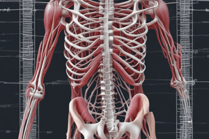
- Skeletal muscles attach to bones, enabling voluntary movements.
- Tendons are connective tissues linking muscles to bones.
- Muscle synergists aid joint movement.
- Muscle antagonists oppose movement, coordinating with synergists.
- Muscle fibers are cells constituting muscles, categorized as slow-twitch or fast-twitch.
- Motor neurons send signals from the spinal cord to muscles.
- A motor unit consists of a motor neuron and its connected muscle fibers.
- The Pyramidal Motor System is a neural pathway for directing voluntary movements.
- The Primary Motor Cortex (M1) in the frontal lobe plans and executes voluntary movements.
- The Non-Primary Motor Cortex assists in motor control.
- The Extrapyramidal Motor System coordinates movement and posture.
- The Nigrostriatal Pathway, a dopamine pathway, regulates movement.
- Dopamine release affects motor control and reward-related movement.
- Parkinson's Disease is a neurodegenerative condition with motor symptoms.
- Sensory Processing Systems detect and interpret environmental stimuli.
- Receptor cells detect and respond to specific sensory stimuli.
- Sensory transduction converts stimuli into electrical signals.
- Receptive fields define the area where a sensory receptor responds.
- Sensory adaptation decreases sensitivity to constant stimuli over time.
- Phasic receptors respond to changing stimuli and adapt quickly.
- Tonic receptors provide continuous signals and adapt slowly.
- The sensory cortex processes sensory information; associated area usually receives information from the thalamus.
- The somatosensory cortex processes bodily sensory input.
- The auditory cortex processes auditory information
- Increased use of a body part can expand its representation in the somatosensory cortex
- Plasticity is how the brain changes to adapt to new experiences.
- Pacinian corpuscles sense pressure and vibration.
- Dermatomes are skin areas supplied by a single spinal nerve root.
- Sound is pressure waves detectable by the ear.
- The external ear gathers sound waves.
- The middle ear transmits vibrations to the inner ear via ossicles.
- The inner ear contains the cochlea for hearing and balance.
- The cochlea converts sound vibrations into neural signals.
- The organ of Corti in the cochlea houses hair cells for hearing.
- Hair cells convert vibrations to electrical signals.
- Stereocilia are hair-like projections crucial for hearing.
- Generator potential is the change in membrane potential in response to stimuli.
- The spiral ganglion transmits auditory information to the brain.
- EPSPs from glutamate can trigger action potentials in neurons.
- Sound localization is the ability to determine a sound's origin.
- Auditory pathways carry sound information from the ear to the brain.
- The primary auditory cortex processes auditory information (A1).
- Tinnitus is ringing or buzzing in the ears.
- The vestibular system contributes to balance and spatial orientation.
- The ampulla in the inner ear contains balance receptors.
- The cupula moves in response to fluid motion.
- The utricle detects horizontal acceleration.
- The saccule detects vertical acceleration.
- Vestibular hair cells release glutamate when stimulated.
- The vestibular-ocular reflex stabilizes vision during head movements.
- Flavor combines taste and smell.
- Papillae on the tongue contain taste buds.
- Filiform papillae do not contain taste buds.
- Microvilli on taste receptor cells increase surface area.
- Taste pores are openings for tastants to interact with receptor cells.
- The five types of tastes are sweet, sour, salty, bitter, and umami.
- Salt dissolves into sodium and chloride ions, triggering ATP release.
- Sour taste results from hydrogen ions, leading to ATP release.
- T1R receptors detect sweet and umami tastes.
- Savory (umami) taste is associated with amino acids.
- Bitter tastes, often from toxic substances, are detected by T2R receptors.
- The gustatory pathway transmits taste information to the brain.
- Olfactory receptor cells detect odor molecules.
- The olfactory bulb processes odor information.
- Glomeruli are structures in the olfactory bulb for olfactory receptor cells to synapse.
- Mitral cells relay information in the olfactory bulb to the brain.
- The primary olfactory cortex processes olfactory information.
- The vomeronasal organ detects pheromones.
- Pheromones are chemical signals affecting same-species behavior.
- Visible light spans electromagnetic wavelengths detectable by the human eye (400-700 nm).
- The retina is the light-sensitive layer at the back of the eye.
- The cornea is the transparent front part of the eye.
- The lens focuses light onto the retina.
- Photoreceptors convert light into electrical signals.
- Bipolar cells transmit signals from photoreceptors to ganglion cells.
- Retinal ganglion cells send visual information to the brain.
- Visual acuity is clarity of vision.
- The optic disc is the blind spot where the optic nerve exits the eye.
- Light level adaptation is the eye's adjustment to light levels.
- Cones mediate color vision.
- Agnosia is the inability to recognize objects or sounds.
- The law of specific nerve energies states that sensation depends on the type of nerve activated.
- Skeletal muscles contract and pull on the skeleton to produce voluntary bodily motion.
- Muscles work together (muscle synergists) or against each other (muscle antagonists).
- Fast-twitch muscle fibers contract quickly but tire easily, whereas slow-twitch fibers contract slowly and tire slowly.
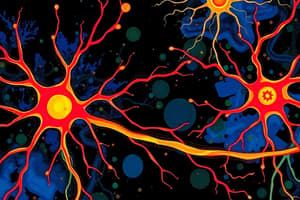
- Motor neurons have cell bodies in the CNS, axons forming peripheral nerves, and axon terminals synapsing on muscle fibers.
- The neuromuscular junction is where motor neurons synapse on muscle fibers (forms on muscle fibers).
- A motor unit comprises a motor neuron and all the muscle fibers it innervates.
- The Pyramidal Motor System involves cell bodies in M1 with axons that form synapses on motor neurons in the CNS to control cranial nerves.
- The Primary Motor Cortex (M1) is located in the precentral gyrus of the frontal lobe; the amount of space taken up in M1 reflects precision of motor control.
- Activity of neurons in M1 is associated with the direction of movement.
- The Non-Primary Motor Cortex includes the Supplementary Motor Area (pre-planned movement) and Premotor Cortex (reactions to external events); plays role in motor planning.
- The Extrapyramidal Motor System regulates the pyramidal motor system via thalamic nuclei.
- Basal ganglia receive dopamine from the midbrain.
- The cerebellum contributes to precision motor control.
- The nigrostriatal pathway involves cell bodies in the substantia nigra sending axons to the dorsal striatum (basal ganglia) where they release dopamine.
- Dopamine activates neurons with D1 receptors and inhibits neurons with D2 receptors; D1-like receptors send "go" signals and D2-like receptors send "stop" signals.
- D1 like neurons are excitatory
- D2 like neurons are inhibitory
- Akinesia is the inability to initiate voluntary motion.
- Bradykinesia is abnormally slow motion.
- Sensory Transduction is the process of receptor cells converting sensory input into electrical signals to be evaluated by the nervous system.
- Phasic Receptors display adaptation and decrease signal, allowing the brain to ignore harmless stimuli.
- Tonic Receptors respond to the presence of stimuli and remind the brain the stimulus is still present, even if it is harmless.
- Somatotopic map is where receptive fields in adjacent areas of skin processed by adjacent areas in the brain.
- Auditory Cortex processes sound information and has a tonotopic map where similar sound frequencies are processed by neighboring areas.
- A somatotopic map is on the primary somatosensory cortex (PSSC)
- Dermatomes are regions of skin supplied by sensory neurons from a single spinal root.
- Pressure waves can be detected by the ear.
- Amplitude indicates a sound wave's volume/intensity.
- Frequency measures a sound wave's pitch in Hertz (Hz).
- External Ear structures collect sound and send it to the middle ear.
- The Pinna is the fleshy exterior part of the external ear and the ear/auditory canal connects the pinna to the eardrum.
- The Middle Ear connects the outer ear to the inner ear.
- Through the three middle ear bones (ossicles), vibrations pass through the Eardrum/Tympanic Membrane
- The Ossicles are three bones through which vibrations pass.
- Vibrations enter the inner ear through the oval window
- The Inner Ear is made up of cochlea and semicircular canals.
- Vibrations pass through the cochlea, a spiral-shaped fluid-filled space where they become pressure waves from the fluid inside.
- In order to detect waves, the organ of Corti is used.
- The organ of Corti a spiral shaped strip of tissue in the cochlea which detects waves and turns them into a signal the nervous system can understand; contains the basilar membrane.
- The Basilar Membrane ripples and flexes in response to waves traveling in the cochlea, with each region responding to certain sound frequencies from highest (base) to lowest (apex).
- Hair Cells are receptors that transduce electrical activity in the basilar membrane.
- The Stereocilia hairs are in the Tectorial membrane, which causes action potential generation.
- Generator Potential is created when sound causes the basilar membrane to flex, leading to hair cell movement relative to the tectorial membrane, which deflects stereocilia, opening gated ion channels.
- Spiral Ganglion are where each hair cell releases neurotransmitters onto nerve endings of multiple spiral ganglion neurons in their cells bodies along the spiral shape of the cochlea.
- Synapses from the cochlea, are where the neurons form onto cochlear nucleus neurons (medulla).
- Spiral ganglia neurons synapse on cochlear nucleus neurons (medulla).
- Axons from cochlear nucleus cross midline.
- Axons synapse on superior olivary nucleus in the pons.
- Neurons of superior olivary nucleus synapse on medial geniculate nucleus neurons (thalamus).
- The process of conveying sensory information to A1 ends with with the information synapsing in the thalamus
- Primary Auditory Cortex, also called A1, contains a topographic map of sensory inputs.
- The map in the Primary Auditory Cortex known as Tonotopic, similar frequencies are processed by adjacent areas in the cortex from low to high (like basilar membrane).
- Receptors that detect head rotation are within,
- The Ampulla is an importatnt structure within detecting head rotation, for balance.
- Stereocilia displacement causes mechanically gated ion channels to open.
- The Utricle and Saccule both detect linear motion, and are located at the base of the semicircular canals.
- Depolarization causes them to release glutamate onto vestibular ganglion neurons.
- Inputs to Flavor consist of Taste (chemicals in food, gustatory), Smell (airborne chemicals, olfactory), and Somatosensory (texture, pain, temperature).
- Located on the papillae are tastebuds and sensory receptors.
- In order to assist with the amount of sensory receptors on the tongue.
- The location of somatosensory receptors, NOT taste buds, are exclusively found in the the filiform papillae.
- Taste buds connect to the tongues surface via taste pores
- Ions and chemicals in food can be detected through taste buds
- Flavors can be categorized as salty, sour, sweet, savory, bitter, based on the chemicals in food. The two chemicals of slat that dissolve into Na+ and Cl- in saliva.
- A generator potential is created by taste receptor cells containing Na channels cells allow Na into, contributing, depolarizing, and opening voltage calcium channels, leading to ATP release.
- The salt dissolves into Sodium and Chlorine, which depolarizes cell, ATP release.
- Acidic flavors can be classified by their concentration of Hydrogen+ ions.
- T1R Receptors bind to sweet chemicals, forming a dimer.
- Activation from G-proteins occur once T1R Receptors are dimerized
- After activation from G-proteins activated T1R Receptors trigger ATP release and depolarize taste receptor cells
- Another name for Umami is Savory
- Umami or Savory is activated and caused by glutamate
- Bitter tastes are caused by chemicals that bind to T2R receptors
- From the taste receptor cells, ATP are released onto Nerve Endings
- Axons are sent to the olfactory bulb upon creation of the receptor.
- Activity of the olfactory receptor happens as neurotransmitters project to the nucleus of the solitary tract
- Located in the temporal lobe is the primary olfactory cortex and contains a olfactory map for processing for each gomerulus.
- Close to the Olfactory Epithelium lies pheromes, as well as chemicals
- Between 400 and 700 nanometers is the range of wavelengths for visible lights
- A wavelength is a key indicator to determine/measure brightness of light.
- The purpose of the Retina tissue is in order to detect and translate information.
- To bend light, a fixed and transparent piece of tissue called cornea is needed to have said effect; that is known as refractional effect
- Photoreceptors that detect light from the back of the eye.
- When there is little or no light, the special cells known as rods are used
- The cells used during day time are know as cones due that they sensitive to bright light. They also translate information from light to color in a higher acuity of vision.
- Bipolar cells can take information from both ends, where as, Ganglion cells outputs information
- Sharp is used to describe the information gathered from cones and transmitted along the cones.
- There is a visual ability due to reflection of light by the tapetum lucidum
- Lack of ability for photoreception causes a blind spot as no photo receptors can be detected.
- An adjustment in change sensitivity from intensity of light is due to light level adaptation. This alters through pupil, allowing more or less light that the organ can handle. There are three sub categories, including Short (S-light), Medium (M-light) and Long (L-light)
- One can be color blind if there is lack in ability to to discern light. Women are less likely, where as, men are more likely to experience this due to red lights and green lights differences.
- Prey animals are able to have eyes located on the sides that allows enhanced/increased view
- The opposite effect is due to Predator animal's eyes being in fron of the head, allowing enhanced depth perception
- Axons, blood vessels and other cells located in both nasal retinas are able to cross at the Optic Chiasm. However, the cells located on temporal do not.
- After being created/processed in RGC/Retinal area -> lateral of thalamus -> Synapses along G Nucleus, or LGN is involved in pathway/travel
- Circadian Rhythm heavily relies on the area of the brain called Suprachiasmatic Nucleus
- There is a hierarchical model of visual processing, which allows to combine information with complex representations.
- There isn't a overall visual center for V processing as fragmented processors work independently
- Visual cortex or V1 is located in the Occidental area, with a map of the work that both eyes can see
- The response of simple cells is dependent on if they are dark or light in a narrow line
- A destruction or deficit created is due from damaged parts of the cortex and in some cases are permanent. To describe, this neurological effect/condition = Agonsia
- A visual cortex made up of feature responders like the Non-Primary V Cortex. This is found in both the second(V2) and the motion of both colors and information from orientation as well
- The route can be the Dorsal stream when traveling along the spinal cord (V2 -> V5)
- In cases where both visual and spatial are processed with the control of motion, the posterior parietal lobe is the area associated with that occurrence
- Another route possible is the Ventral Stream where in this case, the area from V2 -> and V4 -> to the Temporal lobe
- In the recognition of visual stimuli, the temporal lobe area is important for recognizing and labeling
- V5 or the middle temporal, MT, specializes in visual motion
- Known as Motion blindness is due to where action, continuous transitions seems instantaneous jump locations
- Achromatopsia is unable to have color vision
- One of the main functions is to incorporate/integrate with the brain and combine information and details, especially from earlier processing. Allows stand-out basic components of what is processing.
- From areas like PPA. parahippocampal place area, landscape details can be understood. (scenes that object that the system can recognize)
- Faces and similar features fall into area of understanding for FFA or fusiform face area
- A hallucination is a condition that delusional believes in others replaced
- 1835: The theory of specific parts of the nervous system activated determines what information can be absorbed appropriately (Sensory/Neurons)
Studying That Suits You
Use AI to generate personalized quizzes and flashcards to suit your learning preferences.


