Podcast
Questions and Answers
What is the primary neurotransmitter released at the neuromuscular junction by vertebrate lower motor neurons?
What is the primary neurotransmitter released at the neuromuscular junction by vertebrate lower motor neurons?
- GABA
- Dopamine
- Acetylcholine (correct)
- Glutamate
Which characteristic is typical of slow-twitch muscle fibers (Type S)?
Which characteristic is typical of slow-twitch muscle fibers (Type S)?
- Fast, medium-sized twitches with intermediate fatigue resistance
- Small twitches with slow rise and fall times, resistant to fatigue (correct)
- Few mitochondria and reliance on anaerobic metabolism
- Large, brief twitches with high force production
Duchenne's muscular dystrophy is caused by a lack of what?
Duchenne's muscular dystrophy is caused by a lack of what?
- Dystrophin (correct)
- Acetylcholine receptors
- Glycogen
- Mitochondria
What is the significance of the pyramidal decussation in the context of motor control?
What is the significance of the pyramidal decussation in the context of motor control?
What is the functional consequence of lower motor neuron destruction?
What is the functional consequence of lower motor neuron destruction?
How does the CNS adjust the level of contraction in a skeletal muscle on a moment-to-moment basis?
How does the CNS adjust the level of contraction in a skeletal muscle on a moment-to-moment basis?
What is a motor unit?
What is a motor unit?
Which feature of the organization of motor units allows for smooth increases in muscle force?
Which feature of the organization of motor units allows for smooth increases in muscle force?
What is the functional role of Golgi tendon organs in motor control?
What is the functional role of Golgi tendon organs in motor control?
What is meant by the term 'final common pathway' in the context of the motor system?
What is meant by the term 'final common pathway' in the context of the motor system?
The corticobulbar tract primarily controls muscles of the:
The corticobulbar tract primarily controls muscles of the:
What is spasticity, commonly observed after upper motor neuron damage?
What is spasticity, commonly observed after upper motor neuron damage?
Which descending tract is primarily responsible for making postural adjustments in response to vestibular stimulation?
Which descending tract is primarily responsible for making postural adjustments in response to vestibular stimulation?
Which statement describes the general function of upper motor neurons?
Which statement describes the general function of upper motor neurons?
What is the effect of curare on muscle contraction?
What is the effect of curare on muscle contraction?
How do the motor neurons for distal muscles compare somatotopically to those of proximal muscles in the spinal cord?
How do the motor neurons for distal muscles compare somatotopically to those of proximal muscles in the spinal cord?
Which of the following best describes the function of the supplementary motor area (SMA)?
Which of the following best describes the function of the supplementary motor area (SMA)?
What is the Babinski sign, and what does its presence typically indicate in adults?
What is the Babinski sign, and what does its presence typically indicate in adults?
What is the primary mechanism of action of baclofen in treating spasticity?
What is the primary mechanism of action of baclofen in treating spasticity?
A patient exhibits weakness, muscle atrophy, and fasciculations. Where is the most likely site of damage?
A patient exhibits weakness, muscle atrophy, and fasciculations. Where is the most likely site of damage?
Which of the following does NOT originate in motor cortex?
Which of the following does NOT originate in motor cortex?
Which of the following explains why a small woman might have the same absolute amount of type S fibers as a very large man?
Which of the following explains why a small woman might have the same absolute amount of type S fibers as a very large man?
Which of the following is considered to be only involved in voluntary movement?
Which of the following is considered to be only involved in voluntary movement?
Which of the following accurately describes lower motor neuron connections?
Which of the following accurately describes lower motor neuron connections?
The size principle means that lower motor neurons innervating smaller type S units will:
The size principle means that lower motor neurons innervating smaller type S units will:
Damage in the LMN or UMN affect strength? What are some differences?
Damage in the LMN or UMN affect strength? What are some differences?
Reticulospinal tract...
Reticulospinal tract...
When describing upper control, what is the main difference between the parietal lobe and SMA?
When describing upper control, what is the main difference between the parietal lobe and SMA?
What is a jacksonian seizure?
What is a jacksonian seizure?
Damage to corticobulbar affect all motor neurons EXCEPT:
Damage to corticobulbar affect all motor neurons EXCEPT:
Axons descending from motor cortical areas also reach the reticular formation and vestibular nuclei; reaching out to smack a handball, for example,...
Axons descending from motor cortical areas also reach the reticular formation and vestibular nuclei; reaching out to smack a handball, for example,...
Quadriplegic cats can be induced to walk on a treadmill, but inducing humans this way is...
Quadriplegic cats can be induced to walk on a treadmill, but inducing humans this way is...
Lower motor neurons extend from the anterior horn of the spinal cord (or from cranial nerve motor nuclei) directly to...
Lower motor neurons extend from the anterior horn of the spinal cord (or from cranial nerve motor nuclei) directly to...
Damage to facial muscles due to motor cortex, the internal capsule, or the cerebral peduncle on one side causes...
Damage to facial muscles due to motor cortex, the internal capsule, or the cerebral peduncle on one side causes...
The motor cortex is responsible for controlling movements that...
The motor cortex is responsible for controlling movements that...
With upper motor neuron damage, which of the following is correct?
With upper motor neuron damage, which of the following is correct?
Upper motor neurons that are damaged...
Upper motor neurons that are damaged...
What cells live in cigar-shaped clusters in the anterior horn?
What cells live in cigar-shaped clusters in the anterior horn?
Flashcards
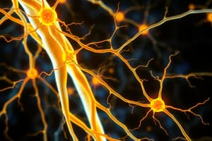
Lower Motor Neurons
Lower Motor Neurons
Neurons that leave the CNS and terminate on skeletal muscle fibers.
Muscle fiber
Muscle fiber
Elongated, multinucleated cell packed with contractile myofibrils, the basic force-producing unit of skeletal muscle.
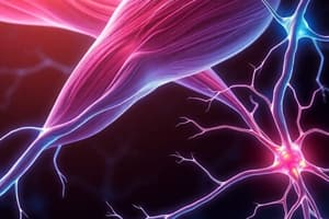
Neuromuscular Junction
Neuromuscular Junction
The location where lower motor neurons form excitatory connections with skeletal muscle targets, releasing acetylcholine.
Dystrophin
Dystrophin
Signup and view all the flashcards
Nicotinic Acetylcholine Receptor
Nicotinic Acetylcholine Receptor
Signup and view all the flashcards
Type S Muscle Fibers
Type S Muscle Fibers
Signup and view all the flashcards
Type FF Muscle Fibers
Type FF Muscle Fibers
Signup and view all the flashcards
Motor Unit
Motor Unit
Signup and view all the flashcards
Reflexes
Reflexes
Signup and view all the flashcards
Muscle Spindle Ia Afferents
Muscle Spindle Ia Afferents
Signup and view all the flashcards
Central Pattern Generators
Central Pattern Generators
Signup and view all the flashcards
Upper Motor Neurons
Upper Motor Neurons
Signup and view all the flashcards
Lower Motor Neurons
Lower Motor Neurons
Signup and view all the flashcards
Corticospinal Tract
Corticospinal Tract
Signup and view all the flashcards
Spasticity
Spasticity
Signup and view all the flashcards
Babinski Sign
Babinski Sign
Signup and view all the flashcards
Amyotrophic Lateral Sclerosis (ALS)
Amyotrophic Lateral Sclerosis (ALS)
Signup and view all the flashcards
Study Notes
- The central nervous system (CNS) manages human movements by coordinating the contraction of numerous muscles.
- This process uses around a million lower motor neurons with axons leaving the CNS and ending on skeletal muscle fibers.
- Some movements such as reflexes, happen automatically while others are adapted for particular situations.
- Automatic movements are controlled by lower motor neurons in the spinal cord or brainstem.
- Less automatic movements need input from the cerebral cortex, alongside the basal ganglia and cerebellum.
- Spinal cord and brainstem overlap for movements, higher centers access them as needed instead of creating every movement from scratch.
Skeletal Muscle
- The fundamental unit for force production in skeletal muscle is the individual muscle fiber which is an elongated, multinucleated cell filled with contractile myofibrils.
- Muscle fiber membranes are excitable and when depolarized to the threshold an action potential spreads across and into the fiber, releasing calcium ions from internal stores.
- Subsequently, calcium ion concentration rises transiently and activates the myofibrils' contractile parts which causes the muscle fiber to twitch.
Neuromuscular Transmission
- Vertebrate lower motor neurons produce excitatory connections to skeletal muscle at neuromuscular junctions, similar to synapses.
- Here, they release acetylcholine onto postjunctional muscle fibers' nicotinic receptors, opposite to invertebrates having both excitatory and inhibitory lower motor neurons.
- Usually, each adult skeletal muscle fiber contains a single neuromuscular junction with multiple release sites.
- As a result, a single action potential in a lower motor neuron leads to significant acetylcholine release, subsequently creating an excitatory postsynaptic potential (EPSP) in the postjunctional muscle fiber.
- The EPSP will be large enough and drives it past the threshold, leading to a solitary muscle action potential and a twitch.
Genetics and Physiology of Muskel Contraction
- Duchenne's muscular dystrophy arises from a deficiency in dystrophin and mechanical instability can lead to tears in the cell membrane, calcium ion entry and cell death.
- Dystrophin links actin filaments inside muscle fibers to a complex of membrane proteins and the extracellular matrix.
- As lower motor neurons’ firing rate increases, twitch frequency and the force produced by muscle fiber increases because successive twitches will summate, also known as temporal summation.
- The sum of excitatory and inhibitory inputs to a lower motor neuron decides the contraction level of muscle fibers innervated by it.
Muscle Fiber Types
- Skeletal muscle fibers come in three types such as slow-twitch (S), fast-twitch (FF) and intermediate (FR) fibers.
- S fibers produce twitches with slow rise and fall times, generate less force but contain many mitochondria, use aerobic metabolism and will maintain force levels for extended periods without tiring.
- FF fibers produce large, short twitches but tire quickly because they contain few mitochondria.
- FR fibers are an intermediate type, producing rapid, medium-sized twitches and have some aerobic capacity and fatigue at an intermediate pace.
- Different species have different proportions of each fiber type and bird that fly long distances contain a high proportion of S fibers and resulting in more "dark meat."
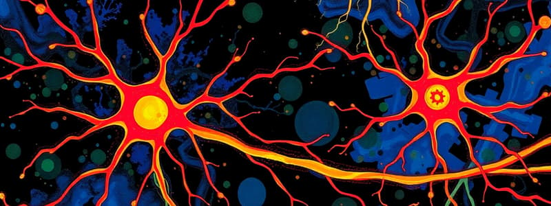
Lower Motor Neurons
- Spinal cord lower motor neurons, also known as alpha motor neurons, reside in cigar-shaped anterior horn clusters.
- Each cluster contains interspersed alpha and gamma motor neurons for one muscle, spanning one to three spinal cord segments.
- Gamma motor neurons for the same muscle are also interspersed and clusters are arranged somatotopically with distal muscles lateral and flexors dorsal to extensors.
- Similarly, neurons for cranial nerve-innervated muscles form nuclei extending through a short piece of the brainstem.
Motor Units
- Each muscle fiber has a neuromuscular junction but each lower motor neuron has terminal branches innervating multiple muscle fibers.
- A motor unit combines a single lower motor neuron and all muscle fibers it innervates which acts as the motor control system's functional output unit.
- The degree of precision needed to control each muscle is reflected in motor unit sizes, which vary within a muscle.
- Large antigravity muscles can have motor units averaging 1000 fibers, in contrast to extraocular muscles containing only 10 fibers.
- All muscle fibers in a unit are of the same type leading to S, FR, and FF types of motor units.
- Larger lower motor neuron cell bodies are present in larger FF units and a size principle dictates motor units are recruited when a muscle contracts more strongly.
- In small neurons, a certain amount of synaptic current creates a larger voltage change than in large neurons and synaptic drive increases, S units reach the threshold first.
- The orderly recruitment to threshold occurs when S fire faster, then FR and then FF units.
- This correlates to force in humans as small forces are produced for long periods for example, and maximal force briefly.
- The force also increases smoothly since the increment to the current force from adding another unit is proportional, increasing weak contraction requires less force, so it adds S units.
Inputs to Lower Motor Neurons
- Each muscle receives input from a pool of lower motor neurons that can be used across multiple movements.
- Arm muscles are used stereotypically such as in reflexes or customized such as in careful manipulations.
- Each lower motor neuron receives inputs from multiple sources.
Pathology and Microbiology
- Viruses travel the CNS by retrograde transport along peripheral nerve fibers which allows some viruses to target a specific cell type like sensory or motor neurons.
- Poliovirus and enteroviruses can target motor neurons causing flaccid paralysis and muscle atrophy at the spinal cord or brainstem infection level.
Reflexes
- Reflexes are involuntary and stereotyped responses to certain sensory stimulation using CNS circuitry and are composed of primary afferent and lower motor neurons.
- The stretch/deep tendon reflex is simple in that muscle spindle afferents from tapping a tendon synapse directly on motor neurons innervating the same muscle.
- All other reflexes use at least one interneuron and as tension in a tendon increases the organ causes reflex inhibition/excitation of neurons supplying said muscle.
- Inhibitory components were though to prevent excessive muscle contraction, but play a subtle role, like in fine motor control required to grasp rocks and eggs.
- The third type of spinal reflex is the withdrawal response to a painful stimulus and is rapid and automatic, involving flexion and multiple muscles and spinal levels.
- Reflexes are adapted in each moment by the CNS as stretch reflexes are suppressed when someone sits down and withdrawal reflexes are also facilitated or suppressed based on the phase someone is in.
Pattern Generators
- Interneurons in reflex circuitry produce parts of complex movements.
- Networks of interneurons in the spinal cord or brainstem create the machinery necessary for rhythmic movements such as walking, breathing and chewing and nonrhythmic movements such facial expressions.
- Specifically, descending pathways from the brainstem can trigger activity in the spinal pattern generator and its rate can be controlled to switch between walking, running and galloping.
- Navigating around obstacles uses descending pathways from the cortex.
- Sensory inputs are required for adjusting the movements to the environment to uneven surfaces.
- The dependency on descending projections from the cortex rises and spinal cord injury patients can have their rhythmic activity improved allowing for better treatment modalities.
Upper Motor Neurons
- Neurons that project directly to lower motor neurons/interneurons that influence lower motor neurons are named upper motor neurons.
- Their cell bodies originate in the brainstem and cerebral cortex allowing them to descend through the brainstem and spinal cord.
Lower Motor Neuron Damage
- Lower motor neuron’s axons act as the route for the CNS to contact skeletal muscles and destruction results in muscle contraction and the resting level of contraction is decreased or abolished.
- Chemical signaling between muscle and the lower motor neurons is disrupted, leading to muscle atrophy and motor units contract spontaneously, early on.
- Later on, muscle fibers produce more acetylcholine receptors, become supersensitive and contract in response to small amounts that cause spontaneous contractions.
Upper Motor Neurons
- Voluntary movement uses planning by the basal ganglia, cerebellum, and cortical areas, needing conscious purposes, along with inputs from frontal areas to the motor cortex.
- The cerebellum also provides signals to ensure accuracy following initiation and damage to association cortex/basal ganglia/cerebellum leads to disorders while weakness is not prominent if association areas aren't damaged.
Descending Pathways
- Upper motor neurons are located in several places and their axons descend different tracts, mainly the corticospinal tract.
- Axons originating in cortical areas descend through the internal capsule, peduncle, pons, and medullary pyramids and 85-90% cross at the pyramids to form the lateral corticospinal tract which mediates voluntary movement.
- A small minority will not cross over or will travel through the anterior funiculus within the anterior corticospinal tract.
- Upper motor neurons traveling through cranial nerve motor nuclei travel through the corticobulbar tract, however the corticobulbar fibers distribute bilaterally, which corresponds with both sides of the head being used together for these muscles.
- Only lower facial muscles which are used in asymmetric facial expressions receive predominantly crossed fibers from corticobulbar fibers causing contralateral weakness if the motor cortex is damaged.
- Reticulospinal tracts from the reticular formation act as important alternatives to corticospinal tracts.
- The lateral vestibulospinal tract projects to any level in the spinal cord and assists in making postural adjustments in response from vestibular stimulation.
- The medial vestibulospinal tract helps stabilize a head as it moves.
- The tectospinal tract orients the individual to periphery stimuli via superior colliculus projections.
- The rubrospinal tract stands in as an alternative to corticospinal in many mammals.
Motor Cortex
- Axons in the corticospinal tract originate in the primary motor cortex (M1) and additional axons from premotor cortex and the supplementary motor area (SMA).
- Different body parts are represented systematically in these motor areas and the map is parallel to that in the somatosensory cortex, with the head lateral and the foot near the longitudinal fissure.
- Corticospinal axons from motor areas affect interneurons that synapse on lower motor neurons.
- Projections mostly reach lower motor neurons to control precise movements.
- Also areas from motor cortical areas reach the reticular formation for background preparatory work.
- Reaching to smack something requires contracting muscles because the center of gravity shifts, requiring postural adjustments being made by the reticulospinal and vestibulospinal tracts from the corticospinal tract's orders beginning before the arm movement.
Upper Motor Neuron Damage
- The effects from damage will depend on where it occurs along the body but can be the cerebral cortex or posterior limb of the internal capsule in the case of arterial occlusion.
- The cortex, basal ganglia, brainstem, spinal cord, and projections are disrupted, resulting in weakness in the distal muscles that depend on corticospinal input.
- This specific weakness is from a syndrome different from lower motor neuron damage like spasticity and unmasked reflexes like the Babinski sign.
- Spasticity is velocity-dependent and worse against flexors of the upper extermity and extensors of the lower extremeties and presents as stretch reflexes and hyperreflexia.
- If a limb is forcibly flexed or extended, resistance provides a clasp-knife response.
- The Babinski sign is dorsiflexion caused by something scratching the foot.
- Spinal cord and brainstem reflexes exist at birth through corticobulbar axons however reflexes get suppressed with development of axons.
- Upper motor neuron damage then allows these connections to persist.
Upper and Lower Motor Neuron Damage Example
- Amyotrophic lateral sclerosis (ALS) attacks both motor neurons which leads to an individual experiencing variable amounts of atrophy and increased reflexes or weakness.
Pharmacology to Treat Spasticity
- By activating metabotropic receptors, stretch reflexes sensitivity is controlled by spinal interneurons to make inhibitory synapses on the central terminals in stretch receptors to reduce amount of glutamate released onto motor neurons.
- Spasticity is effectively reduced with a GABA agonist.
Unusual Upper Motor Neuron Damage
- Damage at the medullary pyramids leads to sparing of projections from the cortex to the basal ganglia, with some upper motor neurons and connections are functional.
- There can still be a Babinski sign but less weakness/spasticity and the largest effect would be on functions normally subserved by cortical projections causing less precise movements.
Studying That Suits You
Use AI to generate personalized quizzes and flashcards to suit your learning preferences.