Podcast
Questions and Answers
The ______ joint allows rotation due to its uniaxial nature.
The ______ joint allows rotation due to its uniaxial nature.
Pivot
The ______ joint allows movement in multiple directions due to its rounded head fitting into a concavity.
The ______ joint allows movement in multiple directions due to its rounded head fitting into a concavity.
Ball and Socket
The ______ joint allows gliding or sliding movements and is usually uniaxial.
The ______ joint allows gliding or sliding movements and is usually uniaxial.
Plane
The ______ joint permits flexion and extension movements only.
The ______ joint permits flexion and extension movements only.
The ______ joint is a biaxial joint that allows movement in two different planes.
The ______ joint is a biaxial joint that allows movement in two different planes.
The most superficial layer of the facial anatomy is the ______ layer.
The most superficial layer of the facial anatomy is the ______ layer.
Beneath the skin layer is the ______ layer, which consists of superficial fat.
Beneath the skin layer is the ______ layer, which consists of superficial fat.
The ______ is the layer responsible for providing structural support and contains muscles.
The ______ is the layer responsible for providing structural support and contains muscles.
The deepest layer in the facial anatomy is made up of ______.
The deepest layer in the facial anatomy is made up of ______.
The ______ layer is absent on the forehead, highlighting its unique anatomical features.
The ______ layer is absent on the forehead, highlighting its unique anatomical features.
Circular muscles have fibers arranged in concentric ______.
Circular muscles have fibers arranged in concentric ______.
The biceps brachii is an example of a ______ muscle.
The biceps brachii is an example of a ______ muscle.
Flat parallel muscles are often accompanied by a fibrous sheet called an ______.
Flat parallel muscles are often accompanied by a fibrous sheet called an ______.
Bipennate muscles have fibers that attach obliquely to a central ______.
Bipennate muscles have fibers that attach obliquely to a central ______.
Unipennate muscles have fibers attached obliquely to a tendon on ______ side only.
Unipennate muscles have fibers attached obliquely to a tendon on ______ side only.
The deltoid muscle is an example of a ______ muscle.
The deltoid muscle is an example of a ______ muscle.
Convergent muscles are wide at one end and narrow at the ______.
Convergent muscles are wide at one end and narrow at the ______.
The digastric muscle has two ______ separated by a tendon.
The digastric muscle has two ______ separated by a tendon.
In the anatomical position, the body is ______.
In the anatomical position, the body is ______.
In the anatomical position, the palms are facing ______.
In the anatomical position, the palms are facing ______.
The thumbs point ______ from the body in the anatomical position.
The thumbs point ______ from the body in the anatomical position.
When a person is laying down face up, they are in the ______ position.
When a person is laying down face up, they are in the ______ position.
The position where a person is lying face down is called ______.
The position where a person is lying face down is called ______.
The biceps brachii is an example of a ______ muscle.
The biceps brachii is an example of a ______ muscle.
Circular muscles have fibers arranged in concentric ______.
Circular muscles have fibers arranged in concentric ______.
Bipennate muscles have fibers that attach obliquely to a central ______.
Bipennate muscles have fibers that attach obliquely to a central ______.
The ______ bone is a flat, thin bone found in the skull, specifically the frontal bone.
The ______ bone is a flat, thin bone found in the skull, specifically the frontal bone.
In the anatomical position, the body is ______.
In the anatomical position, the body is ______.
A ______ bone is a small, flat bone located within the sutures of the skull.
A ______ bone is a small, flat bone located within the sutures of the skull.
The position where a person is lying face down is called ______.
The position where a person is lying face down is called ______.
The ______ bone is cube-shaped and is found in the wrist, such as the carpal bones.
The ______ bone is cube-shaped and is found in the wrist, such as the carpal bones.
An ______ bone, like a vertebra, has a complex shape that allows for flexibility in the spine.
An ______ bone, like a vertebra, has a complex shape that allows for flexibility in the spine.
The ______ bone, such as the patella, is a small, round bone located within a tendon.
The ______ bone, such as the patella, is a small, round bone located within a tendon.
The ______ nerve is found in the posterior part of the leg.
The ______ nerve is found in the posterior part of the leg.
The tibia is located in the ______ compartment of the right leg.
The tibia is located in the ______ compartment of the right leg.
Evertor muscles are located in the ______ compartment of the leg.
Evertor muscles are located in the ______ compartment of the leg.
The ______ flexor muscles are found in the medial section of the leg.
The ______ flexor muscles are found in the medial section of the leg.
Dorsiflexor muscles are located in the ______ compartment of the right leg.
Dorsiflexor muscles are located in the ______ compartment of the right leg.
The diagram depicts the structure of the human ______ and spinal cord.
The diagram depicts the structure of the human ______ and spinal cord.
Cervical nerves originate from the upper ______ area.
Cervical nerves originate from the upper ______ area.
The spinal cord runs down the ______ of the human body.
The spinal cord runs down the ______ of the human body.
Lumbar nerves originate from the lower-______ area.
Lumbar nerves originate from the lower-______ area.
The ______ nerves are located in the lower-back (sacral) region.
The ______ nerves are located in the lower-back (sacral) region.
The lowest nerve pair emanating from the coccyx is called the ______ nerve.
The lowest nerve pair emanating from the coccyx is called the ______ nerve.
The spinal ganglion contains clusters of neuron cell bodies along the ______ cord.
The spinal ganglion contains clusters of neuron cell bodies along the ______ cord.
The cervical enlargement of the spinal cord is located in the ______ area.
The cervical enlargement of the spinal cord is located in the ______ area.
Flashcards are hidden until you start studying
Study Notes
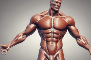
Muscle Types
- Circular muscles have fibers arranged in concentric rings like a circle. The orbicularis oculi (muscle around the eye) is an example.
- Fusiform muscles are spindle-shaped with a thick center and tapering ends, like the biceps brachii (upper arm muscle).
- Flat parallel muscles have fibers arranged parallel to each other and are often accompanied by a fibrous sheet called an aponeurosis. The external oblique muscles in the abdomen are an example.
- Bipennate muscles have fibers attaching obliquely to a central tendon. The rectus femoris (thigh muscle) is an example.
- Unipennate muscles have fibers attached obliquely to a tendon on one side only. The extensor digitorum longus (lower leg muscle) is an example.
- Multipennate muscles have fibers attaching to a central tendon from multiple angles. The deltoid (shoulder muscle) is an example.
- Convergent muscles are wide at one end and narrow at the other (the fibers converge at the tendon). An example is the pectoralis major (chest muscle).
- Some muscles have tendinous intersections where tendons cross over or merge within the muscle. The rectus abdominis is an example.
- Thin parallel muscles have muscle fibers arranged in a parallel pattern but are slender and flat. The sartorius (thigh muscle) is an example.
- Digastric muscles have two bellies separated by a tendon. The omohyoid is an example.
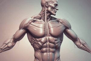
Facial Anatomy Layers
- The skin (epidermis and dermis) layer is the most superficial.
- The superficial fat (subcutaneous) layer is the second layer from the surface.
- The SMAS (superficial musculoaponeurotic system) layer is the third layer from the surface.
- The retaining ligaments and spaces are the fourth layer from the surface.
- The deep fat layer is the fifth layer from the surface and is absent on the forehead.
- The periosteum and deep fascia are the sixth layer from the surface.
- The bones are the deepest layer of facial anatomy.
Joint Types and their Characteristics
- Pivot Joints are uniaxial, allowing rotation. The atlanto-axial joint is an example.
- Ball and Socket Joints are multiaxial, allowing movement in multiple directions. The hip joint is an example.
- Plane Joints are usually uniaxial, allowing gliding or sliding movements. The acromioclavicular joint is an example.
- Hinge Joints are uniaxial, permitting flexion and extension movements only. The elbow joint is an example.
- Saddle Joints are biaxial, allowing movement in two different planes. The carpometacarpal joint is an example.
- Condyloid Joints are biaxial, permitting flexion, extension, abduction, adduction, and circumduction. The metacarpophalangeal joint is an example.
Anatomical Position
-
The anatomical position is:
- Body erect
- Feet slightly apart
- Palms facing forward
- Thumbs point away from the body
- Similar to "standing at attention"
-
Other Positions:
- Supine: Lying down in anatomical position, face up.
- Prone: Face down.
Classification of Bones by Shape
- Flat bones are flat and thin, like the frontal bone of the skull.
- Sutural bones are small, flat bones found within the sutures (joints) of the skull.
- Short bones are small and cube-shaped, like carpal bones in the wrist.
- Irregular bones are complex-shaped, like vertebrae in the spine.
- Sesamoid bones are small, round bones located within a tendon, like the patella (kneecap).
- Long bones are long and cylindrical, like the femur (thigh bone).
Nervous System - Spinal Cord and Brain Diagram
- The brain is the top section of the nervous system, connected to the spinal cord by the brainstem.
- The spinal cord is a long thin structure running down the back.
- Cervical nerves (C1 to C8) originate from the neck area.
- Thoracic nerves (T1 to T12) originate from the upper back.
- Lumbar nerves (L1 to L5) originate from the lower back.
- Sacral nerves (S1 to S5) originate from the sacral region.
- Coccygeal nerve is the lowest nerve pair emanating from the coccyx.
- Cranial nerves (12 pairs) are directly connected to the brain.
- Spinal ganglion is a cluster of neuron cell bodies along the spinal cord containing sensory neurons.
- The cervical enlargement is a wider section of the spinal cord in the neck area.
- The lumbar enlargement is a wider section of the spinal cord in the lower back area.
Anterosuperior View of Right Leg
-
Posterior Compartment:
- Cutaneous nerve
- Fibula
- Deep fascia (outer, circumferential layer)
- Plantar flexor muscles
- Superficial vein
-
Lateral Compartment:
- Subcutaneous tissue (superficial fascia)
- Intermuscular septa
- Evertor muscles
- Neurovascular sheath
-
Medial Compartment:
- Long flexor muscles of foot and ankle
- Interosseous membrane
- Tibia
- Deep fascia blended with periosteum of bone
- Investing fascia of muscle
-
Anterior Compartment:
- Dorsiflexor muscles
- Skin
Studying That Suits You
Use AI to generate personalized quizzes and flashcards to suit your learning preferences.