Podcast
Questions and Answers
Which muscle type is characterized by a striated appearance and is under voluntary control?
Which muscle type is characterized by a striated appearance and is under voluntary control?
- Smooth Muscle
- Skeletal Muscle (correct)
- Cardiac Muscle
- Multinucleated Muscle
What unique feature is associated with cardiac muscle that aids in contraction synchronization?
What unique feature is associated with cardiac muscle that aids in contraction synchronization?
- Nuclei
- Striations
- Intercalated discs (correct)
- Z-discs
Which connective tissue surrounds individual fascicles in skeletal muscle?
Which connective tissue surrounds individual fascicles in skeletal muscle?
- Endomysium
- Sarcolemma
- Perimysium (correct)
- Epimysium
What property of muscle tissue allows it to stretch without damage?
What property of muscle tissue allows it to stretch without damage?
What is the primary function of smooth muscle?
What is the primary function of smooth muscle?
Which component is NOT a part of the skeletal muscle structure?
Which component is NOT a part of the skeletal muscle structure?
What is the primary role of the Z-discs in a sarcomere?
What is the primary role of the Z-discs in a sarcomere?
Which type of muscle tissue contains spindle-shaped fibers and is typically involved in involuntary functions?
Which type of muscle tissue contains spindle-shaped fibers and is typically involved in involuntary functions?
What is the role of the Z-discs in muscle contraction?
What is the role of the Z-discs in muscle contraction?
Which proteins form the troponin complex in thin filaments?
Which proteins form the troponin complex in thin filaments?
What characterizes the A-band in a sarcomere?
What characterizes the A-band in a sarcomere?
What is the primary function of myosin heads during contraction?
What is the primary function of myosin heads during contraction?
What happens to the I-band during muscle contraction?
What happens to the I-band during muscle contraction?
What is the structural role of the M-line in the sarcomere?
What is the structural role of the M-line in the sarcomere?
Which of the following statements is true regarding the H-zone?
Which of the following statements is true regarding the H-zone?
What is the function of tropomyosin in muscle contraction?
What is the function of tropomyosin in muscle contraction?
What happens to the H-zone during muscle contraction?
What happens to the H-zone during muscle contraction?
What role do T-tubules play in muscle contraction?
What role do T-tubules play in muscle contraction?
During the cross-bridge cycle, what occurs during the power stroke phase?
During the cross-bridge cycle, what occurs during the power stroke phase?
What initiates depolarization of the muscle fiber at the neuromuscular junction?
What initiates depolarization of the muscle fiber at the neuromuscular junction?
Which mechanism leads to muscle relaxation?
Which mechanism leads to muscle relaxation?
What is required for continuous muscle contraction?
What is required for continuous muscle contraction?
What condition can lead to muscle fatigue?
What condition can lead to muscle fatigue?
What structure is formed by a T-tubule and two adjacent terminal cisternae of the SR?
What structure is formed by a T-tubule and two adjacent terminal cisternae of the SR?
Flashcards are hidden until you start studying
Study Notes
Muscle Types
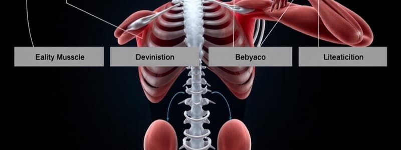
- Skeletal Muscle: Voluntary control, striated appearance, multinucleated fibers, attached to bones via tendons
- Cardiac Muscle: Involuntary control, striated, branched fibers, one to two nuclei per cell, located in heart walls, contains intercalated discs for synchronized contractions
- Smooth Muscle: Involuntary control, non-striated, spindle-shaped fibers, one nucleus, found in walls of hollow organs
Common Muscle Properties
- Contractility: Ability to shorten forcefully
- Extensibility: Capacity to stretch without damage
- Elasticity: Ability to return to resting length after stretching
Skeletal Muscle as an Organ
- Composed of muscle fibers, blood vessels, nerve fibers, and connective tissues
- Connective tissue layers provide support and structure:
- Epimysium: Surrounds entire muscle
- Perimysium: Surrounds individual fascicles
- Endomysium: Encloses individual muscle fibers
Muscle Fibers (Cells)
- Long and cylindrical, up to 100 μm in diameter and 30 cm in length
- Sarcolemma: Plasma membrane
- Sarcoplasm: Cytoplasm
- Sarcoplasmic Reticulum (SR): Specialized ER that stores and regulates calcium ions
The Sarcomere: Functional Unit
- Composed of actin and myosin filaments
- Approximately 2 μm long
- Z-Discs: Attachment points for thin (actin) filaments
- Thin Filaments: Primarily made of actin (G-actin) molecules forming filamentous actin (F-actin)
- Regulatory Proteins:
- Troponin: Binds Ca++ to expose myosin-binding sites on actin
- Tropomyosin: Wraps around actin, blocking myosin-binding sites when relaxed
- Regulatory Proteins:
- Thick Filaments: Made up of myosin proteins with a long tail and globular head
- Myosin Heads: Have ATPase activity for energy during contraction
Sarcomere Components
- M-Line: Central line where thick filaments are anchored
- A-Band: Contains thick filaments and overlapping thin filaments, appears dark under a microscope
- I-Band: Contains only thin filaments, appears light under a microscope
- H-Zone: Central region of the A-band with only thick filaments, appears lighter than the rest of the A-band
Neuromuscular Junction and Excitation-Contraction Coupling
- Neuromuscular Junction: Connection between a motor neuron and a muscle fiber
- Action Potential: Neurotransmitter acetylcholine (ACh) binds to receptors, initiating depolarization and action potential generation
- T-Tubules: Invaginations of the sarcolemma carrying action potential into the muscle fiber
- Triad Structure: T-tubule and two terminal cisternae of the SR, allowing rapid calcium release
Sliding Filament Model of Contraction
- Cross-Bridge Formation: Myosin heads attach to exposed binding sites on actin
- Cross-Bridge Cycle:
- Binding: Myosin heads attach to actin
- Power Stroke: Myosin pulls actin towards the M-line, shortening the sarcomere
- Release: ATP binds to myosin, causing detachment from actin
- Re-cocking: ATP is hydrolyzed, re-energizing the myosin head
- Continuous contraction occurs as long as Ca++ and ATP are available
Muscle Relaxation
- ACh Breakdown: Acetylcholinesterase breaks down ACh, stopping action potential generation
- Ca++ Re-uptake: Calcium is pumped back into SR, restoring tropomyosin covering on actin-binding sites
Muscle Fatigue
- Depletion of ATP or accumulation of metabolic byproducts (e.g., lactic acid)
- Neural factors, such as reduced motor neuron firing can also contribute
Studying That Suits You
Use AI to generate personalized quizzes and flashcards to suit your learning preferences.


