Podcast
Questions and Answers
What is a key feature of smooth muscle cells that distinguishes them from skeletal muscle cells?
What is a key feature of smooth muscle cells that distinguishes them from skeletal muscle cells?
- They are spindle shaped and non-striated. (correct)
- They contain well-organized sarcomeres.
- They have multiple nuclei.
- They possess a striated appearance.
Which of the following triggers contraction in smooth muscle cells?
Which of the following triggers contraction in smooth muscle cells?
- Only electrical impulses.
- Mechanical, electrical, and chemical stimuli. (correct)
- Only chemical stimuli.
- Only mechanical stimuli.
What structural feature is primarily responsible for the striated appearance of skeletal muscle fibers?
What structural feature is primarily responsible for the striated appearance of skeletal muscle fibers?
- Dense bodies.
- Myofibrils arranged in series. (correct)
- Multinucleation of cells.
- Actin filaments surrounding myosin.
How do smooth muscle cells differ in their regenerative capacity compared to other muscle types?
How do smooth muscle cells differ in their regenerative capacity compared to other muscle types?
Which statement accurately describes the nuclei in skeletal muscle fibers?
Which statement accurately describes the nuclei in skeletal muscle fibers?
What is the primary functional unit of a skeletal muscle fiber?
What is the primary functional unit of a skeletal muscle fiber?
Which function is primarily associated with vascular smooth muscle?
Which function is primarily associated with vascular smooth muscle?
The rhythmic contractions of smooth muscle in the digestive tract serve which primary purpose?
The rhythmic contractions of smooth muscle in the digestive tract serve which primary purpose?
What process enables skeletal muscle fibers to attain their large size?
What process enables skeletal muscle fibers to attain their large size?
What distinguishes smooth muscle tissue's contraction compared to skeletal muscle contraction?
What distinguishes smooth muscle tissue's contraction compared to skeletal muscle contraction?
What is the main role of the actomyosin cross-bridge cycle in muscle contraction?
What is the main role of the actomyosin cross-bridge cycle in muscle contraction?
Which connective tissue structure surrounds individual skeletal muscle fibers?
Which connective tissue structure surrounds individual skeletal muscle fibers?
How does cardiac muscle differ from skeletal muscle in terms of regeneration capacity?
How does cardiac muscle differ from skeletal muscle in terms of regeneration capacity?
What characterizes Type I 'red' muscle fibers?
What characterizes Type I 'red' muscle fibers?
What is the composition of the peritendineum in tendons?
What is the composition of the peritendineum in tendons?
What is a key feature of intercalated discs in cardiac muscle tissue?
What is a key feature of intercalated discs in cardiac muscle tissue?
What is the main role of synovial bursae?
What is the main role of synovial bursae?
Which statement about cardiac myocytes is true?
Which statement about cardiac myocytes is true?
What defines the structural feature of fascia in relation to muscle?
What defines the structural feature of fascia in relation to muscle?
What is primarily found in the dark-staining anisotropic A band of a sarcomere?
What is primarily found in the dark-staining anisotropic A band of a sarcomere?
Flashcards
What is a sarcomere?
What is a sarcomere?
The smallest contractile unit of skeletal muscle, responsible for muscle contraction. It's made up of overlapping thick (myosin) and thin (actin) filaments.
What is Perimysium?
What is Perimysium?
A dense connective tissue layer that surrounds a group of muscle fibers (fascicle).
What is an intercalated disc?
What is an intercalated disc?
A specialized cell-to-cell junction in cardiac muscle that allows for the rapid transmission of electrical impulses, creating synchronized contractions.
What is a red muscle fiber?
What is a red muscle fiber?
Signup and view all the flashcards
What are tendinocytes?
What are tendinocytes?
Signup and view all the flashcards
What is a fascia?
What is a fascia?
Signup and view all the flashcards
What are satellite cells?
What are satellite cells?
Signup and view all the flashcards
What is a synovial bursa?
What is a synovial bursa?
Signup and view all the flashcards
What is regeneration capacity?
What is regeneration capacity?
Signup and view all the flashcards
What is the structure of cardiac muscle cells?
What is the structure of cardiac muscle cells?
Signup and view all the flashcards
What is the shape and structure of smooth muscle cells?
What is the shape and structure of smooth muscle cells?
Signup and view all the flashcards
How does smooth muscle contract?
How does smooth muscle contract?
Signup and view all the flashcards
What is the function of smooth muscle in blood vessels?
What is the function of smooth muscle in blood vessels?
Signup and view all the flashcards
What is the function of smooth muscle in the digestive tract?
What is the function of smooth muscle in the digestive tract?
Signup and view all the flashcards
How does smooth muscle regenerate?
How does smooth muscle regenerate?
Signup and view all the flashcards
What is the size and shape of skeletal muscle fibers?
What is the size and shape of skeletal muscle fibers?
Signup and view all the flashcards
Describe the nuclei of a skeletal muscle fiber.
Describe the nuclei of a skeletal muscle fiber.
Signup and view all the flashcards
How are skeletal muscle fibers formed?
How are skeletal muscle fibers formed?
Signup and view all the flashcards
What are myofibrils and what are they composed of?
What are myofibrils and what are they composed of?
Signup and view all the flashcards
What gives skeletal muscle its striated appearance and how is it organized?
What gives skeletal muscle its striated appearance and how is it organized?
Signup and view all the flashcards
Study Notes
Smooth Muscle Tissue
-
Shape and Structure: Small, elongated, spindle-shaped cells with tapered ends; lack striations due to absence of sarcomeres. Actin and myosin filaments are arranged less organized. Dense bodies anchor actin filaments.
-
Function: Specialized for slow, prolonged contractions.
-
Stimulation: Contraction triggered by mechanical (stretching), electrical (nerve impulses), and chemical (hormones) stimuli.
-
Location and Function Examples:
- Vascular smooth muscle regulates blood vessel diameter, impacting blood pressure.
- Digestive tract smooth muscle facilitates peristalsis, moving food through the tract.
- Urinary tract smooth muscle facilitates urination.
-
Regeneration: Possesses high capacity for regeneration; cells can divide and increase in number. Pericytes can differentiate into smooth muscle cells. Cells can also hypertrophy.
Skeletal Muscle Tissue
-
Shape and Structure: Largest cells in the body; single, multinucleated fibers; nuclei located at periphery. Approximately one nucleus every 3μm along the fiber length.
-
Formation: Formed by the fusion of multiple myoblasts during development and growth.
-
Subunit: Myofibril, the structural and functional subunit, composed of precisely aligned myofilaments (myosin-thick and actin-thin filaments).
-
Striations: Exhibit striated appearance due to repeating sarcomeres (contractile units) in longitudinal sections.
-
Sarcomere Structure:
- ~2.5 μm in length in skeletal muscle.
- A fiber (30 cm long) contains a staggering 120,000 sarcomeres.
- I band - light staining, primarily thin filaments.
- A band - dark staining, primarily thick filaments.
- Z line - anchors thin filaments.
-
Contraction: Contraction achieved through the actomyosin cross-bridge cycle.
-
Differentiation and Repair: Muscle fibers (myofibers) are terminally differentiated and don't undergo mitosis. Satellite cells, skeletal muscle stem cells, repair damaged fibers.
Cardiac Muscle Tissue
-
Shape and Structure: Striated, short, cylindrical cells; centrally located single nucleus; connected by intercalated discs. Intercalated discs are specialized cell-to-cell junctions.
-
Size: Cardiomyocytes are smaller than skeletal muscle fibers (~80-100 μm long and ~15 μm in diameter).
-
Sarcomere Length: Resting sarcomere length (about 2.2 μm) is slightly shorter than in skeletal muscle.
-
Function: Specialized cardiac conducting muscle cells rhythmically generate and transmit action potentials.
-
Contraction: Cells can hypertrophy or hypotrophy but cannot divide.
-
Repair: Limited ability to regenerate; heart attack damage results in scar tissue formation.
Muscle Tissue Regeneration Capacity
- Smooth Muscle: High regeneration capacity.
- Skeletal Muscle: Limited regeneration capacity; satellite cells facilitate repair.
- Cardiac Muscle: Minimal regeneration capacity; damage results in scar tissue.
Connective Tissue Layers in Muscle
-
Skeletal Muscle:
- Endomysium: surrounds individual fibers.
- Perimysium: surrounds groups of fibers (fascicles).
- Epimysium: surrounds the entire muscle.
-
Tendon:
- Epitendineum: surrounds the entire tendon.
- Peritendineum: divides fascicles within the tendon.
- Endotendineum: surrounds individual fibers within a fascicle.
Red and White Muscle Fibers
-
Red Muscle Fibers (Type I):
- Rich in capillaries, mitochondria, and myoglobin.
- Adapted for aerobic metabolism, slow-twitch with prolonged contractions and low force.
- Dark colored.
-
White Muscle Fibers (Type II):
- Less dense in capillaries, mitochondria, and myoglobin (pale colored).
- Fast-twitch, high force, but fatigue quickly.
- Adapted for short bursts of anaerobic activity.
Fascia
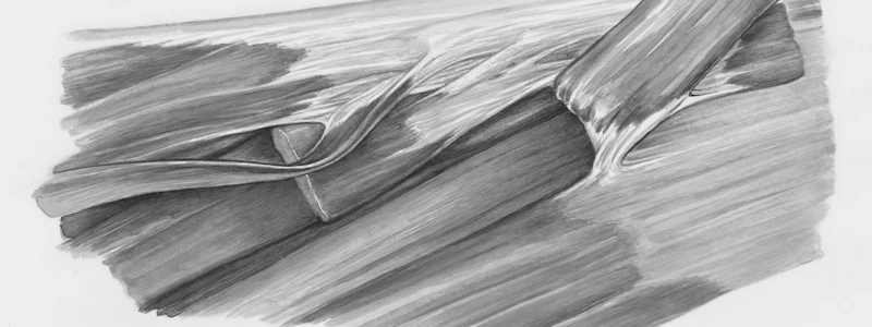
- Dense regular connective tissue with wavy collagen fibers oriented parallel to the direction of pull.
- Surrounds muscles, muscle groups, blood vessels, and nerves.
- Flexible but highly resistant to unidirectional tension.
Tendon Sheath
- Dense regular connective tissue;
- Parallel collagen fibers;
- Fewer blood vessels;
- Tendinocytes (fibroblasts) positioned between collagen bundles.
Synovial Bursa
- Sac-like cavity lined with synovial membrane.
- Secretes lubricating synovial fluid.
- Interposed between tendons and bony prominences, or other friction points.
Studying That Suits You
Use AI to generate personalized quizzes and flashcards to suit your learning preferences.


