Podcast
Questions and Answers
Which of the following connective tissues surrounds bundles of muscle fibers (fascicles)?
Which of the following connective tissues surrounds bundles of muscle fibers (fascicles)?
- Epimysium
- Endomysium
- Perimysium (correct)
- Sarcolemma
What action directly allows myosin to bind to actin?
What action directly allows myosin to bind to actin?
- The binding of calcium to troponin (correct)
- The release of ATP
- The breakdown of ATP by ATPase
- The re-uptake of calcium into the sarcoplasmic reticulum
What is the primary role of the motor end plate?
What is the primary role of the motor end plate?
- To generate ATP for muscle contraction
- To insulate the muscle fiber
- To store acetylcholine for later use
- To form a pocket around the motor neuron by the sarcolemma (correct)
During muscle contraction, what change occurs in the sarcomere?
During muscle contraction, what change occurs in the sarcomere?
What causes the release of calcium ions ($Ca^{++}$) from the sarcoplasmic reticulum (SR)?
What causes the release of calcium ions ($Ca^{++}$) from the sarcoplasmic reticulum (SR)?
What event directly leads to the 'cocking' of the myosin head?
What event directly leads to the 'cocking' of the myosin head?
After the power stroke, what causes the myosin head to detach from actin?
After the power stroke, what causes the myosin head to detach from actin?
What is the role of satellite cells in skeletal muscle?
What is the role of satellite cells in skeletal muscle?
Which of the following is the correct order of events during muscle contraction?
Which of the following is the correct order of events during muscle contraction?
Which of the following actions is NOT a function of skeletal muscle?
Which of the following actions is NOT a function of skeletal muscle?
Flashcards
Epimysium
Epimysium
Outer layer of connective tissue around a skeletal muscle.
Perimysium
Perimysium
Connective tissue that surrounds bundles of muscle fibers (fascicles).
Endomysium
Endomysium
Connective tissue surrounding individual muscle fibers within fascicles.
Basement membrane
Basement membrane
Signup and view all the flashcards
Sarcolemma
Sarcolemma
Signup and view all the flashcards
Satellite Cells
Satellite Cells
Signup and view all the flashcards
Neuromuscular Junction
Neuromuscular Junction
Signup and view all the flashcards
Motor Unit
Motor Unit
Signup and view all the flashcards
Motor End Plate
Motor End Plate
Signup and view all the flashcards
Neuromuscular Cleft
Neuromuscular Cleft
Signup and view all the flashcards
Study Notes
- Topic 8 is about muscle structure and contraction, referring to Chapter 8.
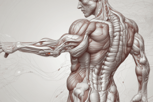
Skeletal Muscle
- The human body contains over 600 skeletal muscles
- Skeletal muscle accounts for 40-50% of total body mass
- Skeletal muscle functions to produce force for locomotion and breathing
- Skeletal muscle functions to produce force for postural support
- Skeletal muscle functions in heat production during cold stress
Connective Tissue Covering Skeletal Muscle
- Epimysium is the outer layer
- Perimysium is connective tissue that surrounds bundles of muscle fibers called fascicles
- Endomysium is connective tissue that surrounds each muscle fiber within the fascicles
- The basement membrane is protective tissue around every fiber under the endomysium
- Sarcolemma is a cell membrane of muscle cells
Satellite Cells
- Located above the sarcolemma but below the basement membrane
- Play a role in muscle growth and repair
- The addition of nuclei increases capacity for protein synthesis
- Important for adaptation to strength training
- Important for adaptation to myonuclear domain
Microstructure of Skeletal Muscle
- Includes the myofibril, satellite cells, and skeletal muscle fiber
- Includes Z line and sarcomere
Microstructure of Skeletal Muscle
- Includes the H zone, M line, Z line, I band, and A band
- Myosin filaments are thick
- Actin filaments are thin
Sarcoplasmic Reticulum and Transverse Tubules
- Includes the sarcolemma, triad of the reticulum, terminal cisternae, transverse tubule, myofibrils, A band, I band, sarcoplasmic reticulum, mitochondria, Z line, and the Nucleus
Neuromuscular Junction
- Includes mitochondria, synaptic vesicles, synaptic cleft, folded sarcolemma, motor end plate, motor neuron fiber, nerve fiber branches, muscle fiber nucleus, and myofibril of muscle fiber
Neuromuscular Junction
- The neuromuscular junction: Area where the motor neuron and muscle fiber connect
- A motor unit is a motor neuron and all the fibers it innervates
- Motor end plate: pocket formed around the motor neuron by the sarcolemma
- The neuromuscular cleft is a gap between the neuron and muscle fiber
- Acetylcholine is released from the motor neuron
- Acetylcholine causes end-plate potential (EPP)
- EPP results in depolarization of the muscle fiber
Muscular Contraction
- Muscular contraction is complex, involving proteins and energy systems
- The end result is actin sliding over the myosin, which causes the muscle to shorten and develop tension
- The sliding filament theory explains muscular contraction
Sliding Filament Theory
- Muscle fibers contract by a shortening of their myofibrils because actin slides over myosin
- The distance between the Z lines reduces
- Cross-bridges between actin and myosin form
- Myosin heads point towards the actin, bind, and pull the actin towards the center causing shortening and force generation
Sliding Filament Theory
- Includes sarcomere, A band, Z line, actin filaments, and myosin filaments
Energy for Contraction
- Energy is required at several steps of the process
- ATP breakdown is done via ATPase on the myosin head
- Energy release energizes the myosin cross-bridge, resulting in actin being pulled towards the center
- A single contraction cycle, or power stroke, of all the cross-bridges in a muscle would only shorten the muscle ~1%, some muscles can shorten up to 60%, shortening must be done many times
Excitation
- Process begins when a nerve impulse arrives at the neuromuscular junction
- Depolarization goes down the transverse tubules deep into the muscle fiber
- Results in the release of Ca++ from the SR
Contraction
- Released Ca++ diffuses into the muscle and binds to troponin
- Troponin lies directly on tropomyosin in the grooves of the actin double strand
- In a relaxed muscle, cross-bridges cannot form because tropomyosin covers binding sites
Contraction
- Stored Ca++ released from the lateral sac of the SR binds to troponin which causes a position change in tropomyosin that reveals the binding sites for cross-bridge formation
- Binding starts energy release leading which causes shortening/contraction
- When a "fresh" ATP attaches, the actin-myosin cross-bridge breaks
- The ATPase hydrolyzes ATP again and provides energy for a new cocking of the myosin head where the cross-bridge reattaches at a new site to generate another power stroke
- Repeats as long as there is Ca available to bind to troponin
Steps of Skeletal Muscle Contraction
- Nerve impulse reaches the neuromuscular junction
- Depolarization sweeps through the transverse (T) tubules
- Ca++ is released from SR
- Ca++ binds to troponin
- Tropomyosin rolls and reveals the site of cross-bridge formation
- ATP binds to myosin, energy is released, and myosin is cocked and energized
- Myosin binds to actin when tropomyosin is gone
- Pi is released, Myosin rotates toward midline and shortening occurs
- ADP is released and myosin remains bound to actin until another ATP binds to myosin
- ATP is broken down by myosin ATPase and shortening occurs
Relaxation
- Signal to stop contraction is the absence of a nerve impulse
- Absence of nerve impulses triggers an energy-requiring Ca++ pump within the SR to resequestor Ca++ back into the SR
- Removing Ca++ from troponin will cause tropomyosin to cover binding sites on actin
Steps of Skeletal Muscle Contraction
- Nerve impulse is gone triggering the re-sequestration of Ca++ into the SR
- Once Ca++ is pulled back into the SR the contraction is over and myosin can't bind anymore because tropomyosin is in the way
Muscle Excitation, Contraction, and Relaxation
- Muscle excitation involves the muscle action potential being propagated along the muscle plasma membrane
- Ca2+ is released during Contraction from the lateral sac of SR and leads to Ca2+ binding to troponin.
- The removal of Ca++ from troponin restores the tropomyosin blocking action during relaxation.
- Cross-bridges move to facilitate the event for the thick filament to bind with actin
Studying That Suits You
Use AI to generate personalized quizzes and flashcards to suit your learning preferences.