Podcast
Questions and Answers
How does the arrangement of fascicles in pennate muscles maximize force production?
How does the arrangement of fascicles in pennate muscles maximize force production?
- Fascicles run parallel to the muscle's long axis, allowing for greater speed of contraction.
- Fascicles are short and attach perpendicularly to the tendon, reducing the overall muscle length.
- Fascicles converge on a single tendon, enabling a large number of fibers to contribute to the pulling force. (correct)
- Fascicles are uniformly distributed across the muscle, providing even force distribution.
Which connective tissue layer contains stretch receptors (muscle spindles) that detect changes in muscle length?
Which connective tissue layer contains stretch receptors (muscle spindles) that detect changes in muscle length?
- Fascia
- Perimysium (correct)
- Endomysium
- Epimysium
Considering the arrangement of muscles around joints, what biomechanical principle necessitates the presence of antagonistic muscle pairs?
Considering the arrangement of muscles around joints, what biomechanical principle necessitates the presence of antagonistic muscle pairs?
- Joints require constant stabilization, provided by the opposing tension of agonist and antagonist muscles.
- Muscles operate using lever systems, requiring opposing forces for efficient movement.
- The nervous system requires feedback from opposing muscles to coordinate movement accurately.
- Individual muscles can only contract to shorten, thus requiring an opposing muscle to restore the initial position. (correct)
How do fixator muscles contribute to the coordinated movement of a joint?
How do fixator muscles contribute to the coordinated movement of a joint?
What determines whether a muscle is classified as intrinsic or extrinsic?
What determines whether a muscle is classified as intrinsic or extrinsic?
Why is the terminology of 'origin' and 'insertion' considered imperfect and potentially misleading when describing muscle attachments?
Why is the terminology of 'origin' and 'insertion' considered imperfect and potentially misleading when describing muscle attachments?
How do intramuscular fat content and glycogen storage in skeletal muscles influence the stabilization of blood glucose concentration?
How do intramuscular fat content and glycogen storage in skeletal muscles influence the stabilization of blood glucose concentration?
How does the structural arrangement of collagen fibers contribute to force transmission in indirect muscle attachments?
How does the structural arrangement of collagen fibers contribute to force transmission in indirect muscle attachments?
Considering the innervation patterns, why is damage to a specific spinal nerve likely to produce a more regional and less specific pattern of muscular deficit than damage to a cranial nerve?
Considering the innervation patterns, why is damage to a specific spinal nerve likely to produce a more regional and less specific pattern of muscular deficit than damage to a cranial nerve?
What is the functional significance of the galea aponeurotica in the context of scalp muscle actions?
What is the functional significance of the galea aponeurotica in the context of scalp muscle actions?
How does the unique course and points of attachment of the platysma muscle influence its role in facial expression and potentially in respiration?
How does the unique course and points of attachment of the platysma muscle influence its role in facial expression and potentially in respiration?
Why is the suprahyoid group of muscles considered essential for both chewing and swallowing?
Why is the suprahyoid group of muscles considered essential for both chewing and swallowing?
How does the coordinated contraction of the pharyngeal constrictor muscles contribute to the swallowing process?
How does the coordinated contraction of the pharyngeal constrictor muscles contribute to the swallowing process?
What is the functional significance of the lumbar region’s iliocostalis, longissimus, and spinalis muscle columns working collectively as the erector spinae?
What is the functional significance of the lumbar region’s iliocostalis, longissimus, and spinalis muscle columns working collectively as the erector spinae?
What mechanical advantage is conferred by the iliotibial tract in stabilizing the knee, especially when the opposite foot is lifted?
What mechanical advantage is conferred by the iliotibial tract in stabilizing the knee, especially when the opposite foot is lifted?
Considering the actions of the gluteus medius and minimus, how do these muscles coordinate to ensure stability during the gait cycle?
Considering the actions of the gluteus medius and minimus, how do these muscles coordinate to ensure stability during the gait cycle?
How does the structural integration of the fascia lata with the tendons of the gluteus maximus and tensor fasciae latae facilitate efficient knee stabilization during locomotion?
How does the structural integration of the fascia lata with the tendons of the gluteus maximus and tensor fasciae latae facilitate efficient knee stabilization during locomotion?
How do the origins and insertions of the gastrocnemius contribute to its dual actions of plantar flexion and knee flexion?
How do the origins and insertions of the gastrocnemius contribute to its dual actions of plantar flexion and knee flexion?
What is the functional significance of the crural muscles being tightly bound by deep fasciae in the leg?
What is the functional significance of the crural muscles being tightly bound by deep fasciae in the leg?
What is the functional implication of the flexor digitorum longus having insertions on the distal phalanges of digits II-V?
What is the functional implication of the flexor digitorum longus having insertions on the distal phalanges of digits II-V?
How do the extrinsic muscles of the hand contribute to the actions of the intrinsic muscles of the hand?
How do the extrinsic muscles of the hand contribute to the actions of the intrinsic muscles of the hand?
Explain how the specific attachments of each muscle within the orbital and nasal region contribute to their distinct functions.
Explain how the specific attachments of each muscle within the orbital and nasal region contribute to their distinct functions.
Fusiform muscles, like the biceps brachii, create more force than parallel muscles due to a higher number of muscle fibers. Agree or Disagree?
Fusiform muscles, like the biceps brachii, create more force than parallel muscles due to a higher number of muscle fibers. Agree or Disagree?
The origin of the frontalis muscle is...
The origin of the frontalis muscle is...
What two actions are performed by the flexor carpi ulnaris?
What two actions are performed by the flexor carpi ulnaris?
The pectoralis minor is known to do what?
The pectoralis minor is known to do what?
What are actions of the buccinator?
What are actions of the buccinator?
The genoglossus performs what actions?
The genoglossus performs what actions?
What are three groups of muscles inside the foot?
What are three groups of muscles inside the foot?
True or False: the levator ani is divided into three portions that are sometimes regarded as separate muscles: the ischiococcygeus, the iliococcygeus, and the pubococcygeus.
True or False: the levator ani is divided into three portions that are sometimes regarded as separate muscles: the ischiococcygeus, the iliococcygeus, and the pubococcygeus.
The Semispinalis capitis and semispinalis cervicis perform what actions?
The Semispinalis capitis and semispinalis cervicis perform what actions?
If the knee is fixed and the popliteus rotates the femur laterally on the tibia, is the body in the sitting position or standing position?
If the knee is fixed and the popliteus rotates the femur laterally on the tibia, is the body in the sitting position or standing position?
At the hip when the trunk is fixed, what is the action of the iliacus muscle?
At the hip when the trunk is fixed, what is the action of the iliacus muscle?
The external abdominal oblique supports which structure?
The external abdominal oblique supports which structure?
Actions of the latissimus dorsi includes what?
Actions of the latissimus dorsi includes what?
How do muscles assist in stability?
How do muscles assist in stability?
The spinal nerves are identified by what?
The spinal nerves are identified by what?
Flashcards
Stability (muscle tissue)
Stability (muscle tissue)
Muscles maintain posture and hold joints in place.
Endomysium
Endomysium
Thin sleeve of loose connective tissue around each muscle fiber.
Perimysium
Perimysium
Thicker connective tissue sheath wrapping muscle fibers into fascicles.
Epimysium
Epimysium
Signup and view all the flashcards
Fascia
Fascia
Signup and view all the flashcards
Fusiform Muscles
Fusiform Muscles
Signup and view all the flashcards
Muscle Compartment
Muscle Compartment
Signup and view all the flashcards
Muscle Origin
Muscle Origin
Signup and view all the flashcards
Muscle Insertion
Muscle Insertion
Signup and view all the flashcards
Prime Mover (Agonist)
Prime Mover (Agonist)
Signup and view all the flashcards
Synergist
Synergist
Signup and view all the flashcards
Antagonist
Antagonist
Signup and view all the flashcards
Fixator
Fixator
Signup and view all the flashcards
Intrinsic Muscle
Intrinsic Muscle
Signup and view all the flashcards
Extrinsic Muscle
Extrinsic Muscle
Signup and view all the flashcards
Frontalis
Frontalis
Signup and view all the flashcards
Occipitalis
Occipitalis
Signup and view all the flashcards
Mentalis
Mentalis
Signup and view all the flashcards
Genioglossus
Genioglossus
Signup and view all the flashcards
Temporalis
Temporalis
Signup and view all the flashcards
Masseter
Masseter
Signup and view all the flashcards
digastric
digastric
Signup and view all the flashcards
geniohyoid
geniohyoid
Signup and view all the flashcards
sternocleidomastoid
sternocleidomastoid
Signup and view all the flashcards
splenius capitis and splenius cervicis
splenius capitis and splenius cervicis
Signup and view all the flashcards
extensors of the neck
extensors of the neck
Signup and view all the flashcards
external intercostals
external intercostals
Signup and view all the flashcards
Mylohyoid
Mylohyoid
Signup and view all the flashcards
Quadratus Lumborum
Quadratus Lumborum
Signup and view all the flashcards
External Abdominal Oblique
External Abdominal Oblique
Signup and view all the flashcards
Rectus Abdominis
Rectus Abdominis
Signup and view all the flashcards
Erector Spinae
Erector Spinae
Signup and view all the flashcards
Ischiocavernosus
Ischiocavernosus
Signup and view all the flashcards
Levator Ani
Levator Ani
Signup and view all the flashcards
pectoralis minor
pectoralis minor
Signup and view all the flashcards
serratus anterior
serratus anterior
Signup and view all the flashcards
extensor carpi radialis longus
extensor carpi radialis longus
Signup and view all the flashcards
extensor carpi radialis brevis
extensor carpi radialis brevis
Signup and view all the flashcards
extensor pollicis longus
extensor pollicis longus
Signup and view all the flashcards
iliacus
iliacus
Signup and view all the flashcards
Study Notes
Structural and Functional Organization of Muscles
- Muscle cells produce force on organs and tissues for movement.
- Tissue types include skeletal, cardiac, and smooth, with at least five main functions:
- Movement
- Stability
- Control of openings/passages
- Heat production (85% of body heat)
- Glycemic control (stabilizing blood glucose levels)
- Skeletal muscles contain connective tissue, nerves, and blood vessels.
- A muscle contains four connective tissue components:
- The endomysium surrounds each muscle fiber
- The perimysium wraps muscle fibers into fascicles
- The epimysium surrounds the entire muscle
- The fascia separates muscles/groups and subcutaneous tissue.
Fascicle Orientation
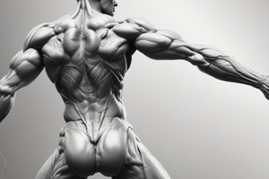
- The orientation of fascicles impacts muscle strength and pull direction and are classified into five categories:
- Fusiform muscles are thick in the middle and tapered at ends
- Parallel muscles have a uniform width and parallel fascicles (strap-like or quadrilateral)
- Triangular (convergent) muscles are fan-shaped (broad origin, narrow insertion)
- Pennate muscles are feather-shaped, with oblique fascicle insertion on a tendon running the muscle's length, including:
- Unipennate: fascicles approach tendon from one side
- Bipennate: fascicles approach tendon from both sides
- Multipennate: shaped like a bunch of feathers with quills converging
- Circular muscles (sphincters) form rings around openings.
- Muscle compartments are groups of related muscles separated by connective tissue fascia
- Intermuscular septa are thick fascia separating one compartment from another.
Skeletal Muscle Attachments
- Skeletal muscles attach to bones via connective tissue extensions:
- Indirect attachment: muscle ends short, gap bridged by a tendon
- Direct (fleshy) attachment: muscle tissue appears to emerge directly from bone
- Most skeletal muscles attach to a different bone at each end, crossing at least one joint.
- The muscle's origin is at the stationary end, while the insertion is at the mobile end, although this terminology is imperfect.
- The belly is the thicker middle region of the muscle.
- Some muscles insert on fascia, tendon, or collagen fibers of the dermis instead of bone.
Muscle Actions
- Skeletal muscle groups coordinate joint control.
- Muscles are classified into these categories based on action:
- Prime mover (agonist): produces most of the force
- Synergist: aids the prime mover by steadying or modifying direction
- Antagonist: opposes the prime mover to control speed/range, and prevent injury
- Fixator: steadies a bone to allow another muscle to pull on something else
- Intrinsic muscles are contained within a region, while extrinsic muscles act on a region from elsewhere.
- Innervation is the identity of the nerve that stimulates a muscle.
Nerve Types
- There are two nerve types:
- Spinal: emerge from the spinal cord and innervate below the neck, identified by letter-number corresponding to adjacent vertebrae and each branch.
- Cranial: emerge from the brain base and innervate head and neck, identified by roman numerals, but not all innervate skeletal muscle.
- Working muscles require good blood supply for glucose, fatty acids, and oxygen.
- Muscle names use Latin to describe structure, location, or action
- Learning the muscular system requires various tactics.
Muscles of the Head and Neck
- Muscles of the head and neck can be grouped as:
- Muscles of facial expression
- Muscles of chewing and swallowing
- Muscles that move the head as a whole
- Facial muscles perform facial expressions and aid oral functions.
- All but one facial muscle is innervated by the facial nerve (CN VII).
- The muscles of the scalp (frontalis and occipitalis) are connected by the galea aponeurotica.
- Other facial and nasal muscles of the orbital region include:
- Orbicularis oculi muscle
- Levator palpebrae superioris
- Corrugator supercilii
- Nasalis
- The oral region contains eight muscles, including:
- Orbicularis oris
- Levator labii superioris
- Levator anguli oris
- Zygomaticus major
- Zygomaticus minor
- Risorius
- Depressor anguli oris
- Depressor labii inferioris
- The mental (chin) and buccal (cheek) regions include:
- Mentalis muscle
- Buccinator muscle
- The platysma is located in the cervical and mental region.
- Muscles of chewing and swallowing aid in food manipulation and tongue movement.
Tongue Muscles
- The tongue includes intrinsic and extrinsic muscles.
- Four extrinsic muscles connect the tongue to the rest of the head.
- Three of the four extrinsic tongue muscles are innervated by the hypoglossal nerve (CN XII).
- There are four extrinsic muscles of the tongue:
- The genioglossus
- The hyoglossus
- The styloglossus
- The palatoglossus
- Four pairs of muscles produce biting and chewing movements:
- Temporalis
- Masseter
- The medial pterygoid
- The lateral pterygoid
- The suprahyoid group contains four pairs of hyoid muscles assisting in chewing and swallowing, including the:
- Digastric
- Geniohyoid
- Mylohyoid
- Stylohyoid
- The infrahyoid group contains four pairs of muscles inferior to the hyoid bone aiding in swallowing, including the:
- Omohyoid
- Sternohyoid
- Thyrohyoid
- Sternothyroid
- The muscles of the pharynx consist of three pairs that act as pharyngeal constrictors.
Muscles of the Head
- Muscles that move the head originate on the vertebral column, thoracic cage, and pectoral girdle and insert on the cranial bones.
- Different contractions of the head can cause contralateral or ipsilateral movements.
- The flexors of the neck include four muscles whose prime mover is the sternocleidomastoid, innervated by the accessory nerve and spinal nerves.
- The three Scalenes (anterior, middle, and posterior), when contracted unilaterally, tilts head toward the same shoulder or rotates face away.
- The extensors of the neck are in the nuchal region.
- The trapezius extends and laterally flexes the neck.
- The splenius capitis and splenius cervicis produce ipsilateral flexion and extend the head.
- The semispinalis capitis and semispinalis cervicis extend and contralaterally rotate the head.
Muscles of the Trunk
- Trunk muscles fall into four categories:
- Respiration
- Abdominal wall support
- Vertebral column movement
- Pelvic floor support
- Muscles of respiration include the diaphragm, external intercostal, internal intercostal, and innermost intercostal muscles.
- The diaphragm separates the thoracic and abdominal cavities.
- Eleven pairs of external and internal intercostal muscles aid with respiration and are innervated by intercostal nerves.
- Muscles of the anterior abdominal wall also include layers of broad flat muscles.
- Abdominal muscles are innervated by the anterior rami of spinal nerves.
- Tendons of oblique and transverse muscles are aponeuroses that enclose the rectus abdominis in the rectus sheath.
- Three lateral abdominal muscles assist with posture and movements of the vertebral column:
- External abdominal oblique supports the abdominal viscera against the pull of gravity, stabilizes the vertebral column, maintains posture, and compresses abdominal organs. Unilateral contraction causes contralateral rotation of the spine.
- Internal abdominal oblique action is the same as the external oblique except that unilateral contraction causes ipsilateral rotation of the waist.
- Transverse abdominal compresses abdominal contents but does not contribute to movements of the vertebral column.
- Rectus abdominis muscles are divided into segments by transverse tendinous intersections.
- Muscles of the back extend, rotate, and laterally flex the vertebral column.
- The most prominent superficial back muscles, such as the latissimus dorsi and trapezius, are concerned with upper limb movements.
- Deeper back muscles include the:
- Iliocostalis
- Longissimus
- Spinalis
- Major deep muscles that assist with back movement include:
- Semispinalis thoracis
- Quadratus lumborum
- Erector spinae muscles
- Multifidus
Pelvic and Shoulder Muscles
- The floor of the pelvic cavity is mainly formed by the levator ani.
- The levator ani can also be divided into three portions:
- Ischiococcygeus
- Iliococcygeus
- Pubococcygeus
- Upper and lower limbs contain numerous muscles for body movement and object manipulation, organized into compartments separated by fasciae.
- The upper limb has anterior and posterior compartments, while the lower limb has anterior, posterior, medial, and lateral compartments.
- Muscles acting on the pectoral girdle originate on the axial skeleton; they are divided into anterior and posterior groups.
- Major muscles of the anterior group are responsible for the abduction of the arm.
- Major muscles making up the posterior group include the superficial trapezius and three deep muscles: the levator scapulae, rhomboid minor, and rhomboid major.
Muscles of the Arm
- Muscles acting on the arm are made up of muscles that cross the shoulder joint and insert on the humerus.
- Two of the nine muscles are considered axial muscles because they originate in the axial skeleton; the other seven comprise the scapular muscles, which originate on the scapula
- These muscles include the prime movers of the shoulder-joint and axilla.
- Two axial muscles include the pectoralis major and the latissimus dorsi.
- Of the seven scapular muscles, three flex, extend, and rotate the arm, and four make up the rotator cuff.
- Three scapular muscles include the:
- Deltoid
- Teres major
- Coracobrachialis
Rotator Cuff Muscles
- The tendons of the four remaining scapular muscles form the rotator cuff
- The first three muscles lie on the posterior side of the scapula, and the fourth occupies the subscapularis fossa on the anterior surface of the scapula
- The supraspinatus
- The infraspinatus
- The teres minor
- The subscapularis
Muscles of the Forearm
- Muscles acting on the forearm are in both the arm and the forearm itself
- The elbow and forearm can perform four motions: flexion, extension, pronation, and supination.
- Muscles with bellies in the arm (brachium) include elbow flexors in the anterior compartment and the elbow extender in the posterior compartment.
- Most forearm muscles (antebrachium) act on the wrist and hand, synergists in elbow flexion/extension and function in pronation/supination.
- Extrinsic muscles are in the forearm, and intrinsic muscles are in the hand itself
- Forearm muscles are divided into anterior and posterior compartments, each further divided into superficial and deep layers.
- The superficial layer of the anterior (flexor) compartment are wrist and finger flexors and arise from a common tendon on the humerus
- Muscles in the superficial layer of the anterior (flexor) compartment include the:
- The flexor carpi radialis
- The flexor carpi ulnaris
- The flexor digitorum superficialis
- The palmaris longus
- The deep layer of the anterior (flexor) compartment includes two flexors: The flexor digitorum profundus and the flexor pollicis longus
- The superficial layer of the posterior (extensor) compartment has five muscles: The extensor carpi radialis longus, the extensor carpi radialis brevis, The extensor digitorum, The extensor digiti minimi, and the extensor carpi unlaris
- The deep layer of the posterior (extensor) compartment: the abductor pollicis longus the extensor pollicis brevis, the extensor pollicis longus, and the extensor indicis
Intrinsic Muscles
- Intrinsic muscles of the hand assist flexors and extensors in the forearm and make finger movements more precise.
- The three groups of intrinsic muscles of the hand are:
- The thenar group
- The hypothenar group
- The midpalmar group
- The hypothenar group muscles form a fleshy mass and is concerned with movement of the little finger. There are also palmar interosseous muscles.
- The muscles are organized into four layers.
- Ventral 1 is the layer encountered beneath the plantar aponeurosis.
- The flexor digitorum brevis is a stout muscle on the midline that flexes digits II-IV and supports the arches. Ventral 2 assists in the extensor hallucis longus Ventral layer 3 contains muscles that mostly serve solely to bend particular toes or to bend digits rather than the joints where the digits join the tarsus.
Muscles of the Hip and Lower Limb
- The largest muscles make actions on the femur and hip joint, the leg and knee joint, extrinsic (leg) muscles acting on the foot and ankle joint, and intrinsic (foot) muscles acting on arches and toes.
- Muscles acting on the hip and femur mostly originate in the hip bone
- Anterior muscles that operate in the hip are mainly the iliacus and psoas major.
- Lateral and posterior hip assist the gluteal muscles.
- There are a total of six lateral hip muscles.
- Medial muscles assist with the hip to aid movement.
- The muscles acting in the knee and leg create actions on the knee joint. Some cross both the hip and knee joints; the other muscles make it easier to move. There are certain muscles known as the hamstrings. The popliteus also unlocks the knees and dislocation of the tibia
- Muscle actions that operate on the foot also aid in the return of blood from the legs. Anterior also prevents the toes on scuffing the ground.
- The crural and peroneal are two leg muscles assisting the foot.
- The intrinsic of the foot act to help support arches. Ventral layer 2,3,4.
Studying That Suits You
Use AI to generate personalized quizzes and flashcards to suit your learning preferences.