Podcast
Questions and Answers
What is the functional unit of a muscle fiber?
What is the functional unit of a muscle fiber?
- Myosin
- Myofiber
- Fascicle
- Sarcomere (correct)
Which of the following protein is responsible for contracting muscles?
Which of the following protein is responsible for contracting muscles?
- Myosin (correct)
- Troponin
- Actin
- Tropomyosin
What is the role of the regulatory proteins in the sarcomere?
What is the role of the regulatory proteins in the sarcomere?
- Provide structure and support to the sarcomere.
- Act as a catalyst for the breakdown of ATP.
- Control the interaction between actin and myosin. (correct)
- Transport calcium ions within the sarcomere.
What is the H zone in the sarcomere?
What is the H zone in the sarcomere?
Which of the following is NOT a characteristic of the light band (I band) of the sarcomere?
Which of the following is NOT a characteristic of the light band (I band) of the sarcomere?
Why is the A band of the sarcomere considered "dark" when viewed under a light microscope?
Why is the A band of the sarcomere considered "dark" when viewed under a light microscope?
Which of the following statements BEST describes the relationship between the sarcomere and the muscle fiber?
Which of the following statements BEST describes the relationship between the sarcomere and the muscle fiber?
What is the function of the Z line in the sarcomere?
What is the function of the Z line in the sarcomere?
What happens to the H zone during muscle contraction?
What happens to the H zone during muscle contraction?
During muscle relaxation, which ion is actively pumped into the sarcoplasmic reticulum?
During muscle relaxation, which ion is actively pumped into the sarcoplasmic reticulum?
What is the role of troponin and tropomyosin in the resting state of muscle contraction?
What is the role of troponin and tropomyosin in the resting state of muscle contraction?
What is the direct consequence of a muscle cell being excited by a nerve impulse?
What is the direct consequence of a muscle cell being excited by a nerve impulse?
What is the role of the T-tubules in muscle contraction?
What is the role of the T-tubules in muscle contraction?
Which of the following is NOT a characteristic of the dihydropyridine receptor (DHPR)?
Which of the following is NOT a characteristic of the dihydropyridine receptor (DHPR)?
What is the primary function of the ryanodine receptor (RyR) in muscle contraction?
What is the primary function of the ryanodine receptor (RyR) in muscle contraction?
What is the role of ATP in the cross-bridge cycling process?
What is the role of ATP in the cross-bridge cycling process?
Which of the following events occurs directly after calcium binds to troponin?
Which of the following events occurs directly after calcium binds to troponin?
What is the primary reason for the loss of tension during muscle relaxation?
What is the primary reason for the loss of tension during muscle relaxation?
Choose the event that directly triggers the power stroke in muscle contraction.
Choose the event that directly triggers the power stroke in muscle contraction.
What is the primary function of smooth muscle?
What is the primary function of smooth muscle?
Which type of muscle is responsible for the movement of food through the digestive system?
Which type of muscle is responsible for the movement of food through the digestive system?
What is the main characteristic of skeletal muscle that makes it suitable for voluntary movement?
What is the main characteristic of skeletal muscle that makes it suitable for voluntary movement?
What feature makes smooth muscle different from skeletal muscle?
What feature makes smooth muscle different from skeletal muscle?
Which type of muscle is responsible for the beating of the heart?
Which type of muscle is responsible for the beating of the heart?
Which of the following agents directly affects the release of acetylcholine?
Which of the following agents directly affects the release of acetylcholine?
The toxin produced by Clostridium botulinum causes which of the following effects?
The toxin produced by Clostridium botulinum causes which of the following effects?
Which of the following agents irreversibly inhibits acetylcholinesterase?
Which of the following agents irreversibly inhibits acetylcholinesterase?
What is the primary cause of respiratory failure in individuals affected by organophosphate poisoning?
What is the primary cause of respiratory failure in individuals affected by organophosphate poisoning?
Which of the following processes is NOT involved in muscle relaxation?
Which of the following processes is NOT involved in muscle relaxation?
What is the mechanism by which curare causes muscle paralysis?
What is the mechanism by which curare causes muscle paralysis?
Which of the following statements about myasthenia gravis is TRUE?
Which of the following statements about myasthenia gravis is TRUE?
What is the role of acetylcholinesterase in neuromuscular transmission?
What is the role of acetylcholinesterase in neuromuscular transmission?
What are the similarities between the neuromuscular junction (NMJ) and a synapse?
What are the similarities between the neuromuscular junction (NMJ) and a synapse?
Which of the following conditions would be most likely to result in muscle spasms and paralysis?
Which of the following conditions would be most likely to result in muscle spasms and paralysis?
What is the difference between an excitatory postsynaptic potential (EPSP) and an excitatory postsynaptic potential (EPP)?
What is the difference between an excitatory postsynaptic potential (EPSP) and an excitatory postsynaptic potential (EPP)?
What is the primary function of the dihydropyridine (DHP) receptors in muscle contraction?
What is the primary function of the dihydropyridine (DHP) receptors in muscle contraction?
Which of the following is NOT a similarity between the NMJ and a synapse?
Which of the following is NOT a similarity between the NMJ and a synapse?
Which of the following statements BEST describes the mechanism of action of Botox®?
Which of the following statements BEST describes the mechanism of action of Botox®?
Which of these describes the immediate energy source for muscle contraction?
Which of these describes the immediate energy source for muscle contraction?
Which energy system is responsible for a fraction of the energy needed for sustained muscle activity?
Which energy system is responsible for a fraction of the energy needed for sustained muscle activity?
Which of the following events occurs FIRST in the process of muscle relaxation?
Which of the following events occurs FIRST in the process of muscle relaxation?
What is the main difference in the energy production between the immediate and the nonoxidative energy systems?
What is the main difference in the energy production between the immediate and the nonoxidative energy systems?
Which of the following conditions is characterized by an inability to relax muscles after contraction?
Which of the following conditions is characterized by an inability to relax muscles after contraction?
Which of the following statements is TRUE regarding the nonoxidative energy system?
Which of the following statements is TRUE regarding the nonoxidative energy system?
What is the primary function of the sarcolemma in muscle contraction?
What is the primary function of the sarcolemma in muscle contraction?
Identify the enzyme responsible for creatine phosphate's conversion to ATP.
Identify the enzyme responsible for creatine phosphate's conversion to ATP.
How does the release of calcium from the sarcoplasmic reticulum lead to muscle contraction?
How does the release of calcium from the sarcoplasmic reticulum lead to muscle contraction?
Which of the following statements about the neuromuscular junction is TRUE?
Which of the following statements about the neuromuscular junction is TRUE?
Which type of muscle fiber is primarily responsible for powerful, coarse movements?
Which type of muscle fiber is primarily responsible for powerful, coarse movements?
What is the primary factor responsible for the initial gains in strength during a strength training program?
What is the primary factor responsible for the initial gains in strength during a strength training program?
What is the relationship between the size of a motor unit and the force it generates?
What is the relationship between the size of a motor unit and the force it generates?
Which of the following statements correctly describes the size principle of motor neuron recruitment?
Which of the following statements correctly describes the size principle of motor neuron recruitment?
Which of the following muscle fiber types has the highest oxidative capacity?
Which of the following muscle fiber types has the highest oxidative capacity?
Which of the following best describes the function of myoglobin in muscle tissue?
Which of the following best describes the function of myoglobin in muscle tissue?
Which of the following is NOT a characteristic of Type II fibers?
Which of the following is NOT a characteristic of Type II fibers?
What is the primary event that triggers muscle hypertrophy?
What is the primary event that triggers muscle hypertrophy?
Which of the following scenarios would illustrate the concept of motor unit recruitment?
Which of the following scenarios would illustrate the concept of motor unit recruitment?
Which of the following is the most accurate description of a motor unit?
Which of the following is the most accurate description of a motor unit?
Flashcards
Muscle Cells
Muscle Cells
Specialized cells responsible for contraction and movement in the body.
Skeletal Muscle
Skeletal Muscle
A type of muscle that contracts voluntarily for movement of the skeleton.
Cardiac Muscle
Cardiac Muscle
Involuntary muscle found only in the heart, responsible for pumping blood.
Smooth Muscle
Smooth Muscle
Signup and view all the flashcards
Muscle Contraction Functions
Muscle Contraction Functions
Signup and view all the flashcards
Muscle Tissue Types
Muscle Tissue Types
Signup and view all the flashcards
Skeletal Muscle Composition
Skeletal Muscle Composition
Signup and view all the flashcards
Muscle Fiber
Muscle Fiber
Signup and view all the flashcards
Sarcomere
Sarcomere
Signup and view all the flashcards
Myofilaments
Myofilaments
Signup and view all the flashcards
A Band
A Band
Signup and view all the flashcards
I Band
I Band
Signup and view all the flashcards
Z Line
Z Line
Signup and view all the flashcards
M Line
M Line
Signup and view all the flashcards
H Zone
H Zone
Signup and view all the flashcards
Ca2+ Influx
Ca2+ Influx
Signup and view all the flashcards
T-Tubules
T-Tubules
Signup and view all the flashcards
Dihydropyridine Receptors (DHPR)
Dihydropyridine Receptors (DHPR)
Signup and view all the flashcards
Troponin
Troponin
Signup and view all the flashcards
Cross-bridge Cycle
Cross-bridge Cycle
Signup and view all the flashcards
Power Stroke
Power Stroke
Signup and view all the flashcards
Actin Binding Site
Actin Binding Site
Signup and view all the flashcards
ATP Hydrolysis
ATP Hydrolysis
Signup and view all the flashcards
NMJ
NMJ
Signup and view all the flashcards
Excitatory Postsynaptic Potential (EPP)
Excitatory Postsynaptic Potential (EPP)
Signup and view all the flashcards
Graded Potential
Graded Potential
Signup and view all the flashcards
Synapse vs. NMJ
Synapse vs. NMJ
Signup and view all the flashcards
Nonoxidative Energy Sources
Nonoxidative Energy Sources
Signup and view all the flashcards
Glycogenolysis
Glycogenolysis
Signup and view all the flashcards
Immediate Energy System
Immediate Energy System
Signup and view all the flashcards
Lactic Acid
Lactic Acid
Signup and view all the flashcards
Type I Muscle Fibers
Type I Muscle Fibers
Signup and view all the flashcards
Type II Muscle Fibers
Type II Muscle Fibers
Signup and view all the flashcards
Motor Unit
Motor Unit
Signup and view all the flashcards
Motor Unit Recruitment
Motor Unit Recruitment
Signup and view all the flashcards
Size Principle
Size Principle
Signup and view all the flashcards
Neural Adaptations
Neural Adaptations
Signup and view all the flashcards
Type IIa Muscle Fibers
Type IIa Muscle Fibers
Signup and view all the flashcards
Type IIx Muscle Fibers
Type IIx Muscle Fibers
Signup and view all the flashcards
Muscle Contraction Force
Muscle Contraction Force
Signup and view all the flashcards
Skeletal Muscle Mechanics
Skeletal Muscle Mechanics
Signup and view all the flashcards
Excitation-Contraction Coupling
Excitation-Contraction Coupling
Signup and view all the flashcards
Role of ATP
Role of ATP
Signup and view all the flashcards
ACh Breakdown
ACh Breakdown
Signup and view all the flashcards
SERCA Pump
SERCA Pump
Signup and view all the flashcards
Actin's Binding Sites
Actin's Binding Sites
Signup and view all the flashcards
Rigor Mortis
Rigor Mortis
Signup and view all the flashcards
Black Widow Spider Venom
Black Widow Spider Venom
Signup and view all the flashcards
Clostridium Botulinum Toxin
Clostridium Botulinum Toxin
Signup and view all the flashcards
Curare
Curare
Signup and view all the flashcards
Myasthenia Gravis
Myasthenia Gravis
Signup and view all the flashcards
Organophosphates
Organophosphates
Signup and view all the flashcards
Respiratory Failure
Respiratory Failure
Signup and view all the flashcards
Dihydropyridine Channels
Dihydropyridine Channels
Signup and view all the flashcards
Return to Resting State
Return to Resting State
Signup and view all the flashcards
Acetylcholine (ACh)
Acetylcholine (ACh)
Signup and view all the flashcards
Study Notes
Skeletal Muscle Thought Questions Answers
-
- h. Actin
-
- e. Myosin
-
- b. T-tubule
-
- b. T-tubule
-
- d. Sarcoplasmic reticulum
-
- f. Troponin
-
- g. Tropomyosin
-
- c. ATP
Muscle
- Almost all cells have intracellular machinery for movement
- The contraction specialists of the body are muscle cells
- Skeletal
- Cardiac
- Smooth Muscle Cells
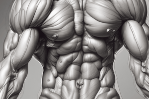
- Skeletal muscle fibers have a highly developed ability to contract, develop tension, and do work.
Contraction of Muscle Allows
- Purposeful movement of the body in relation to the environment
- Manipulation of external objects
- Propulsion of contents through hollow organs
- Emptying the contents of organs to the environment
Muscle Composition
- Muscle comprises the largest group of tissues in the body
- ~ 40% body weight in men, ~32% in women
- Skeletal muscle: 660 in the adult human
- Smooth and Heart = 10%
Muscle Classification
- Functional vs Structural
- Skeletal Muscle - Voluntary
- Cardiac Muscle - Involuntary
- Smooth Muscle - Involuntary
Organization of the Nervous System
- The nervous system is comprised of the central nervous system (CNS) and the peripheral nervous system (PNS)
- The CNS includes the brain and spinal cord.
- The PNS consists of all the nerve fibers branching out from the CNS, connecting it to the rest of the body, transmitting sensory stimuli.
- Different divisions branch out from the CNS and PNS to control different functions, and effector organs will carry out orders.
Levels of Organization in Skeletal Muscle
- Whole muscle = an organ
- Muscle fiber = a cell
- Relatively large (up to 2.5 ft)
- Multinucleated to maintain high protein production
- Myofibril = intracellular structure
- Thick and thin filaments = myofilaments
- Myosin and actin = contractile proteins
Contractile Proteins
- Thick filaments: several hundred myosin (heads and tails)
Thin Filaments
- Mostly actin (helix)
- Regulatory proteins: tropomyosin and troponin
Changes in Banding Pattern During Shortening
- Light microscope: light and dark bands
- Relaxed: H zone, I band, A band
- Contracted: H zone shorter, I band shorter, A band same width
- Sarcomere shorter
Medical Mnemonic
- Muscle sarcomere: Only one vowel in dark and light (dark = A band, light = I band)
- Muscle sarcomere bands: "Zee Intelligent Animal Has Muscle" (ZIAHM) --> From the Z line working inward (ZIAHM)
Questions
-
Which of the following is the light band of the sarcomere?
-
I band
-
Which of the following remains the same width during contraction?
-
A band
-
Which of the following describes a sarcomere?
-
All of the above
Molecular Basis of Muscle Contraction
- Resting State: Ca2+ absent in sarcoplasm; SERCA actively pumps Ca2+ into the SR; no attachment between actin & myosin filaments; muscle cell is relaxed
Excitation-Contraction Coupling
- Excitation of Muscle Cell: Propagated action potential; Acetylcholine is released in the cleft; Acetylcholine binds to receptors on the motor end plate; cation channels open; Na+ moves in EPP
- The Result of Excitation: Specialized membranes take AP from the surface to the center of the cell; SR = modified ER, consists of interconnecting tubules surrounding each myofibril. T-tubule runs perpendicular to surface.
Excitation-Contraction Coupling: Spread of AP down T-tubules, activates receptors, DHP receptor, voltage-gated Ca2+ channel.
Excitation-Contraction Coupling: Steric Block Model
- Ca2+ binds to troponin
- Troponin changes its shape
- Troponin-tropomyosin complex is physically pulled aside
- Actin's binding sites uncovered
Excitation-Contraction Coupling: Cross-bridge Cycling
- Before X-bridge ever links: ATP hydrolyzed; ATP and P remain attached to myosin; Energy stored in X-bridge (“cocked” and ready to be fired). Steps: Energized, Resting, Binding, Bending, Detachment
Excitation-Contraction Coupling – Cross-bridge Cycling (Continued)
- Binding of fresh ATP breaks linkage between actin and myosin
Which is responsible for removing the steric inhibition for the act of contraction?
- Ca2+
Which is responsible for detachment?
- ATP
Excitation-Contraction Coupling – Return to Resting State
- Neural excitation stops: - Acetylcholine released by axon of motor neuron crosses cleft and binds to receptors on motor end plate
- Previously released ACh is broken down by AChase
- SERCA pumps Ca2+ back
- Dihydropyridine channels close; diffusion of Ca2+ out of SR stops
- Actin's binding sites are covered; actin slides back to relaxed position away from center of sarcomere
All of the following result in muscle relaxation EXCEPT:
- No more ATP or Rigor Mortis
Agents & Diseases that Affect the NMJ
- Alters the Release of ACh: Black widow spider venom, Clostridium botulinum toxin
- Blocks ACh Receptor: Curare, Myasthenia Gravis
- Prevents Inactivation of ACh: Organophosphates (certain pesticides and nerve gases)
Synapse vs NMJ
- Similarities: 2 excitable cells separated by a narrow cleft; axon terminals store NT released by Ca2+ induced exocytosis; NT binds to receptors on the membrane, which open channels
- Differences: Synapse – junction between 2 neurons, NMJ – junction between a motor neuron and a skeletal muscle fiber; NMJ is always excitatory (EPP); Synapse is excitatory (EPSP) or inhibitory (IPSP)
Skeletal Muscle has 3 Energy Systems
-
- Immediate: ATP and CP, available immediately to drive muscle contraction
-
- Nonoxidative: breakdown of glucose and glycogen
-
- Oxidative: breakdown of carbohydrates, fats, & certain amino acids
Muscle Fiber Types
- Based on: Speed of contraction (myosin ATPase activity), Type of metabolic pathway
- Type I: slow twitch, slow oxidative (ST), SO
- Type IIa: fast twitch, fast oxidative/glycolytic (FOG), FTa
- Type IIx: fast twitch, fast glycolytic (FG), FTx
Type I vs Type II
- Type I: Slow twitch, oxidative metabolism, ~50% of fibers, used for endurance-type activities, Soleus muscle
- Type II: Fast twitch, Type IIa: moderate high ox capacity, high glycolytic capacity, ~25% fibers, used for higher intensity movements
- Type IIx: low ox capacity, highest glycolytic capacity ~25% fibers, used for explosive movements
Motor Units
- Motor unit = 1 motor neuron + all the muscle fibers it innervates
- When a motor neuron is activated, all fibers it supplies contract simultaneously
- For stronger contractions, more motor units are recruited
Motor Unit Recruitment
- Method for altering force production: Less force production - smaller motor units (type I); More force production - larger motor units (type II)
Size Principle of Motor Neuron Recruitment
- The CNS increases muscle force by activating additional motor units in the order of their increasing size, starting with the smallest. Recruitment order: type I, type Ila, type IIx
Increased use: strength training
- Neural adaptations must take place before changes in muscle size
- Early gains in strength due to neural factors. Later, increasing cross-sectional area (hypertrophy) becomes more important
Skeletal Muscle Mechanics: contraction of whole muscles
- The force exerted by the same muscle can vary depending on the load.
- Examples: piece of paper, book, 50lb weight
Two major factors determine gradation of whole muscle tension
- The # of muscle fibers contracting within a muscle
-
of motor units recruited; Size of the motor unit
-
- The tension developed by each contracting fiber
- Frequency of stimulation; Length-tension relationship
Frequency of Stimulation
- Twitch summation: fiber stimulated a second time before it relaxes = greater tension
- Tetanus: fiber stimulated so rapidly no relaxation
Length of Muscle fiber
- Length-tension relationship: The relationship between initial length and tension can be explained by the # of cross-bridges that can be formed during the contraction
- Tension is greatest when the muscle is at a certain length; too short or too long cause less force, as the availability of overlapping myofilaments is less.
Muscle Contraction Types
- Isometric: Muscle produces force but does not change length; Joint angle does not change
- Isotonic: Muscle produces force and changes length; Joint movement produced
Isotonic Contraction Subtypes:
- Concentric: Muscle shortens while producing force; Sarcomere shortens
- Eccentric: Muscle lengthens while producing force; Cross-bridges form, but sarcomere lengthens
Smooth Muscle
- Location: Walls of hollow organs (gallbladder, uterus, bladder); Tubes (GI tract, blood vessels)
- Characteristics: tone (basal tension); contractions superimposed on tone; maintains shape and pushes contents along.
- Contraction: Slower & longer; Generates comparable force using less ATP; Responds to variety of stimuli like nerves, hormones, stretch ; latch state is possible allowing prolonged contraction w/o input of ATP
- Structure compared to skeletal muscle: lacks sarcomeres (no striations), troponin, and t-tubules; has dense bodies (analogous to Z lines), dense bodies anchor actin, held in place by intermediate filaments; tropomyosin, but role unclear; caveolae: indentations in sarcolemma, may act like T tubules, SR but not well developed.
- Excitation-Contraction Coupling: RMP is relatively low (-50 to -60 mV), long AP = 10-50 ms (vs 2-3 ms in sk muscle); No voltage-gated Na+ channels
- Ca2+ triggers contraction: Role of Ca2+: The state of the thick filaments(not thin filaments) are affected by Ca2+. Where the Ca2+ comes from: Depolarization leads to voltage-gated Ca2+ channel opening. NTs, hormones can also open Ca2+ channels; some SR release
Smooth Muscle Contraction Mechanism
- Extracellular Ca2+ enters: Voltage-gated and ligand-gated channels; Stretch-activated channel
- Chemical activation: Ca2+ binds to calmodulin, leading to activation of myosin light chain kinase (MLCK); MLCK phosphorylates the light chain of myosin; when myosin is phosphorylated, X-bridges can form and break repeatedly. Smooth: myosin is “off”; Skeletal: myosin always “on”
Smooth Muscle Relaxation Mechanism
- Relaxation of smooth muscle is a result of removal of the contractile stimulus (↓ed [Ca2+]) or direct action of a substance that inhibits the contractile mechanism (↑ed myosin phosphatase activity)
Pharmacological Relaxation of Smooth Muscle
- L-type Ca2+ channel blocker ↑ in cAMP or cGMP; cAMP (EPI via β₂) inhibits MLCK; cGMP (nitroglycerin) stimulates myosin phosphatase
Tonic Contraction
- How can vascular smooth muscle (think aorta) endure 60 “insults” per minute & sustain BP w/o expending a lot of ATP?
- Smooth muscle has a way for cross bridges to remain attached, cycle slowly, & consume less ATP
Studying That Suits You
Use AI to generate personalized quizzes and flashcards to suit your learning preferences.