Podcast
Questions and Answers
What is the overall process that transforms the nerve impulse into muscle contraction?
What is the overall process that transforms the nerve impulse into muscle contraction?
- Muscle contraction
- Neuromuscular transmission
- Action potential on muscle cell
- Excitation-contraction coupling (correct)
What is the function of the sarcoplasmic reticulum in muscle contraction?
What is the function of the sarcoplasmic reticulum in muscle contraction?
- To release calcium ions
- To regulate muscle relaxation
- To store calcium ions (correct)
- To inhibit muscle contraction
What triggers the opening of the calcium channels on the sarcoplasmic reticulum membrane?
What triggers the opening of the calcium channels on the sarcoplasmic reticulum membrane?
- Decrease in muscle length
- Action potentials in T-tubules Conformational change in voltage-sensing proteins, known as the RYR’s
- Action potentials in T-tubules cause conformational change in voltage-sensing proteins, known as the DHPR’s (correct)
- Increase in calcium ion concentration
What is the function of the ryanodine receptors (RyRs) in muscle contraction?
What is the function of the ryanodine receptors (RyRs) in muscle contraction?
What is the role of the T tubules in muscle contraction?
What is the role of the T tubules in muscle contraction?
What is the function of SERCA 1a in muscle contraction?
What is the function of SERCA 1a in muscle contraction?
What is the 'Walk Along Theory' of muscle contraction?
What is the 'Walk Along Theory' of muscle contraction?
What is the result of the action potential on the sarcolemma?
What is the result of the action potential on the sarcolemma?
What is the condition necessary for muscle contraction to continue?
What is the condition necessary for muscle contraction to continue?
What is the energy state of myosin heads when they bind with ATP?
What is the energy state of myosin heads when they bind with ATP?
What is the immediate result of ATPase activity in the myosin head?
What is the immediate result of ATPase activity in the myosin head?
What is the role of calcium in muscle contraction?
What is the role of calcium in muscle contraction?
What is the result of the power stroke in muscle contraction?
What is the result of the power stroke in muscle contraction?
What is the function of the troponin-tropomyosin complex in muscle contraction?
What is the function of the troponin-tropomyosin complex in muscle contraction?
What is formed when myosin heads bind to actin filaments?
What is formed when myosin heads bind to actin filaments?
What is the result of the cleavage of ATP by the myosin head?
What is the result of the cleavage of ATP by the myosin head?
What is the source of calcium that binds to troponin?
What is the source of calcium that binds to troponin?
What triggers the power stroke in muscle contraction?
What triggers the power stroke in muscle contraction?
Which of the following is NOT true regarding the SERCA 1a (calcium pump)?
Which of the following is NOT true regarding the SERCA 1a (calcium pump)?
Part 1 of steps: The AP propogates down the (1). The (2)__ senses membrane depolarization, opening (3)__ on the Sarcoplasmic Reticulum. This (4) opens briefly and releases a pulse of (5)_ ions.
Part 1 of steps: The AP propogates down the (1). The (2)__ senses membrane depolarization, opening (3)__ on the Sarcoplasmic Reticulum. This (4) opens briefly and releases a pulse of (5)_ ions.
Part 2: Calcium diffuses into the myofilaments and binds to (1)__, Calcium causes (2) to move away from myosin binding sites.
Part 2: Calcium diffuses into the myofilaments and binds to (1)__, Calcium causes (2) to move away from myosin binding sites.
Part 3: Before muscle contraction begins, myosin heads bind with _____. This is the ______ energy configuration. The ATPase activity cleaves ATP to _____ and ____ causing the head to become _______ and in a ________ position.
Part 3: Before muscle contraction begins, myosin heads bind with _____. This is the ______ energy configuration. The ATPase activity cleaves ATP to _____ and ____ causing the head to become _______ and in a ________ position.
Part 4: Myosin heads bind to the binding sites creating a __________. The release of the ____ triggers the __________. This goes from a ________ energy state to a _______ energy state. The myosin head bends towards the center of the _______, causing the actin to slide toward the ___ line.
Part 4: Myosin heads bind to the binding sites creating a __________. The release of the ____ triggers the __________. This goes from a ________ energy state to a _______ energy state. The myosin head bends towards the center of the _______, causing the actin to slide toward the ___ line.
Part 5: As the power stroke occurs, the ______ is released from the myosin head. The myosin head remains tightly bound to actin, known as the _____ state. The binding of a new ____ causes detachment of the myosin head from the ______ filament.
Part 5: As the power stroke occurs, the ______ is released from the myosin head. The myosin head remains tightly bound to actin, known as the _____ state. The binding of a new ____ causes detachment of the myosin head from the ______ filament.
Match the following terms to their description
Match the following terms to their description
What are the three energy sources for muscle contraction?
What are the three energy sources for muscle contraction?
A muscle fiber either contracts completely or not at all, describes:
A muscle fiber either contracts completely or not at all, describes:
Flashcards are hidden until you start studying
Study Notes
Muscle Contraction
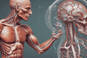
- Excitation-contraction coupling is the process that transforms a nerve impulse into muscle contraction
- Action potentials on the sarcolemma spread from the motor end plate along the length of the fiber and into the cell along the transverse tubules (T tubules)
Excitation-Contraction Coupling
- T tubules are associated with the sarcoplasmic reticulum (SR)
- SR is composed of large chambers called terminal cisternae and long longitudinal tubules that surround the myofibrils
- The action potential causes a conformational change in voltage-sensing proteins (dihydropyridine receptors – DHPRs) on the T tubule
- DHPRs are mechanically coupled to the ryanodine receptors (RyRs) calcium channels on the SR membrane
- Calcium channels open, allowing calcium to rapidly diffuse out of the SR and initiate muscle contraction
Calcium Regulation
- A calcium pump (SERCA 1a) removes calcium ions from the myofibrillar fluid after contraction occurs
- SERCA1a is a continually active calcium pump located in the walls of the SR that pumps Ca back into the SR
- Muscle contraction continues as long as Ca ion concentration remains high
Walk-Along Theory of Muscle Contraction
- Before muscle contraction begins, myosin heads bind with ATP (low energy configuration)
- ATPase activity of the myosin head cleaves the ATP into ADP and phosphate ion
- Cleavage products are kept bound to the head, which becomes energized in a “cocked position”
- Forces are generated by the interaction of the cross-bridges from myosin and actin filaments
- Calcium release from the SR exposes myosin-binding sites on the actin filament, allowing myosin heads to bind and form a cross-bridge
- The release of phosphate triggers the POWER STROKE – a conformational change in the myosin head, which moves from a high-energy to a low-energy state
Studying That Suits You
Use AI to generate personalized quizzes and flashcards to suit your learning preferences.