Podcast
Questions and Answers
In smooth muscle cells, what structural feature connects actin filaments to facilitate contraction?
In smooth muscle cells, what structural feature connects actin filaments to facilitate contraction?
- Dense bodies (correct)
- T-tubules
- Intercalated discs
- Z lines
Skeletal muscle cells are characterized by a single, centrally located nucleus.
Skeletal muscle cells are characterized by a single, centrally located nucleus.
False (B)
What is the functional unit of a myofibril that is responsible for muscle contraction?
What is the functional unit of a myofibril that is responsible for muscle contraction?
sarcomere
The layer of connective tissue that surrounds individual muscle fibers is called ______.
The layer of connective tissue that surrounds individual muscle fibers is called ______.
Match each type of skeletal muscle fiber with its key characteristic:
Match each type of skeletal muscle fiber with its key characteristic:
Which of the following is unique to cardiac muscle tissue?
Which of the following is unique to cardiac muscle tissue?
The epicardium of the heart corresponds to the visceral layer of the serous pericardium.
The epicardium of the heart corresponds to the visceral layer of the serous pericardium.
What is the function of the sinoatrial (SA) node in the heart?
What is the function of the sinoatrial (SA) node in the heart?
The tunica media of medium-sized arteries is composed predominantly of ______.
The tunica media of medium-sized arteries is composed predominantly of ______.
Which type of capillary is characterized by a discontinuous basement membrane and the presence of Kupffer cells?
Which type of capillary is characterized by a discontinuous basement membrane and the presence of Kupffer cells?
Flashcards
Muscle tissue
Muscle tissue
Muscle tissue contracts via actin and myosin filaments, existing in skeletal, smooth, and cardiac forms.
Smooth muscle
Smooth muscle
Tapered cells with central nuclei, gap junctions, actin and myosin filaments, found in organs, stimulated by various impulses.
Myofibrils
Myofibrils
A subunit of muscle fiber, composed of myofilaments, its functional unit is the sarcomere.
Thick filament
Thick filament
Signup and view all the flashcards
Thin filament
Thin filament
Signup and view all the flashcards
Cardiac muscle
Cardiac muscle
Signup and view all the flashcards
Epicardium
Epicardium
Signup and view all the flashcards
Venules
Venules
Signup and view all the flashcards
Tunica media (medium veins)
Tunica media (medium veins)
Signup and view all the flashcards
Tunica adventitia
Tunica adventitia
Signup and view all the flashcards
Study Notes
- The lecture discusses muscle, heart, and blood vessel histology.
Muscle Tissue
- There are four basic tissue types, muscle being one
- Contractions occur via myofilaments (actin and myosin).
- Myofilaments come in thin(actin) and thick(myosin) versions.
- Sarcoplasm exists within muscle tissue.
Types of muscle
-
Smooth Muscle
- Tapered cells
- Nuclei located in the center of the cell
- Gap junctions facilitate intercellular communication.
- Contains thin actin and thick myosin II filaments.
- Usually found in organs
- Can be stimulated by mechanical impulses, electrical depolarization, or chemical stimuli.
- Myosin filaments are side polar
- Actin connects to dense bodies.
- Thick myosin is scattered throughout the sarcoplasm.
- Thin filaments contain actin.
-
Skeletal Muscle
- Striated.
- Syncytium - multinucleated cell.
- Muscle cell = a muscle fiber
- Nucleus is located at the side of the cell.
- Sarcolemma: Plasma membrane.
- Organization of Skeletal Muscle
- Muscle -> fascicle - muscle fiber -> myofibril -> myofilament.
- Epimysium: surrounds fascicles.
- Perimysium: surrounds fascicle
- Endomysium: surrounds muscle fibers.
- Three types of skeletal muscle fibers:
- Type I (slow oxidative) fibers are slow-twitch, resistant to fatigue, and contain many mitochondria and myoglobin.
- Type IIa (fast oxidative glycolytic) fibers- many mitochondria, myoglobin, glycogen, fatigue resistant, and are fast-twitch, high tension
- Type IIb (fast glycolytic) fibers- low mitochondria and myoglobin, fatigue-prone with very high tension, contain high glycogen and are fast-twitch
- Deeply stained indicates Type I.
- Lightly stained indicates Type IIb.
- Stains for oxidative enzymes in mitochondria.
-
Cardiac Muscle
- Found in the heart and is striated.
- Nuclei is found in the center of the cell.
- Branched cells
- Intercalated discs
- Intercalated Discs
- Fascia adherens (where sarcomeres anchor to the plasma membrane).
- Maculae adherentes (desmosomes) bind cells together.
- Gap junctions allow ionic continuity.
- Lots of mitochondria.
Myofibrils and Myofilaments
- Myofibrils are subunits of muscle fiber.
- Composed of myofilaments
- The functional unit of myofibrils is the sarcomere.
- Thick filament: Myosin II.
- Thin filament: F-actin, tropomyosin, and troponin.
Sarcomere
- Contains:
- A band (darker)
- I band (lighter)
- Z line
- M line
- H band
- Troponin covers a myosin binding site on actin, and is uncovered by calcium (by binding to troponin C).
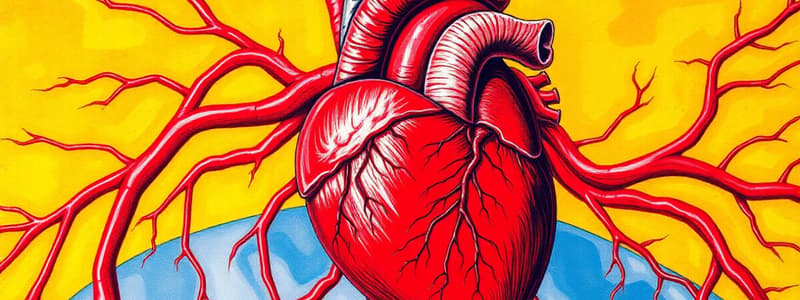
Heart Structure
- A four-chambered organ that facilitates unidirectional blood flow.
- Contains two ventricles and two atria.
- Composed of cardiac muscle, coronary arteries, and cardiac veins.
- Fibrous skeleton of the heart consists of dense irregular connective tissue, four fibrous rings, and two fibrous trigones.
- The membranous portion of the interventricular system is located between the atria and ventricles.
- Heart Wall Layers:
- The epicardium adheres to the outer layer of the heart.
- Myocardium
- Cardiac muscle (thicker in ventricle than in atrium). -Endocardium
- Endothelial and subendothelial connective tissue.
Conducting System
- Sinoatrial (SA) node generates electrical impulses, is called the pacemaker of the heart
- SA node impulse spreads along cardiac muscle fibers of atria
- Impulse is then taken up by atrioventricular (AV) node and then carried along the AV bundle (of His).
- Purkinje fibers carry the generated electrical signals down to muscles
- Purkinje fibers- specialized cardiac muscle cells, are larger than ventricular muscle cells
Blood Vessel Histology
-
General Features of Blood Vessels:
- Tunica intima (inner layer): endothelial cells and connective tissue.
- Tunica media (middle layer): smooth muscle.
- Tunica adventitia (outermost layer): connective tissue.
-
Large (elastic) Arteries:
- E.g Aorta and pulmonary arteries: brachiocephalic, common carotid
- Walls contain multiple sheets of elastic lamellae.
- Tunica intima: endothelium, subendothelial connective tissue, and inconspicuous internal elastic membrane.
- Tunica media: multiple layers of smooth muscle cells separated by elastic lamellae which is the thickest layer
- Tunica adventitia: thin layer of collagen and elastic fibers.
- Vaso vasorum: blood vessels and nervi vascularis.
-
Medium (Muscular) Arteries:
- Contains more smooth muscle and less elastin than elastic arteries.
- Most "named" arteries
- Internal elastic membrane distinguishes from elastic arteries
- Tunica intima is thinner than in elastic arteries.
- Tunica media is almost entirely smooth muscle.
- Tunica adventia is thick and separated by an external elastic membrane.
-
Small Arteries and Arterioles:
- Differ based on amount of smooth muscle layers.
- Tunica adventia is thin and ill-defined for both.
- Small arteries: -Tunica intima contains internal elastic membrane. -Tunica media has up to 8 layers of smooth muscle. -Arterioles: -Tunica intima may or may not have internal elastic membrane. -Tunica media has up to 1-2 layers of smooth muscle.
-
Types of Capillaries:
- Continuous: found in muscle, lung, and CNS where only small molecules can pass
- Pericytes present, surrounding the capillary
- Fenestrated: Endocrine glands, gallbladder, kidney, intestinal tract for fluid, metabolite absorption
- contain 80-100 nm fenestrations
- Discontinuous: found in liver, bone marrow, and spleen, characterized by the presence of Kupffer cells and a discontinuous basement membrane
- Continuous: found in muscle, lung, and CNS where only small molecules can pass
-
Venules:
- Collect blood from capillaries.
- Two types: post-capillary and muscular.
- Post-capillary: Tunica intima with endothelial lining and basal lamina and pericytes.
- No tunica media
- Muscular:
- Tunica intima- endothelial lining with basal lamina.
- Tunica media- 1-2 layers of smooth muscles cells.
- Small veins- continuous with venules.
-
Medium Veins:
- Veins with diameters up to 10 mm (e.g., radial, popliteal vein).
- Tunica intima- endothelium and basal lamina, occasional smooth muscle cells.
- Tunica media- several layers of circularly arranged smooth muscle cells with interspersed collagen and elastic fibers.
- Tunica adventitia- typically thicker than the tunica media and consists collagen fibers and elastic fibers.
-
Large Veins:
- Veins with diameters greater than 10 mm (e.g., SVC, IVC, subclavian).
- Tunica intima- Endothelial lining, basal lamina, connective tissue
- Tunica media- thin, circumferentially arranged smooth muscle cells, collagen fibers and some fibroblasts.
- Tunica adventitia- thickest layer, contains collagen, elastic fibers, fibroblasts, and longitudinally arranged smooth muscle cells.
Studying That Suits You
Use AI to generate personalized quizzes and flashcards to suit your learning preferences.


