Podcast
Questions and Answers
Describe the microscopic arrangement of cardiac muscle fibers, relating it to their function as a coordinated unit.
Describe the microscopic arrangement of cardiac muscle fibers, relating it to their function as a coordinated unit.
Cardiac muscle fibers are connected end-to-end by intercalated discs, which contain gap junctions. These gap junctions allow for rapid ion flow, enabling the muscle fibers to contract as a functional syncytium, crucial for efficient heart contraction.
How does the presence of abundant mitochondria and glycogen granules in cardiac muscle fibers support the heart's continuous activity?
How does the presence of abundant mitochondria and glycogen granules in cardiac muscle fibers support the heart's continuous activity?
Abundant mitochondria provide a continuous supply of ATP through oxidative phosphorylation to meet the high energy demands of continuous contraction. Glycogen granules serve as a readily available glucose reserve for ATP production during increased activity.
Explain how the branching and anastomosing arrangement of cardiac muscle fibers contribute to the heart's ability to withstand varying pressures during contraction.
Explain how the branching and anastomosing arrangement of cardiac muscle fibers contribute to the heart's ability to withstand varying pressures during contraction.
The branching and anastomosing network distributes the contractile force in multiple directions, spreading the load. This reduces stress on individual fibers and allows the heart to contract forcefully against a range of pressures without tearing or damage.
What role do T-tubules play in the contraction of cardiac muscle fibers?
What role do T-tubules play in the contraction of cardiac muscle fibers?
Explain the arrangement of the sarcoplasmic reticulum (SR) and T-tubules in cardiac muscle cells and its functional significance.
Explain the arrangement of the sarcoplasmic reticulum (SR) and T-tubules in cardiac muscle cells and its functional significance.
Describe the structure and function of intercalated discs in cardiac muscle tissue.
Describe the structure and function of intercalated discs in cardiac muscle tissue.
How do fascia adherens in intercalated discs contribute to the mechanical stability of cardiac muscle tissue during contraction?
How do fascia adherens in intercalated discs contribute to the mechanical stability of cardiac muscle tissue during contraction?
Explain how the gap junctions within intercalated discs contribute to the heart's function as a functional syncytium.
Explain how the gap junctions within intercalated discs contribute to the heart's function as a functional syncytium.
How does the histology of the epicardium, the outermost layer of the heart, contribute to the heart's overall function?
How does the histology of the epicardium, the outermost layer of the heart, contribute to the heart's overall function?
Describe the histological components of the myocardium and explain their significance in cardiac function.
Describe the histological components of the myocardium and explain their significance in cardiac function.
What is the significance of the endocardium being composed of simple squamous epithelium and underlying loose connective tissue?
What is the significance of the endocardium being composed of simple squamous epithelium and underlying loose connective tissue?
Describe the key features of the endocardium that help prevent thrombus formation within the heart chambers.
Describe the key features of the endocardium that help prevent thrombus formation within the heart chambers.
How are the valves of the heart histologically adapted to withstand the mechanical stresses of continuous blood flow and pressure changes?
How are the valves of the heart histologically adapted to withstand the mechanical stresses of continuous blood flow and pressure changes?
Explain the histological organization of heart valves and how this organization contributes to their functional integrity.
Explain the histological organization of heart valves and how this organization contributes to their functional integrity.
Describe the structural components of the impulse conducting system of the heart.
Describe the structural components of the impulse conducting system of the heart.
How do Purkinje fibers differ histologically from typical cardiac muscle fibers, and how do these differences relate to their function?
How do Purkinje fibers differ histologically from typical cardiac muscle fibers, and how do these differences relate to their function?
Compare and contrast the histological features and arrangement of cardiac muscle with those of skeletal muscle.
Compare and contrast the histological features and arrangement of cardiac muscle with those of skeletal muscle.
Describe the key features of the moderator band (septomarginal trabecula) and its functional significance in the right ventricle.
Describe the key features of the moderator band (septomarginal trabecula) and its functional significance in the right ventricle.
What is the role of lipochrome pigment in cardiac muscle fibers, and how does its accumulation change over time?
What is the role of lipochrome pigment in cardiac muscle fibers, and how does its accumulation change over time?
Regarding cardiac muscle, describe how light microscopy (LM) and electron microscopy (EM) contribute differently to our understanding of its cellular organization and structures.
Regarding cardiac muscle, describe how light microscopy (LM) and electron microscopy (EM) contribute differently to our understanding of its cellular organization and structures.
Explain how identifying irreversible injury to cardiac muscle cells can be indicative of a heart condition.
Explain how identifying irreversible injury to cardiac muscle cells can be indicative of a heart condition.
What is the significance of cardiac muscle lacking satellite cells in the context of myocardial repair after injury?
What is the significance of cardiac muscle lacking satellite cells in the context of myocardial repair after injury?
How des the arrangement of smooth muscle and capillaries at the base of heart valves contribute to their function?
How des the arrangement of smooth muscle and capillaries at the base of heart valves contribute to their function?
Explain why cardiac muscle fibers appear as a syncytium, but are not. What structures make this apparent?
Explain why cardiac muscle fibers appear as a syncytium, but are not. What structures make this apparent?
Cardiac muscle is involuntary. Where is it located in the heart and what specialized purpose does this serve?
Cardiac muscle is involuntary. Where is it located in the heart and what specialized purpose does this serve?
What are the three main layers of the structure of the heart wall?
What are the three main layers of the structure of the heart wall?
What function does the epicardium serve?
What function does the epicardium serve?
If a patient has a diagnosis of Myocardial Infarction, what might you see histologically in the area of the infarct?
If a patient has a diagnosis of Myocardial Infarction, what might you see histologically in the area of the infarct?
What two purposes does the endocardium lining consist of?
What two purposes does the endocardium lining consist of?
What key components do the intercalated discs contain to maintain structural support and provide connections between the cardiac muscle fiber cells?
What key components do the intercalated discs contain to maintain structural support and provide connections between the cardiac muscle fiber cells?
Explain how the simple squamous epithelium layer in the endocardium contributes to blood flow.
Explain how the simple squamous epithelium layer in the endocardium contributes to blood flow.
What role do T Tubules fill in cardiac tissue?
What role do T Tubules fill in cardiac tissue?
Describe the appearance of the Purkinje fibers.
Describe the appearance of the Purkinje fibers.
What do Purkinje fibers traverse the cavity of?
What do Purkinje fibers traverse the cavity of?
Flashcards
Cardiac Muscle Fibers
Cardiac Muscle Fibers
Involuntary muscle found in the heart wall (myocardium).
Intercalated Discs
Intercalated Discs
Cell junctions joining cardiac muscle fibers end to end.
Caliber of Fiber
Caliber of Fiber
The size of the cardiac muscle fiber.
Cardiac Muscle Nuclei
Cardiac Muscle Nuclei
Signup and view all the flashcards
Granular Cytoplasm
Granular Cytoplasm
Signup and view all the flashcards
Intercalated Disc (EM)
Intercalated Disc (EM)
Signup and view all the flashcards
T-tubules and Sarcoplasmic Reticulum
T-tubules and Sarcoplasmic Reticulum
Signup and view all the flashcards
Intercalated Discs Function
Intercalated Discs Function
Signup and view all the flashcards
Intercalated Discs Transmission
Intercalated Discs Transmission
Signup and view all the flashcards
Fascia Adherence
Fascia Adherence
Signup and view all the flashcards
Desmosome Function
Desmosome Function
Signup and view all the flashcards
Gap Junction Function
Gap Junction Function
Signup and view all the flashcards
Cardiac Muscle Growth
Cardiac Muscle Growth
Signup and view all the flashcards
Cardiac Muscle Repair
Cardiac Muscle Repair
Signup and view all the flashcards
Wall of Heart Layers
Wall of Heart Layers
Signup and view all the flashcards
Epicardium Definition
Epicardium Definition
Signup and view all the flashcards
Epicardium Layer
Epicardium Layer
Signup and view all the flashcards
Myocardium
Myocardium
Signup and view all the flashcards
Endocardium
Endocardium
Signup and view all the flashcards
Layers of the Endocardium
Layers of the Endocardium
Signup and view all the flashcards
Structure of the Valve
Structure of the Valve
Signup and view all the flashcards
Impulse Conducting System Types
Impulse Conducting System Types
Signup and view all the flashcards
Moderator Band
Moderator Band
Signup and view all the flashcards
Purkinji Fibers
Purkinji Fibers
Signup and view all the flashcards
Faster Conduction
Faster Conduction
Signup and view all the flashcards
Glycogen amount in color
Glycogen amount in color
Signup and view all the flashcards
Intercalary Disc
Intercalary Disc
Signup and view all the flashcards
Study Notes
- The lecture focuses on the histology of cardiac muscle fibers and the wall of the heart.
- It aims to enable students to describe the microscopic structure of cardiac muscle fibers, correlate structure to function, differentiate between muscle fiber types, and detail the heart wall's histological structure.
- Professor Nasra Ayuob is the Head of Medical Histology & Cell Biology.
Cardiac Muscle Fibers
- Cardiac muscle fibers are involuntary and found in the heart wall (myocardium).
- The cardiac muscle fiber is not a syncytium.
- Individual muscle cells are joined end to end via cell junctions known as intercalated discs.
Light Microscopy (LM) of Cardiac Muscle
- Cardiac muscle cells have a small caliber, measuring about 25 um.
- They exhibit variable lengths with extensive branching and anastomosing.
- Connective tissue (endomysium) surrounds the muscle and contains blood vessels and lymphatics.
- Irregular striations are present.
- Nuclei are large, oval, central, and can be mono or binucleated.
- The cytoplasm is granular and acidophilic.
Electron Microscopy (EM) of Cardiac Muscle Fibers
- The sarcoplasm of cardiac muscle fibers contains glycogen granules, mitochondria, and Golgi saccules.
- Lipid droplets and lipochrome pigment are also present.
- Lipochrome accumulation increases with age, potentially leading to brown atrophy of the heart.
- Myofibrils are rich, traversed by dark staining intercalated discs.
- The myofibrils exhibit branching and anastomosing.
- They show less regular arrangement compared to light microscopy.
- Myofibrils are variable in diameter.
- Tubules and sarcoplasmic reticulum in cardiac muscle are less developed.
- Cardiac muscle fibers form diads instead of triads composed of a T-tubule and terminal cisterna.
- T-tubules are wider and surround the myofibrils at the Z-line.
- Terminal cisternae are small and incomplete.
Intercalated Discs
- Intercalated discs are specialized interdigitating junctions between cardiac muscle cells.
- They always coincide with Z lines and bind the cells.
- Under light microscopy, intercalated discs appear as straight or step-like structures.
- Specific staining can be achieved using Iron Hematoxylin or Silver.
- Intercalated discs transmit forces of contraction.
- Three types of membrane-to-membrane contact comprise the structure: fascia adherence, desmosomes, and gap junctions
- Fascia adherence performs intracellular adhesion and anchors myofibrils, representing half a Z-line.
- Desmosomes prevent cell separation during contraction.
- Gap junctions, with a 2 nm gap, enable direct communication via interconnecting channels that allow free and rapid transmission of nerve impulses resulting in the fiber contracting as a single unit physiologically.
- The growth of cardiac muscle occurs through hypertrophy, involving an increase in the size of cardiac muscle cells due to the new formation of myofibrils.
- There is no cell division or regeneration.
- Repair occurs with no satellite cells. When muscle fibers are lost, they are replaced with fibrous C.T. resulting in healing by fibrosis.
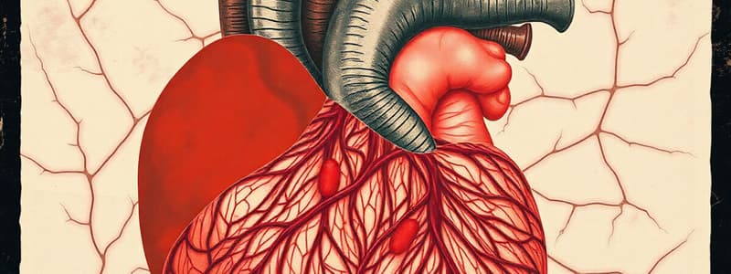
Structure of the Heart Wall
- The heart wall consists of 2 thin atria and 2 thick ventricles.
- Three layers comprise the heart wall: epicardium, myocardium, and the endocardium,.
- Visceral layer of the double layered pericardium. It invests the heart from outside.
- Epicardium is formed of simple squamous epithelium (outer) and a subserous layer of C.T. containing coronary blood vessels and fat cells.
- The C.T. blends with the endomysium of the myocardium.
- The myocardium or main bulk are cardiac muscle fibers running in various directions and separated by loose C.T. endomysium containing blood vessels and lymphatics.
- Covered internally by the endocardium and externally by the epicardium.
- The endocardium lines the chambers and covers the valves.
- It consists of four layers from inside to outside: simple squamous endothelial layer (most inner), subendothelial dense C.T. layer, a layer of dense elastic fibers, and a layer of loose C.T which contains blood vessels and purkinji fibers (outer).
- Valves consist of a fold of endocardium with a supporting middle layer of dense collagen fibers, elastic fibers, phagocytic cells, and thickened at the base (mitral and tricuspid valves) from rings of C.T. around the orifice, and blood vessels, smooth muscle fibers and capillaries at the base.
- The impulse conducting system contains S-A Node (the pace maker), A-V Node, A-V bundle of His, and 2 branches and their ramification including the moderator band.
Moderator Band
- Cardiac muscle fibers form a bundle through which the right branches of the A-V bundle of His traverse into the lateral wall of the right ventricle under the endocardium.
- It appears pale, formed mainly of Purkinje fibers separated by loose C.T. endomysium, and contains fat cells with blood capillaries.
- Paler color is due to large amount of glycogen that dissolve during preparation and few myofibrils arranged peripherally.
- Larger in diameter, with faster conduction due to the gap junction.
- There are no intercalary discs but numerous gap junctions are present for rapid conduction.
- Desmosomes, no t-tubules, and cytoplasm is granulated with bundles of C.T. sheath.
Studying That Suits You
Use AI to generate personalized quizzes and flashcards to suit your learning preferences.


