Podcast
Questions and Answers
What is the primary function of tendons in the skeletal muscle structure?
What is the primary function of tendons in the skeletal muscle structure?
- To facilitate muscle contraction
- To provide energy to muscle fibers
- To connect muscles to bones (correct)
- To protect muscle cells from injury
Which connective tissue surrounds the individual muscle fibers within a fascicle?
Which connective tissue surrounds the individual muscle fibers within a fascicle?
- Myofibril
- Perimysium
- Endomysium (correct)
- Epimysium
What might be implied by the presence of the 'mar/in' pattern for actin and myosin proteins?
What might be implied by the presence of the 'mar/in' pattern for actin and myosin proteins?
- They may interact more efficiently for contraction (correct)
- Their arrangement is based on muscle fiber type
- They are randomly organized within the myofibrils
- They limit the flexibility of muscle fibers
How do muscle strains occur in skeletal muscles?
How do muscle strains occur in skeletal muscles?
Which component of skeletal muscle serves as the protective outer layer of the entire muscle?
Which component of skeletal muscle serves as the protective outer layer of the entire muscle?
Which characteristic correctly describes the bicep brachii?
Which characteristic correctly describes the bicep brachii?
According to the muscle rules, which statement regarding muscles is accurate?
According to the muscle rules, which statement regarding muscles is accurate?
What is the significance of the term 'insertion' in the context of muscle anatomy?
What is the significance of the term 'insertion' in the context of muscle anatomy?
Which muscle rule is violated by a muscle that only has one attachment?
Which muscle rule is violated by a muscle that only has one attachment?
What role do $Ca^{2+}$ ions play in muscle contraction?
What role do $Ca^{2+}$ ions play in muscle contraction?
Which action correctly represents the function of flexors based on muscle rules?
Which action correctly represents the function of flexors based on muscle rules?
Which molecule is primarily responsible for providing energy during muscle contraction?
Which molecule is primarily responsible for providing energy during muscle contraction?
What triggers the release of $Ca^{2+}$ ions in the muscle contraction process?
What triggers the release of $Ca^{2+}$ ions in the muscle contraction process?
During muscle contraction, what happens to myosin heads after they detach from actin?
During muscle contraction, what happens to myosin heads after they detach from actin?
What is the effect of ATP breakdown during muscle contraction?
What is the effect of ATP breakdown during muscle contraction?
What is the primary role of the triceps brachii in relation to the humerus?
What is the primary role of the triceps brachii in relation to the humerus?
Which statement correctly describes the insertion of the pectoralis minor?
Which statement correctly describes the insertion of the pectoralis minor?
What action is primarily performed by the pectoralis major?
What action is primarily performed by the pectoralis major?
From which ribs does the pectoralis minor originate?
From which ribs does the pectoralis minor originate?
Which muscle is responsible for downward rotation of the scapula?
Which muscle is responsible for downward rotation of the scapula?
Which muscle is characterized by its fiber direction as described in the content?
Which muscle is characterized by its fiber direction as described in the content?
What is the primary reason for the naming of the Gluteus Minimus?
What is the primary reason for the naming of the Gluteus Minimus?
Which of the following muscles is described in the content with respect to its origin and insertion?
Which of the following muscles is described in the content with respect to its origin and insertion?
What characteristic is specifically noted for the Deltoid muscle?
What characteristic is specifically noted for the Deltoid muscle?
Which muscle is categorized by a specific action related to its functionality?
Which muscle is categorized by a specific action related to its functionality?
Flashcards are hidden until you start studying
Study Notes
Muscle Contraction Model
- Actin and myosin are the proteins responsible for muscle contraction.
- Tropomyosin covers the binding sites on actin preventing myosin from attaching.
- Troponin binds to calcium ions ($Ca^{2+}$) and moves tropomyosin aside to expose the binding sites on actin.
- Myosin heads bind to actin, forming cross-bridges.
- ATP is broken down to ADP and a phosphate group, providing energy for the myosin heads to pivot and pull the actin filaments.
- The myosin heads detach from actin when a new ATP molecule binds to them.
- This cycle continues as long as calcium ions ($Ca^{2+}$) and ATP are available.
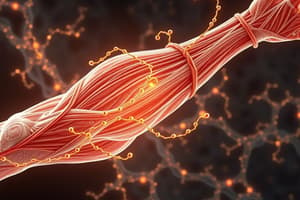
Skeletal Muscle Belly Structure
- Tendon: Connective tissue that attaches muscles to bones.
- Epimysium: The outermost layer of connective tissue surrounding a muscle.
- Perimysium: Connective tissue that surrounds bundles of muscle fibers called fascicles.
- Fascicle: A bundle of muscle fibers.
- Endomysium: Connective tissue that surrounds individual muscle fibers.
- Muscle Fiber/ Muscle cell: Individual muscle cells.
- Myofibril: Thread-like structures within muscle fibers, containing actin and myosin filaments.
Muscle Rules
- Rule 1: Muscles must have at least two attachments and cross at least one joint.
- Rule 2: Muscles always pull and get shorter during contraction.
- Rule 3: The attachment that moves during contraction is called the insertion, while the attachment that stays fixed is called the origin.
- Rule 4: Muscles that decrease the angle between bones are called flexors, while muscles that increase the angle are called extensors.
- Rule 5: Muscles work in opposing pairs (e.g., biceps brachii and triceps brachii).
- Rule 6: When diagramming muscles, show the direction of the pull from the origin to the insertion.
Bicep Brachii
- Origin: Ventral humerus, with two points on the lateral and medial sides.
- Insertion: Proximal ventral ulna.
- Action: Flexes the elbow.
Triceps Brachii
- Origin: Flat tendon from the infraglenoid tubercle of the scapula.
- Insertion: Posterior surface of the olecranon process of the ulna.
- Action: Extends the elbow and assists in stabilizing the humerus in the glenoid cavity.
Pectoralis Minor
- Origin: Margins of the 3rd to 5th ribs.
- Insertion: Coracoid process of the scapula.
- Action: Stabilizes the scapula, contributing to internal and downward rotation.
Pectoralis Major
- Origin: Anterior surface of the medial half of the clavicle.
- Insertion: Lateral lip of the intertubercular sulcus of the humerus.
- Action: Adducts, medially rotates, and transversely adducts the arm at the shoulder joint.
Muscles of the Elbow and Chest
- Trapezius and Rhomboid Minor: Named based on their shape.
- Gluteus Maximus and Gluteus Minimus: Named based on their size.
- Frontalis and Temporalis: Named based on their location.
- Orbicularis Oculi and Transverse Abdominis: Named based on their fiber direction.
- Flexor carpi ulnaris and Extensor digitorum: Named based on their specific functions/actions.
- Longus: Named based on its length.
- Sterno cleidomastoid and Brachioradialis: Named based on their location.
- Biceps Brachii and Triceps Brachii: Described based on their origin and insertion.
Posterior Muscles
- Deltoid: Shown with its lateral and medial heads.
Studying That Suits You
Use AI to generate personalized quizzes and flashcards to suit your learning preferences.