Podcast
Questions and Answers
Which of the following is NOT considered a major component of the brain?
Which of the following is NOT considered a major component of the brain?
- Brain stem
- Cerebellum
- Spinal cord (correct)
- Cerebrum
Which part of the brain is primarily responsible for connecting the cerebrum with the cerebellum and spinal cord?
Which part of the brain is primarily responsible for connecting the cerebrum with the cerebellum and spinal cord?
- Medulla oblongata
- Pons
- Midbrain (correct)
- Cerebral cortex
Where is the cerebellum located?
Where is the cerebellum located?
- In the anterior cranial fossa
- Directly below the midbrain
- In the posterior cranial fossa (correct)
- Anterior to the cerebrum
What is the primary advantage of using MRI over CT for brain imaging in the context of tumors?
What is the primary advantage of using MRI over CT for brain imaging in the context of tumors?
What is a limitation of MRI when compared to other imaging techniques?
What is a limitation of MRI when compared to other imaging techniques?
Which part of the brain stem contains ascending and descending tracts that facilitate communication between the spinal cord and brain?
Which part of the brain stem contains ascending and descending tracts that facilitate communication between the spinal cord and brain?
In what position should a patient be placed for a standard brain MRI?
In what position should a patient be placed for a standard brain MRI?
When comparing MRI and CT for imaging brain trauma, which of the following is most accurate?
When comparing MRI and CT for imaging brain trauma, which of the following is most accurate?
What is the primary purpose of padding the patient during an MRI?
What is the primary purpose of padding the patient during an MRI?
Which component of the brain contains a cortex that appears as grey matter, primarily made of nerve cell bodies?
Which component of the brain contains a cortex that appears as grey matter, primarily made of nerve cell bodies?
For obtaining axial slices of the brain, which anatomical landmark should the sagittal localizer be parallel to?
For obtaining axial slices of the brain, which anatomical landmark should the sagittal localizer be parallel to?
What is a promising application of MR angiography?
What is a promising application of MR angiography?
What is the primary anatomical coverage for all localizer scans of the brain?
What is the primary anatomical coverage for all localizer scans of the brain?
When obtaining coronal slices, a sagittal localizer should be aligned parallel to which anatomical structure?
When obtaining coronal slices, a sagittal localizer should be aligned parallel to which anatomical structure?
Which MRI sequence is particularly useful for identifying areas of edema or inflammation?
Which MRI sequence is particularly useful for identifying areas of edema or inflammation?
In which of the following situations would diffusion-weighted imaging (DWI) be most preferable?
In which of the following situations would diffusion-weighted imaging (DWI) be most preferable?
Flashcards
What is Magnetic Resonance Imaging (MRI)?
What is Magnetic Resonance Imaging (MRI)?
A specialized imaging technique that uses strong magnetic fields and radio waves to create detailed images of the brain and other soft tissues.
What are some neurological conditions that MRI helps diagnose?
What are some neurological conditions that MRI helps diagnose?
MRI is exceptionally effective in diagnosing a variety of conditions affecting the brain, including multiple sclerosis, subcortical arteriosclerotic encephalopathy, gliosis, and syrinx.
How is a patient positioned for an MRI?
How is a patient positioned for an MRI?
The patient is positioned lying on their back, with their head positioned first in the scanner, ensuring proper alignment and comfort.
What is the purpose of the 'Sagittal Localizer' in an MRI scan?
What is the purpose of the 'Sagittal Localizer' in an MRI scan?
Signup and view all the flashcards
What is a 'Flair' image and what is its significance?
What is a 'Flair' image and what is its significance?
Signup and view all the flashcards
What is the purpose of the 'Coronal Localizer' in an MRI scan?
What is the purpose of the 'Coronal Localizer' in an MRI scan?
Signup and view all the flashcards
What is the purpose of the 'Axial Localizer' in an MRI scan?
What is the purpose of the 'Axial Localizer' in an MRI scan?
Signup and view all the flashcards
Why are multiple MRI sequences used during a scan?
Why are multiple MRI sequences used during a scan?
Signup and view all the flashcards
What is the Central Nervous System (CNS)?
What is the Central Nervous System (CNS)?
Signup and view all the flashcards
What is the Peripheral Nervous System (PNS)?
What is the Peripheral Nervous System (PNS)?
Signup and view all the flashcards
What is the cerebrum?
What is the cerebrum?
Signup and view all the flashcards
What is the cerebellum?
What is the cerebellum?
Signup and view all the flashcards
What is the brain stem?
What is the brain stem?
Signup and view all the flashcards
What is one advantage of MRI for brain tumors?
What is one advantage of MRI for brain tumors?
Signup and view all the flashcards
What is MRI's role in diagnosing strokes?
What is MRI's role in diagnosing strokes?
Signup and view all the flashcards
How is MRI useful for brain trauma?
How is MRI useful for brain trauma?
Signup and view all the flashcards
Study Notes
MRI of the Brain
- The presentation covers MRI of the brain, including anatomical overview, indications, MRI procedure, and sequences.
- The nervous system is a complex network of nerves and cells, carrying messages between the brain, spinal cord, and various body parts.
- The nervous system is divided into two main parts:
- Central Nervous System (CNS): Includes the brain and spinal cord.
- Peripheral Nervous System (PNS): Consists of nerves branching from the brain and spinal cord, forming communication pathways.
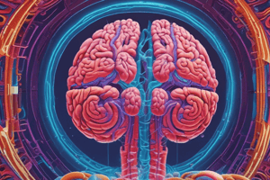
- The brain is composed of three main components:
- Cerebrum: The largest part, consisting of two hemispheres. The cortex has nerve cell bodies (grey matter), with nerve fibers (white matter) connecting it to other areas.
- Cerebellum: The second largest part, located in the posterior cranial fossa, with a layer of grey matter on its outer surface, containing concentrated cell bodies.
- Brain Stem: Composed of:
- Midbrain: Connects the cerebrum to the cerebellum and spinal cord.
- Pons: Located anterior to the cerebellum; connects the cerebrum and cerebellum to the spinal cord, having fibers from the cerebellum and cerebrum.
- Medulla oblongata: Forms the lower brain stem, contains ascending and descending tracts connecting the spinal cord and brain.
Indications of Brain MRI
- MRI is a superior diagnostic tool for detecting lesions in the posterior fossa, at the base of the skull, and pituitary fossa, compared to CT.
- Useful for identifying tumors, and conditions like hemorrhagic and ischemic stroke, by detecting thrombosis/stenosis.
- Advantages in trauma cases include demonstrating the entire extent of extracerebral collections and diffuse axonal injury sequelae. Disadvantages relate to longer scanning times and inability to visualise the bony cranium.
- Effective for diagnosing degenerative diseases like multiple sclerosis, subcortical arteriosclerotic encephalopathy, gliosis, and syrinx.
MRI Procedure: Patient Position
- Patients are positioned supine (lying face up) .
- Placement in a head coil is ensured.
- Proper padding prevents image degradation or malalignment due to head movement.
- If available, ensure patients can see out of the bore during the procedure to alleviate any claustrophobia.
MRI Procedure: Scout Slice Placement
- Sagittal Localizer:
- Alignment: Parallel to a line connecting the splenium and genu of the corpus callosum.
- Coverage: Superior (from top) to inferior (to bottom), from craniocervical junction to vertex. Lateral (from side-to-side) and medial (from the center): Temporal lobes, and Posterior to anterior: Occipital to frontal lobes.
- Coronal Slices:
- Alignment: Parallel to the brainstem.
- Coverage: Superior to inferior: Craniocervical junction to vertex. Lateral to medial: Temporal lobes. Posterior to anterior: Occipital to frontal lobes.
- Axial Slices:
- Alignment: Parallel to the falx (midline). Using a line of best fit should the midline be shifted.
- Coverage: Superior to inferior: Craniocervical junction to vertex. Lateral to medial: Temporal lobes, and Posterior to anterior: Occipital to frontal lobes.
MRI Sequences (Routine Brain)
- A table containing details of MRI sequences, including TR, TE, FA, ETL, and Slice thickness.
- Note: T1WI is good for anatomical details. Flair images help visualize edemas and inflammation, useful in multiple sclerosis. DWI is particularly helpful for strokes, abscesses, and cellular tumors.
Studying That Suits You
Use AI to generate personalized quizzes and flashcards to suit your learning preferences.