Podcast
Questions and Answers
Which part of the brain is primarily responsible for voluntary movements and higher cognitive functions?
Which part of the brain is primarily responsible for voluntary movements and higher cognitive functions?
- Brain Stem
- Cerebellum
- Cerebrum (correct)
- Pons
What comprises the central nervous system (CNS)?
What comprises the central nervous system (CNS)?
- Peripheral nerves
- Brain and spinal cord (correct)
- Nerves and ganglia
- Cerebrum and cerebellum
Which component connects the cerebrum with the cerebellum?
Which component connects the cerebrum with the cerebellum?
- Medulla oblongata
- Cerebellum
- Pons
- Midbrain (correct)
What type of tissue primarily composes the outer layer of the cerebellum?
What type of tissue primarily composes the outer layer of the cerebellum?
Which of the following is NOT an indication for conducting a brain MRI?
Which of the following is NOT an indication for conducting a brain MRI?
What structures form the communication network between the central nervous system and the body?
What structures form the communication network between the central nervous system and the body?
What is the primary function of the medulla oblongata?
What is the primary function of the medulla oblongata?
Which MRI sequence would most likely be used to assess a patient with suspected space-occupying lesions?
Which MRI sequence would most likely be used to assess a patient with suspected space-occupying lesions?
What is a significant advantage of MRI over CT in detecting tumors?
What is a significant advantage of MRI over CT in detecting tumors?
Which condition is NOT easily detected by MRI?
Which condition is NOT easily detected by MRI?
Which procedure is NOT considered an advantage of MRI for assessing trauma?
Which procedure is NOT considered an advantage of MRI for assessing trauma?
What patient position is required for an MRI scan?
What patient position is required for an MRI scan?
What is the purpose of using a coronal localizer in MRI?
What is the purpose of using a coronal localizer in MRI?
Which of the following statements about degenerative diseases is true concerning MRI?
Which of the following statements about degenerative diseases is true concerning MRI?
What is the recommended alignment for obtaining sagittal slices?
What is the recommended alignment for obtaining sagittal slices?
What is a common disadvantage of using MRI for brain imaging?
What is a common disadvantage of using MRI for brain imaging?
Flashcards
Cerebrum
Cerebrum
The largest part of the brain, responsible for higher-level functions like thinking, memory, and language. It has two hemispheres (left and right) and is composed of grey and white matter.
Cerebellum
Cerebellum
The second largest part of the brain, located at the back of the head. Responsible for coordinating movement, balance, and posture.
Brain stem
Brain stem
The part of the brain that connects the cerebrum to the spinal cord. It's crucial for relaying signals and controlling essential functions like breathing, heart rate, and consciousness.
Midbrain
Midbrain
Signup and view all the flashcards
Pons
Pons
Signup and view all the flashcards
Medulla oblongata
Medulla oblongata
Signup and view all the flashcards
MRI of the brain
MRI of the brain
Signup and view all the flashcards
Indications for MRI of the brain
Indications for MRI of the brain
Signup and view all the flashcards
MRI for Tumors
MRI for Tumors
Signup and view all the flashcards
MRI for Stroke
MRI for Stroke
Signup and view all the flashcards
MRI for Trauma
MRI for Trauma
Signup and view all the flashcards
MRI for Degenerative Disease
MRI for Degenerative Disease
Signup and view all the flashcards
Patient Position in MRI
Patient Position in MRI
Signup and view all the flashcards
Scout Slices in MRI
Scout Slices in MRI
Signup and view all the flashcards
Sagittal Localizer for Axial Slices
Sagittal Localizer for Axial Slices
Signup and view all the flashcards
Sagittal Localizer for Coronal Slices
Sagittal Localizer for Coronal Slices
Signup and view all the flashcards
Study Notes
MRI of the Brain
- This presentation details MRI procedures for brain imaging.
- The presenter is Dr. Hayder Jasim Taher, PhD of Medical Imaging, University of Hilla.
Anatomical Overview
- The nervous system is a complex network carrying messages to and from the brain and spinal cord to various body parts.
- It is divided into:
- Central Nervous System (CNS): Includes the brain and spinal cord.
- Peripheral Nervous System (PNS): Consists of nerves branching out from the brain and spinal cord, forming a communication network between CNS and body parts.
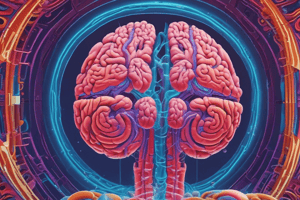
- The brain comprises three main parts:
- Cerebrum: The largest part, consisting of two hemispheres with a cortex (grey matter - nerve cell bodies) and underlying white matter (nerve fibers).
- Cerebellum: The second largest part, located in the posterior cranial fossa. It has an outer layer of grey matter and inner white matter.
- Brainstem: Consists of the midbrain, pons, and medulla oblongata. It connects the cerebrum and cerebellum to the spinal cord. Contains ascending and descending tracts for communication.
Indications of Brain MRI
- MRI is a specialist investigation for neurological conditions.
- It's crucial for diagnosing:
- Tumors (especially those in the posterior fossa, near the skull base, or pituitary fossa), where MRI is superior to CT in detecting these lesions.
- Hemorrhage-Ischemic strokes, with MRI being effective at detecting thrombosis/stenosis (use of MR angiography).
- Traumatic brain injuries (evaluating extracerebral collections and diffuse axonal injury). It can't visualize the bony cranium.
- Degenerative diseases like multiple sclerosis, subcortical arteriosclerotic encephalopathy, gliosis, and syrinx.
MRI Procedure
- Patient Position:
- Supine position (head first).
- Placed in a head coil.
- Well-padded to prevent image degradation from head movement.
- If the coil has a mirror, the patient should be able to see out of the bore to address claustrophobia.
- Scout Slice Placement:
- Sagittal localizer for axial slices (aligned parallel to a line joining the splenium and genu of the corpus callosum). Sagittal is also used for coronal slices (aligned parallel to the brainstem).
- Axial localizers provide sagittal images.
- Coverage for all slice types includes superior to inferior (Craniocervical junction to vertex), lateral to medial (Temporal lobes), and posterior to anterior (Occipital to frontal lobes).
- Coronal and axial slices can also be obtained using coronal localizers.
- MRI Sequences (Routine Brain):
- A table provides details about various sequences (e.g., sagittal T1, coronal T2, axial DWI, different FSE, SWI, post-contrast sequences). Specific timing information (TR, TE, FA, ETL) and slice thickness are included.
MRI Sequences (Routine brain) - Notes
- T1WI: Provides good anatomical images.
- Flair: Useful for identifying areas of edema, inflammation, and multiple sclerosis plaques.
- DWI: Preferable for stroke, abscesses, and cellular tumors due to restricted diffusion in those conditions.
Studying That Suits You
Use AI to generate personalized quizzes and flashcards to suit your learning preferences.