Podcast
Questions and Answers
Which of the following is a limitation of using MRI for brain imaging?
Which of the following is a limitation of using MRI for brain imaging?
- It only shows brain activity, not structure.
- It cannot establish cause-and-effect relationships. (correct)
- It has excellent temporal resolution.
- It uses radioactive substances.
FMRI has a high temporal resolution, allowing real-time brain activity measurement.
FMRI has a high temporal resolution, allowing real-time brain activity measurement.
False (B)
What type of brain scan involves injecting a radioactive tracer to measure glucose metabolism?
What type of brain scan involves injecting a radioactive tracer to measure glucose metabolism?
PET scan
An ________ is a non-invasive technique that uses electrodes on the scalp to measure electrical activity in the brain.
An ________ is a non-invasive technique that uses electrodes on the scalp to measure electrical activity in the brain.
Match each brain imaging technique with its primary strength:
Match each brain imaging technique with its primary strength:
Which of the following is a key strength of using EEG?
Which of the following is a key strength of using EEG?
CAT scans are superior to MRIs in providing detailed images of soft tissues in the brain.
CAT scans are superior to MRIs in providing detailed images of soft tissues in the brain.
In the context of brain scanning techniques, what does NGRI stand for?
In the context of brain scanning techniques, what does NGRI stand for?
The Maguire et al. (2000) study used __________ to measure the volume of grey matter in the hippocampus of taxi drivers.
The Maguire et al. (2000) study used __________ to measure the volume of grey matter in the hippocampus of taxi drivers.
Match the study with its primary finding relating to brain function and behavior:
Match the study with its primary finding relating to brain function and behavior:
Which brain scanning method is most suitable for identifying early signs of stroke?
Which brain scanning method is most suitable for identifying early signs of stroke?
CAT scans are ideal for imaging soft tissues such as the brainstem and spinal cord.
CAT scans are ideal for imaging soft tissues such as the brainstem and spinal cord.
What is one limitation of using fMRI in psychological research concerning the generalizability of results?
What is one limitation of using fMRI in psychological research concerning the generalizability of results?
In the Raine et al. (1997) study, the researchers used a _________ design to compare murderers and non-murderers.
In the Raine et al. (1997) study, the researchers used a _________ design to compare murderers and non-murderers.
Match the key finding from the Luby et al. (2013) study with its implication for early childhood development:
Match the key finding from the Luby et al. (2013) study with its implication for early childhood development:
Which of the following is a significant limitation of PET scans?
Which of the following is a significant limitation of PET scans?
The Aspinall et al. (2015) study used fMRI technology to explore brain activity in urban environments.
The Aspinall et al. (2015) study used fMRI technology to explore brain activity in urban environments.
In the Fisher et al. (2005) study, what neurotransmitter was strongly associated with the brain areas active when participants viewed photos of their loved ones?
In the Fisher et al. (2005) study, what neurotransmitter was strongly associated with the brain areas active when participants viewed photos of their loved ones?
The study by Wei et al. (2015) found that patients who had strokes in the _________ hemisphere showed a higher risk of depression.
The study by Wei et al. (2015) found that patients who had strokes in the _________ hemisphere showed a higher risk of depression.
Match the brain imaging technique with its associated cost range, as given in the evaluated cost effectiveness section:
Match the brain imaging technique with its associated cost range, as given in the evaluated cost effectiveness section:
Which imaging technique involves the use of X-rays to produce horizontal, or axial, images (often called slices) of the brain?
Which imaging technique involves the use of X-rays to produce horizontal, or axial, images (often called slices) of the brain?
During an EEG, clinicians apply a radioactive tracer to the scalp to better detect abnormalities in brain activity.
During an EEG, clinicians apply a radioactive tracer to the scalp to better detect abnormalities in brain activity.
What aspect of brain activity does fMRI measure to infer cognitive processes?
What aspect of brain activity does fMRI measure to infer cognitive processes?
In the Maguire et al. (2000) study, the taxi drivers had a greater volume of grey matter in their __________ hippocampi compared to the control group.
In the Maguire et al. (2000) study, the taxi drivers had a greater volume of grey matter in their __________ hippocampi compared to the control group.
Match each limitation with the brain scanning technique prone to it:
Match each limitation with the brain scanning technique prone to it:
Which of the following is NOT a strength of CAT scans?
Which of the following is NOT a strength of CAT scans?
One strength of fMRI is that it can establish a direct cause-and-effect relationship between brain activity and behavior.
One strength of fMRI is that it can establish a direct cause-and-effect relationship between brain activity and behavior.
What is the primary focus of voxel-based morphometry (VBM) in MRI studies?
What is the primary focus of voxel-based morphometry (VBM) in MRI studies?
The Aspinall et al. (2015) study found that walks in green spaces resulted in higher levels of _________ brain activity and reduced frustration.
The Aspinall et al. (2015) study found that walks in green spaces resulted in higher levels of _________ brain activity and reduced frustration.
Match each study evaluation point with its implication:
Match each study evaluation point with its implication:
Which of the following is a strength of the study conducted by Luby et al. (2013)?
Which of the following is a strength of the study conducted by Luby et al. (2013)?
Colours shown on brain scans accurately represent the exact location of brain activity at a specific time.
Colours shown on brain scans accurately represent the exact location of brain activity at a specific time.
What is a primary ethical advantage of using non-invasive brain scanning techniques like MRI?
What is a primary ethical advantage of using non-invasive brain scanning techniques like MRI?
The Raine et al. (1997) study showed that the murderer group had less activity in the __________, an area linked to impulse-control.
The Raine et al. (1997) study showed that the murderer group had less activity in the __________, an area linked to impulse-control.
Match the evaluation point with its relevance to the Fisher et al. (2005) study:
Match the evaluation point with its relevance to the Fisher et al. (2005) study:
A participant may feel claustrophobic and opt out of the study, leading to:
A participant may feel claustrophobic and opt out of the study, leading to:
In the study by Aspinall et al. (2015), urban settings (shopping/commercial areas) showed reduced engagement and arousal compared to green spaces.
In the study by Aspinall et al. (2015), urban settings (shopping/commercial areas) showed reduced engagement and arousal compared to green spaces.
According to Wei et al. (2015), what type of data did CAT scans provide in determining the influence of stroke location on post-stroke depression?
According to Wei et al. (2015), what type of data did CAT scans provide in determining the influence of stroke location on post-stroke depression?
A limitation of using MRI is disturbance caused by noise, temperature and human error in ___________, which means that they are not always reliable
A limitation of using MRI is disturbance caused by noise, temperature and human error in ___________, which means that they are not always reliable
Match each study's outcome with the scanning method used:
Match each study's outcome with the scanning method used:
Flashcards
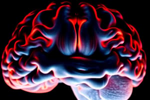
Magnetic Resonance Imaging (MRI)
Magnetic Resonance Imaging (MRI)
Uses a large magnet and radio waves to produce images of brain structures.
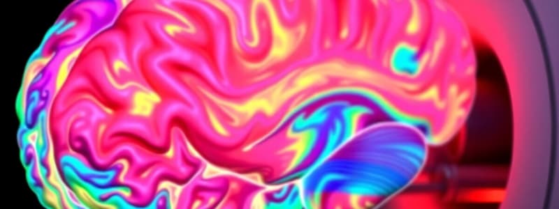
Functional Magnetic Resonance Imaging (fMRI)
Functional Magnetic Resonance Imaging (fMRI)
Measures oxygenated blood flow in the brain to indicate brain activity.
Positron Emission Tomography (PET)
Positron Emission Tomography (PET)
Uses a radioactive tracer to measure glucose metabolism in the brain.
Computerized Axial Tomography (CAT)
Computerized Axial Tomography (CAT)
Signup and view all the flashcards
Electroencephalogram (EEG)
Electroencephalogram (EEG)
Signup and view all the flashcards
Maguire et al. (2000)
Maguire et al. (2000)
Signup and view all the flashcards
Luby et al. (2013)
Luby et al. (2013)
Signup and view all the flashcards
Dopamine
Dopamine
Signup and view all the flashcards
Fisher et al. (2005)
Fisher et al. (2005)
Signup and view all the flashcards
Raine et al. (1997)
Raine et al. (1997)
Signup and view all the flashcards
Prefrontal Cortex (PFC)
Prefrontal Cortex (PFC)
Signup and view all the flashcards
Amygdala
Amygdala
Signup and view all the flashcards
Neuroplasticity
Neuroplasticity
Signup and view all the flashcards
Wei et al. (2015)
Wei et al. (2015)
Signup and view all the flashcards
Aspinall et al. (2015)
Aspinall et al. (2015)
Signup and view all the flashcards
Study Notes
Magnetic Resonance Imaging (MRI)
- Utilizes a large magnet and radio waves to produce images of brain structures.
- Pinpoints specific brain structures that may be damaged or have increased grey matter.
- Identifies links between brain and behavior.
- Identifies blood circulation problems by showing blood flow in the brain.
- Creates high-resolution brain images.
- Considered non-invasive
- Susceptible to disturbances from noise, temperature, and calibration errors, affecting reliability.
- Can be expensive.
- Is affected by patients movement while scanning
- Does not show a cause-and-effect relationship.
- Shows brain structure but not activity.
Functional Magnetic Resonance Imaging (fMRI)
- Measures oxygenated blood flow in the brain to indicate brain activity.
- Measures oxygenated blood in specific brain regions.
- Brain activity can be linked to cognitive processes, such as emotion.
- Does not use radioactive substances.
- Records activity in all parts of the brain.
- Has a 5-second delay between brain activity and measurement, which may miss important information.
- Focuses on localization of brain function and does not consider neuroplasticity.
- Shows no cause-and-effect relationship
General Evaluation of Brain Imaging Techniques
- The unnatural environment may affect findings.
- Colors can exaggerate effects on the brain.
- Brain images are compilations taken over time.
- Scanning is relatively more ethical.
- Researchers use images, creating better triangulation
Positron Emission Tomography (PET)
- Uses a radioactive tracer to measure glucose metabolism in the brain.
- Can highlight abnormalities and illnesses more effectively than other scanning techniques.
- Involves some risk to the patient due to the use of a radioactive tracer.
Computerized Axial Tomography (CAT)
- It is a noninvasive diagnostic imaging procedure that uses X-rays to produce images of the brain.
- Provides detailed information about brain tissue and structures.
- CT scans are fast, making them ideal in emergency situations like stroke, trauma, or head injury.
- Good for visualizing bone fractures and detecting acute intracranial hemorrhages.
- More accessible than MRI, especially in smaller or rural hospitals.
- Less expensive than MRI.
- Involves ionizing radiation, which adds cumulative risk, especially concerning children or repeated scans.
- Not as good as MRI in showing detailed images of soft tissues like the brainstem, spinal cord, or small tumors.
- May miss early signs of stroke within the first few hours of onset (MRI is more sensitive in this case).
- In rare cases, contrast-enhanced CT can trigger allergic reactions or affect kidney function.
Electroencephalogram (EEG)
- Detects abnormalities in brain waves.
- Electrodes are pasted onto the scalp to detect brain cells activity.
- Excellent for detecting real-time brain activity.
- Shows brain activity on a millisecond scale.
- Highly effective for diagnosing conditions like epilepsy, seizure disorders, and monitoring brain function during anesthesia or sleep studies.
- Cannot precisely localize abnormal activity deep within the brain.
Maguire et al. (2000) - MRI
- Aimed to investigate spatial navigation in London black cab taxi drivers using MRI.
- Participants included 16 healthy, right-handed male London black cab taxi drivers aged 32-62.
- MRI measured the volume of grey matter in the hippocampus.
- Taxi drivers showed a greater volume of grey matter in their posterior hippocampi compared to the control group.
- A positive correlation was found between the volume of posterior hippocampal grey matter and time spent as a taxi driver.
- The posterior hippocampus may be linked to spatial navigation skills.
- Highly controlled clinical method obtained objective data for easy comparison and analysis.
- A researcher blind to the conditions counted the pixels on the MRI images, increasing internal validity.
- A correlation cannot show cause-and-effect, so it is impossible to know whether the taxi drivers already had naturally high levels of hippocampal grey matter.
- Only generalizable to male, right-handed London taxi drivers.
Luby et al. (2013) - MRI
- Aimed to investigate whether poverty experienced in childhood is shown in delayed brain development using MRI.
- Participants included145 right-handed children from the USA, categorized as living in poverty.
- Children underwent regular testing to measure cognitive, emotional, and social skills.
- Data was collected on the children's closeness to caregivers and stressful events in their lives.
- Children had two MRI scans of the whole brain or just the hippocampus and amygdala.
- Both the hippocampus and the amygdala showed less white and grey matter in the MRI scans.
- Positive care from adults had a less negative effect on the hippocampus, while stressful life events affected the left hippocampus.
- Poverty appears to have a negative effect on brain development in childhood.
- Behavioral, cognitive, and social measures were checked against MRI results, increasing internal validity.
- Longitudinal design allowed for real changes and comparisons across time.
- The MRI scans may have picked up differences in the brains of the children which were not the result of poverty.
- Difficult to generalize since sample only represents pre-school children living in poverty with depression symptoms.
Fisher et al. (2005) - fMRI
- Aimed to investigate the brain systems involved in early-stage intense romantic love.
- Participants included 10 females and 7 males aged 18-26 who reported being 'in love'.
- Participants were shown a photograph of their loved one, followed by a distraction task, then a neutral photograph.
- When viewing the photograph of their loved one, the right ventral tegmental area and the right caudate nucleus were active.
- These areas are associated with dopamine activation.
- People in the early stages of romantic love access areas of the brain associated with motivation and reward.
- Human beings may have evolved a brain system which ensures that they become addicted to love which increases the study’s validity.
- The use of a standardised procedure means that the study is replicable, which increases its reliability.
- Small sample size of 17 participants means that the results are not very meaningful and may not be robust in terms of statistical analysis.
- The idea that romantic love that can be measured via fMRI is overly reductionist.
Raine et al. (1997) - PET
- Aimed to investigate differences in the brains of impulsive murderers compared to non-murderers.
- Participants included 41 murderers who had pleaded NGRI (not guilty by reason of insanity).
- A matched pairs design was used with a control group of non-murderers.
- Participants were injected with a radioactive tracer and completed cognitive tasks while undergoing a PET scan.
- Impulsive murderers showed less activity in the prefrontal cortex (PFC).
- Murderers also showed asymmetrical activity in the amygdala.
- Brain dysfunction may mean that someone is more prone to violent outbursts.
- The use of a matched pairs design means that individual differences could to some extent be controlled for
- This use of a purposive sample of NGRI murderers increases the validity of the study as the participants had all experienced a lack of control which led to violent behaviour thus they may all share similar features of brain function
- The results of this study may lead to a deterministic bias against offenders guilty of impulsive murder.
- This is an example of a snapshot design: it cannot tell us anything about behaviour across time
Wei et al. (2015) - CAT Scan
- Aim was to determine whether the location of brain damage (via stroke) influences the risk of developing post-stroke depression.
- A meta-analysis reviewed data from studies using CAT scans on 5,507 stroke patients.
- Stroke damage was categorized by hemisphere (right vs. left).
- Patients with right hemisphere strokes had a higher risk of depression.
- Structural damage in certain brain regions is linked to psychological outcomes.
- Large sample sizes from multiple studies gave high statistical power and confidence in the findings.
- Researchers focused on physiological underpinnings of mental health, providing biological insight into post-stroke depression.
- CAT scans do not show brain activity, only structure.
- Meta-analysis may involve variation in data quality and methodology across different studies.
Aspinall et al. (2015) - EEG Scan
- Aimed to explore how different urban environments impact real-time brain activity using mobile EEG technology.
- Participants were fitted with mobile EEG headsets and walked through a busy urban shopping street, a green path in a park, and a crowded commercial district.
- Urban settings showed increased engagement and arousal.
- Green space walks resulted in higher levels of meditative brain activity and reduced frustration.
- Environmental context directly affects brain activity and mood.
- Exposure to natural green environments can reduce stress and promote well-being.
- High ecological validity as mobile EEGs allowed brain activity to be measured in real-life conditions.
- Showed a clear link between environment and mental state.
- Innovative use of technology not possible with fMRI or CAT in mobile settings.
- Lower spatial resolution of mobile EEGs compared to fMRI.
Evaluation of Brain Imaging Techniques
- EEG is non-invasive and relatively low-cost (around $200) .
- It has high temporal resolution, detecting brain activity changes within milliseconds.
- Lacks spatial precision, offering limited localization of brain activity.
- CAT provides high-resolution static images of brain structure.
- Good for identifying physical abnormalities such as tumors or damage.
- Involves exposure to radiation.
- Relatively affordable (approximately $271) and widely available.
- PET allows for dynamic imaging and localization of brain activity during tasks.
- Good for studying functional processes in the brain.
- PET scans are slow in temporal resolution and costly (about $1266).
Studying That Suits You
Use AI to generate personalized quizzes and flashcards to suit your learning preferences.