Podcast
Questions and Answers
The ______ requires a vacuum and specimens should be sliced thinly.
The ______ requires a vacuum and specimens should be sliced thinly.
Transmission Electron Microscope (TEM)
The ______ provides three-dimensional imaging using a computerized microscope.
The ______ provides three-dimensional imaging using a computerized microscope.
Confocal Scanning Laser Microscope (CLSM)
The resolution of a CLSM is ______ μm.
The resolution of a CLSM is ______ μm.
0.1
The high magnification and resolution of a TEM can reach ______ nm.
The high magnification and resolution of a TEM can reach ______ nm.
For a TEM, specimens often require staining with ______ and lead citrate.
For a TEM, specimens often require staining with ______ and lead citrate.
A ______ field microscope visualizes specimens due to differences in contrast between the specimen and surrounding.
A ______ field microscope visualizes specimens due to differences in contrast between the specimen and surrounding.
The ______ contrast microscope allows for live samples to be observed without the use of dyes.
The ______ contrast microscope allows for live samples to be observed without the use of dyes.
In a dark field microscope, light reaches the specimen from the ______.
In a dark field microscope, light reaches the specimen from the ______.
A fluorescence microscope visualizes specimens that ______ when they absorb other wavelengths of light.
A fluorescence microscope visualizes specimens that ______ when they absorb other wavelengths of light.
The maximum magnification of a bright field microscope is approximately ______X.
The maximum magnification of a bright field microscope is approximately ______X.
The dark field microscope is best for observing ______.
The dark field microscope is best for observing ______.
Differential Interference Contrast (DIC) microscopy uses a ______ to create two distinct beams of polarized light.
Differential Interference Contrast (DIC) microscopy uses a ______ to create two distinct beams of polarized light.
Fluorescence can be induced through ______, such as using DAPI.
Fluorescence can be induced through ______, such as using DAPI.
The ______ Microscope uses beams of electrons to view specimens.
The ______ Microscope uses beams of electrons to view specimens.
A common heavy metal coat used in Electron Microscopes is ______.
A common heavy metal coat used in Electron Microscopes is ______.
The magnification range of Scanning Electron Microscopes is between ______ to 100,000 x.
The magnification range of Scanning Electron Microscopes is between ______ to 100,000 x.
Electrons that scatter in an SEM hit the ______ to render an image.
Electrons that scatter in an SEM hit the ______ to render an image.
A ______ was observed using an SEM in the provided content.
A ______ was observed using an SEM in the provided content.
A microscope is a lab instrument used to view objects that are too small to be seen by the ______ eye.
A microscope is a lab instrument used to view objects that are too small to be seen by the ______ eye.
Light coming from a light source will travel through the ______.
Light coming from a light source will travel through the ______.
The focused light will travel through the tube/body to the ______, which further increases the magnification.
The focused light will travel through the tube/body to the ______, which further increases the magnification.
The final image that an observer sees when using a microscope is called the ______ image.
The final image that an observer sees when using a microscope is called the ______ image.
The degree to which a microscope can enlarge an object is known as ______.
The degree to which a microscope can enlarge an object is known as ______.
The properties or size of the object being observed can affect the quality of the ______ seen through the microscope.
The properties or size of the object being observed can affect the quality of the ______ seen through the microscope.
Different types of ______ are used in microscopes to focus light and create clear images.
Different types of ______ are used in microscopes to focus light and create clear images.
Contrast is one of the factors that can be manipulated to get better quality ______ in microscopy.
Contrast is one of the factors that can be manipulated to get better quality ______ in microscopy.
The total magnification is calculated using the formula (Objective magnification) x (Eyepiece magnification) = Total ______
The total magnification is calculated using the formula (Objective magnification) x (Eyepiece magnification) = Total ______
The ability of a microscope to differentiate two adjacent objects as discrete ______ is known as resolution.
The ability of a microscope to differentiate two adjacent objects as discrete ______ is known as resolution.
The numerical ______ dictates the light gathering ability of the objective and the condenser.
The numerical ______ dictates the light gathering ability of the objective and the condenser.
The ______ index measures the bending of light as it passes from one medium to another.
The ______ index measures the bending of light as it passes from one medium to another.
A ______ is used to collect light from the light source and form it into a cone of light that is focused on the specimen.
A ______ is used to collect light from the light source and form it into a cone of light that is focused on the specimen.
The ______ adjusts the size of the opening through which the light passes before reaching the condenser.
The ______ adjusts the size of the opening through which the light passes before reaching the condenser.
Parfocal lenses maintain ______ even when the focal length is changed during observation.
Parfocal lenses maintain ______ even when the focal length is changed during observation.
Adding ______ between the specimen and the background can aid in viewing the details of the specimen.
Adding ______ between the specimen and the background can aid in viewing the details of the specimen.
Flashcards
Microscope
Microscope
A laboratory instrument used to view extremely small objects.
Magnification
Magnification
The degree to which an object appears larger.
Compound Microscope
Compound Microscope
A microscope with multiple lenses to magnify an object.
Virtual Image
Virtual Image
Signup and view all the flashcards
Light Microscope
Light Microscope
Signup and view all the flashcards
Resolution
Resolution
Signup and view all the flashcards
Electromagnetic Spectrum
Electromagnetic Spectrum
Signup and view all the flashcards
Specimen
Specimen
Signup and view all the flashcards
Microscope Magnification
Microscope Magnification
Signup and view all the flashcards
Microscope Resolution
Microscope Resolution
Signup and view all the flashcards
Resolution Power
Resolution Power
Signup and view all the flashcards
Numerical Aperture
Numerical Aperture
Signup and view all the flashcards
Refractive Index
Refractive Index
Signup and view all the flashcards
Light Dispersion
Light Dispersion
Signup and view all the flashcards
Microscope Illumination
Microscope Illumination
Signup and view all the flashcards
Parfocality
Parfocality
Signup and view all the flashcards
Confocal Scanning Laser Microscope (CLSM)
Confocal Scanning Laser Microscope (CLSM)
Signup and view all the flashcards
CLSM Resolution
CLSM Resolution
Signup and view all the flashcards
Transmission Electron Microscope (TEM)
Transmission Electron Microscope (TEM)
Signup and view all the flashcards
TEM Resolution
TEM Resolution
Signup and view all the flashcards
Preparation for TEM
Preparation for TEM
Signup and view all the flashcards
Bright Field Microscope
Bright Field Microscope
Signup and view all the flashcards
Phase Contrast Microscope
Phase Contrast Microscope
Signup and view all the flashcards
Dark Field Microscope
Dark Field Microscope
Signup and view all the flashcards
Fluorescence Microscope
Fluorescence Microscope
Signup and view all the flashcards
Microbial Ecology
Microbial Ecology
Signup and view all the flashcards
Differential Interference Contrast (DIC) Microscopy
Differential Interference Contrast (DIC) Microscopy
Signup and view all the flashcards
Two-dimensional Imaging
Two-dimensional Imaging
Signup and view all the flashcards
Three-dimensional Imaging
Three-dimensional Imaging
Signup and view all the flashcards
SEM
SEM
Signup and view all the flashcards
Heavy metal coating
Heavy metal coating
Signup and view all the flashcards
Electron scattering
Electron scattering
Signup and view all the flashcards
SEM magnification
SEM magnification
Signup and view all the flashcards
Spirochete
Spirochete
Signup and view all the flashcards
Study Notes
Microscopy Overview
- Microscopy is a laboratory technique used to view objects too small to be seen by the naked eye.
Microscopy Objectives
- Understand the basic principles of microscopy and how they work.
- Identify different types of microscopes and their applications and limitations.
- Understand key terms in microscopy such as magnification and resolution.
What is a Microscope?
- A laboratory instrument used to view and examine objects too small to see with the naked eye.
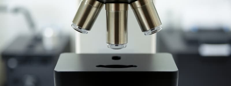
- A compound microscope has multiple lenses that magnify the image.
- It includes parts like ocular lenses, revolving nosepiece, objective lenses, slide holder, stage, condenser, light, coarse adjustment, fine adjustment, and light intensity control.
How Does It Work?
- Light from a light source travels through the specimen.
- Objective lenses focus the light onto the specimen
- The focused light then travels through the tube/body to the eyepiece.
- Magnification occurs as the light passes through the eyepiece.
The Virtual Image
- The final image seen by the observer.
- Image quality is affected by factors like the type of light (electromagnetic spectrum), the properties and size of the object, lens type and placement and contrast.
Key Principles of Microscopy: Magnification
- The degree to which a device enlarges an object/specimen, provided by the lens.
- Expressed as "X" followed by a number, e.g., 40X, 10x.
- Total magnification = (Objective Magnification) x (Eyepiece Magnification).
Key Principles of Microscopy: Resolution
- The ability of a microscope to distinguish between two adjacent objects as separate entities.
- Determined by the wavelength of light and numerical aperture (NA).
- Resolving power= (Wavelength of light) / (2 x NA)
- Numerical aperture dictates the light-gathering ability of the objective and condenser.
Key Principles of Microscopy: Refractive Index
- Measures how light bends as it passes from one medium to another.
- Light bends as it passes through glass.
- Higher magnification, oil can be used to lessen dispersion as it has a refractive index similar to glass.
Key Principles of Microscopy: Illumination
- Light source: e.g., mirror or light bulb.
- Condenser: collects light from the source and focuses it on the specimen, forming a cone of light.
- Diaphragm: used to adjust the size of the opening (aperture) of light before reaching the condenser.
Key Principles of Microscopy: Parfocality
- During the microscope's operation, most microscope objectives allow you to quickly switch to the objective lenses.
- Parfocal lenses maintain focus even when switching objectives.
Key Principles of Microscopy: Contrast
- Interaction between light and the specimen.
- Adding contrast helps view specimen detail.
Types of Microscopes: Two-Dimensional Imaging
- Bright Field Microscope: Specimens are visualized due to differences in density between specimen and surroundings. Uses two sets of lenses (objective and ocular). Maximum magnification is approximately 2000X.
- Phase Contrast Microscope: Uses a phase ring to induce phase shifts due to brightness variations, creating contrast without dyes. Live samples can be observed and dark cells appear on a light background.
- Dark Field Microscope: The light source is blocked, making light reach the specimen only from the side. Observing motile samples.
- Fluorescence Microscope: Visualizes specimens that fluoresce (emit light) when they absorb light of other wavelengths. Can be used for natural or induced fluorescence (through staining). Used in microbiology for bacterial enumeration.
Types of Microscopes: Three-Dimensional Imaging
- Differential Interference Contrast (DIC) Microscopy: Uses polarizers and polarized light to create two distinct beams. This emphasizes cell structures.
- Confocal Scanning Laser Microscope (CLSM): Uses a computer-controlled laser light to isolate and view different layers of the specimen. Resolution is 0.1 μm.
Types of Microscopes: Electron Microscope
- Transmission Electron Microscope (TEM): Electromagnets act as lenses to provide high magnification and resolution (0.2 nm). Specimens are sliced thinly and stained with heavy metals.
- Scanning Electron Microscope (SEM): Beams of electrons hit a coated specimen. Scattered electrons are captured to render an image. Resolution is 15x to 100,000x
Studying That Suits You
Use AI to generate personalized quizzes and flashcards to suit your learning preferences.


