Podcast
Questions and Answers
Which component of a microscope directly regulates the amount of light passing through the condenser?
Which component of a microscope directly regulates the amount of light passing through the condenser?
- Coarse adjustment knob
- Condenser adjustment knob
- Objective lens
- Iris diaphragm (correct)
A student is having trouble bringing a specimen into sharp focus. They have already used the coarse adjustment knob. Which of the following should they adjust next?
A student is having trouble bringing a specimen into sharp focus. They have already used the coarse adjustment knob. Which of the following should they adjust next?
- Condenser adjustment knob
- Fine adjustment knob (not mentioned in text but implied) (correct)
- Ocular lens
- Revolving nosepiece
What is the primary function of the condenser in a light microscope?
What is the primary function of the condenser in a light microscope?
- To focus a cone of light onto the specimen (correct)
- To control the amount of light entering the objective lens
- To magnify the image of the specimen
- To adjust the distance between the objective lens and the specimen
In dark-field microscopy, what causes the specimen to appear bright against a dark background?
In dark-field microscopy, what causes the specimen to appear bright against a dark background?
A researcher needs to view living, unstained spirochetes. Which type of microscopy would be most suitable?
A researcher needs to view living, unstained spirochetes. Which type of microscopy would be most suitable?
What is the main purpose of the opaque area in the dark field condenser?
What is the main purpose of the opaque area in the dark field condenser?
A microscope has an ocular lens with 10x magnification and an objective lens with 40x magnification. What is the total magnification of the microscope?
A microscope has an ocular lens with 10x magnification and an objective lens with 40x magnification. What is the total magnification of the microscope?
Which of the following sequences accurately describes path of light in a light microscope?
Which of the following sequences accurately describes path of light in a light microscope?
A researcher is studying bacteria at a magnification that allows detailed observation of internal structures. Which objective lens is MOST likely being used, and what is the approximate total magnification assuming a standard 10x ocular lens?
A researcher is studying bacteria at a magnification that allows detailed observation of internal structures. Which objective lens is MOST likely being used, and what is the approximate total magnification assuming a standard 10x ocular lens?
Which of the following BEST describes the relationship between refractive index and resolution in microscopy?
Which of the following BEST describes the relationship between refractive index and resolution in microscopy?
A student is having trouble focusing a specimen under a microscope. They have adjusted the coarse adjustment knob but the image remains blurry. What should they do NEXT to achieve a clearer image?
A student is having trouble focusing a specimen under a microscope. They have adjusted the coarse adjustment knob but the image remains blurry. What should they do NEXT to achieve a clearer image?
In a compound microscope, what is the PRIMARY function of the objective lens?
In a compound microscope, what is the PRIMARY function of the objective lens?
Why is it generally NOT recommended to use the coarse adjustment knob when using high power objective lenses (40x or higher)?
Why is it generally NOT recommended to use the coarse adjustment knob when using high power objective lenses (40x or higher)?
A researcher needs to quickly scan a large area of a tissue sample to identify regions of interest. Which objective lens should they use INITIALLY, and what is the main advantage of using this lens for the initial scan?
A researcher needs to quickly scan a large area of a tissue sample to identify regions of interest. Which objective lens should they use INITIALLY, and what is the main advantage of using this lens for the initial scan?
A microscope has a 15x ocular lens and a 60x objective lens. What is the total magnification of the microscope?
A microscope has a 15x ocular lens and a 60x objective lens. What is the total magnification of the microscope?
Which component of a microscope is responsible for holding the slide in place on the stage?
Which component of a microscope is responsible for holding the slide in place on the stage?
Flashcards
Microscope
Microscope
An instrument used to see objects too small for the naked eye.
Microscopic
Microscopic
Invisible to the eye unless aided by a microscope.
Microscopy
Microscopy
The science of investigating small objects using a microscope.
Simple Microscope
Simple Microscope
Signup and view all the flashcards
Compound Microscope
Compound Microscope
Signup and view all the flashcards
Inclination Joint
Inclination Joint
Signup and view all the flashcards
Base (Microscope)
Base (Microscope)
Signup and view all the flashcards
Head (Microscope)
Head (Microscope)
Signup and view all the flashcards
Body Tube
Body Tube
Signup and view all the flashcards
Draw Tube
Draw Tube
Signup and view all the flashcards
Revolving Nosepiece
Revolving Nosepiece
Signup and view all the flashcards
Coarse Adjustment Knob
Coarse Adjustment Knob
Signup and view all the flashcards
Condenser Adjustment Knob
Condenser Adjustment Knob
Signup and view all the flashcards
Iris Diaphragm Lever
Iris Diaphragm Lever
Signup and view all the flashcards
Objective Lens
Objective Lens
Signup and view all the flashcards
Dark-Field Microscope
Dark-Field Microscope
Signup and view all the flashcards
Study Notes
- A microscope is used to see small objects invisible to the naked eye.
- Microscopy is the science of investigating small objects with a microscope.
- Microscopic means invisible without a microscope.
- A good microscope has resolution, contrast, and magnification properties.
Resolution
- Resolution power is the ability to produce separate images of closely placed objects.
- The resolution power of an unaided human eye is about 0.2mm (200µm).
- The resolution of a light microscope is about 0.2µm.
- The resolution of an electron microscope is about 0.5µm.
- Immersion oil enhances resolution due to its higher refractive index than air.
Contrast
- Contrast is improved by staining the specimen.
- Staining enhances cell contrast.
Magnification
- Ocular lens magnification: 10x.
- Objective lens magnifications: Scanning (4x), Low Power (10x), High Power (40x), Oil Immersion (100x).
- Total magnification is calculated by multiplying the objective lens magnification by the ocular lens magnification.
- Scanning field: 40x.
- Low power field: 100x.
- High power field: 400x.
- Oil immersion field: 1000x.
Types of Microscopes
- Simple microscopes use a single lens.
- Compound microscopes use multiple lenses.
Simple Light Microscope
- A magnifying glass is a simple light microscope creating a magnified, virtual, upright image.
Compound Microscope
- Compound scopes are commonly used in research and school labs
Anatomy of a Compound Microscope
- Mechanical parts facilitate adjustment and support
- Inclination Joint: Allows tilting for user convenience.
- Base: Holds the light source, adjustment knobs, and other parts.
- Head: Contains the eyepiece tube and prisms.
- Monocular: One eyepiece.
- Binocular: Two eyepieces.
- Dual head: Two eyepieces, not together.
- Trinocular: Three eyepieces, usually for camera connection.
- C-shaped arm: Connects the ocular and objective lenses.
- Mechanical stage: Holds slides with clips and has control knobs to move the slide for viewing; includes an aperture for light.
- Stage clips secure the specimen.
- Body tube: Attached to the arm and bears the lenses
- Draw tube: Holds ocular lenses.
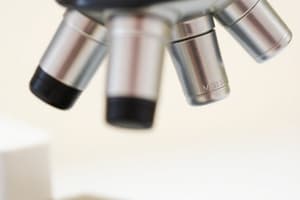
- Revolving/rotating nosepiece: Disc holding objectives.
- Coarse adjustment knob: Elevates or lowers the body tube for approximate focus.
- Condenser adjustment knob: Regulates light intensity.
- Iris diaphragm lever: Controls diaphragm opening.
Magnifying Parts
- Ocular lens: 10x magnification.
- Microscopes with two eyepieces are binocular.
- Objective lens: 3-5 lenses with varying power (4x, 10x, 40x, 100x).
Illuminating Parts
- Provide light
- Condenser: Focuses light on the slide.
- Iris diaphragm: Controls light through the condenser.
- Light source: Mirror or electric bulb.
Principle of Light Microscopy
- Light passes through the iris diaphragm and specimen.
- The objective gathers the light and forms a magnified image.
- Further magnification by the ocular lens produces the final virtual image.
Dark-Field Microscope
- In dark field microscopy, objects appear bright against a dark background.
- It requires a special dark field condenser.
Dark-Field Microscopy - Principle
- The condenser blocks direct light, allowing only oblique light to pass through the specimen.
- Only reflected light enters the objective lens.
- The specimen is brightly illuminated against a dark background.
Dark-Field Microscopy - Application
- Used to identify living, unstained cells and thin bacteria like spirochetes.
Studying That Suits You
Use AI to generate personalized quizzes and flashcards to suit your learning preferences.


