Podcast
Questions and Answers
What is the total magnification of the microscope with the High Power objective in position? (Select all that apply)
What is the total magnification of the microscope with the High Power objective in position? (Select all that apply)
- 100X
- 40X
- 400X (correct)
- 1000X
- unable to be determined
What is the total magnification of the microscope with the Scanning objective in position? (Select all that apply)
What is the total magnification of the microscope with the Scanning objective in position? (Select all that apply)
- 40X (correct)
- 100X
- 400X
- unable to be determined
- 1000X
What is the total magnification of the microscope with the Low Power objective in position? (Select all that apply)
What is the total magnification of the microscope with the Low Power objective in position? (Select all that apply)
- unable to be determined
- 1000X
- 400X
- 100X (correct)
- 40X
What is the total magnification of the microscope with the Oil Immersion objective in position? (Select all that apply)
What is the total magnification of the microscope with the Oil Immersion objective in position? (Select all that apply)
If you put a slide with the letter 'e' on the stage as you would normally read it, and then you observed it under the microscope, how would it appear? (Select all that apply)
If you put a slide with the letter 'e' on the stage as you would normally read it, and then you observed it under the microscope, how would it appear? (Select all that apply)
Of the three ways we can control light, which one will we use almost exclusively in this class?
Of the three ways we can control light, which one will we use almost exclusively in this class?
What is the term for the phenomenon when you switch from one objective to the next, and the microscope should be pretty close to in focus?
What is the term for the phenomenon when you switch from one objective to the next, and the microscope should be pretty close to in focus?
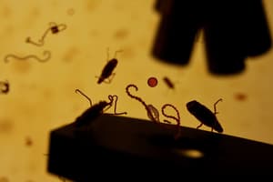
What is the cell morphology and cell arrangement of Staphylococcus epidermidis?
What is the cell morphology and cell arrangement of Staphylococcus epidermidis?
What is the cell morphology and cell arrangement of Escherichia coli?
What is the cell morphology and cell arrangement of Escherichia coli?
What is the cell morphology and cell arrangement of Bacillus subtilis?
What is the cell morphology and cell arrangement of Bacillus subtilis?
In the wet mount of E.coli, what bacterial appendage is responsible for movement of the bacteria around the liquid?
In the wet mount of E.coli, what bacterial appendage is responsible for movement of the bacteria around the liquid?
What is the cell morphology and cell arrangement of Neisseria gonorrhoeae (causative agent of gonorrhea)?
What is the cell morphology and cell arrangement of Neisseria gonorrhoeae (causative agent of gonorrhea)?
What is the cell morphology of Treponema pallidum (causative agent of syphilis)?
What is the cell morphology of Treponema pallidum (causative agent of syphilis)?
What part of the yeast is highlighted by the negative stain and protects the yeast from harm inside its host?
What part of the yeast is highlighted by the negative stain and protects the yeast from harm inside its host?
Is Staphylococcus epidermidis Gram positive or Gram negative?
Is Staphylococcus epidermidis Gram positive or Gram negative?
What is the cell morphology and cell arrangement of Staphylococcus epidermidis?
What is the cell morphology and cell arrangement of Staphylococcus epidermidis?
Is Escherichia coli Gram positive or Gram negative?
Is Escherichia coli Gram positive or Gram negative?
What is the cell morphology and cell arrangement of Escherichia coli?
What is the cell morphology and cell arrangement of Escherichia coli?
Is Bacillus subtilis Gram positive or Gram negative?
Is Bacillus subtilis Gram positive or Gram negative?
What is the cell morphology and cell arrangement of Bacillus subtilis?
What is the cell morphology and cell arrangement of Bacillus subtilis?
Suppose that in the process of doing your very first Gram stain you forgot to add the decolorizer (alcohol). What color would the Gram positive bacteria appear?
Suppose that in the process of doing your very first Gram stain you forgot to add the decolorizer (alcohol). What color would the Gram positive bacteria appear?
Suppose that in the process of doing your second Gram stain you forgot to add the decolorizer (alcohol) again. What color would Gram negative bacteria appear and why?
Suppose that in the process of doing your second Gram stain you forgot to add the decolorizer (alcohol) again. What color would Gram negative bacteria appear and why?
Since the Gram stain allows us to tell two groups of bacteria apart from each other, we can refer to this kind of test as a ______ test.
Since the Gram stain allows us to tell two groups of bacteria apart from each other, we can refer to this kind of test as a ______ test.
If we had to simplify the Gram Stain procedure into one sentence, it would be 'The Gram stain separates bacterial species based on the thickness of the ______ layer surrounding the bacteria.'
If we had to simplify the Gram Stain procedure into one sentence, it would be 'The Gram stain separates bacterial species based on the thickness of the ______ layer surrounding the bacteria.'
Which term describes the highly resistant form of a select group of bacteria?
Which term describes the highly resistant form of a select group of bacteria?
Which of the following endospore-forming bacteria is incorrectly matched with the disease it causes?
Which of the following endospore-forming bacteria is incorrectly matched with the disease it causes?
Botox, the chemical that is used to decrease wrinkles and lines in the face, is a toxin extracted from the spore-forming bacteria Clostridium botulinum. What is Botox most likely doing to decrease wrinkles?
Botox, the chemical that is used to decrease wrinkles and lines in the face, is a toxin extracted from the spore-forming bacteria Clostridium botulinum. What is Botox most likely doing to decrease wrinkles?
What disease caused by spore-forming bacteria is commonly observed after treatment with antibiotics?
What disease caused by spore-forming bacteria is commonly observed after treatment with antibiotics?
If you gram stain spore-forming bacteria, you see many rod-shaped, dark purple colored bacteria. However, some of them have what appears to be a clear colored hole in the middle of the bacterial cell. Why do these bacteria have this clear colored hole?
If you gram stain spore-forming bacteria, you see many rod-shaped, dark purple colored bacteria. However, some of them have what appears to be a clear colored hole in the middle of the bacterial cell. Why do these bacteria have this clear colored hole?
In microbiology, ______ is another term for cloudiness, which is the result of bacteria accumulating in liquid growth media.
In microbiology, ______ is another term for cloudiness, which is the result of bacteria accumulating in liquid growth media.
In Microbiology, we are frequently required to ______, or introduce microbes into another culture medium.
In Microbiology, we are frequently required to ______, or introduce microbes into another culture medium.
According to your results, which bacterial species grows only at the top of the broth culture?
According to your results, which bacterial species grows only at the top of the broth culture?
According to your results, which bacterial species grows all over the broth culture?
According to your results, which bacterial species grows all over the broth culture?
According to your results, which bacterial species grows with a dried out appearance on the agar slant?
According to your results, which bacterial species grows with a dried out appearance on the agar slant?
Examine your streak plate from yesterday. In which quadrant did you get single colonies? (There is no right or wrong answer to this as long as you got single colonies)
Examine your streak plate from yesterday. In which quadrant did you get single colonies? (There is no right or wrong answer to this as long as you got single colonies)
How many different species are represented on your streak plate?
How many different species are represented on your streak plate?
What is the Gram reaction/morphology of the red colored colony?
What is the Gram reaction/morphology of the red colored colony?
What would you expect to observe if you forgot to flame your loop between quadrant streaks?
What would you expect to observe if you forgot to flame your loop between quadrant streaks?
Overall, how would you grade your streak plate technique?
Overall, how would you grade your streak plate technique?
According to your results, which bacterial species is capable of fermenting all three sugars, lactose and glucose, to produce acid?
According to your results, which bacterial species is capable of fermenting all three sugars, lactose and glucose, to produce acid?
According to your results, which bacterial species is not capable of producing acid at all from sucrose, lactose, or glucose?
According to your results, which bacterial species is not capable of producing acid at all from sucrose, lactose, or glucose?
According to your results, which bacterial species is capable of fermenting fewer than three of the sugars?
According to your results, which bacterial species is capable of fermenting fewer than three of the sugars?
According to your results, which bacterial species is capable of degrading DNA?
According to your results, which bacterial species is capable of degrading DNA?
According to your results, which bacterial species is capable of degrading hydrogen peroxide?
According to your results, which bacterial species is capable of degrading hydrogen peroxide?
According to your results, which bacterial species is capable of producing hydrogen sulfide?
According to your results, which bacterial species is capable of producing hydrogen sulfide?
According to your results, which bacterial species is capable of degrading urea?
According to your results, which bacterial species is capable of degrading urea?
According to your results, which bacterial species can break down the amino acid tryptophan?
According to your results, which bacterial species can break down the amino acid tryptophan?
According to your results, which bacterial species does not propel itself around with flagella?
According to your results, which bacterial species does not propel itself around with flagella?
According to your results, which bacteria species is capable of producing amylase?
According to your results, which bacteria species is capable of producing amylase?
On which of the plates would you expect to find bacteria most like the original non-transformed E.coli colonies you initially observed?
On which of the plates would you expect to find bacteria most like the original non-transformed E.coli colonies you initially observed?
If there are any genetically transformed bacterial cells, on which plate(s) would they most likely be located?
If there are any genetically transformed bacterial cells, on which plate(s) would they most likely be located?
Which plates should be compared to determine if any genetic transformation has occurred?
Which plates should be compared to determine if any genetic transformation has occurred?
Examine the plate labeled '-pGLO LB only' after the overnight incubation. You should see a lawn of growth. What would you suspect if nothing grew on your plate?
Examine the plate labeled '-pGLO LB only' after the overnight incubation. You should see a lawn of growth. What would you suspect if nothing grew on your plate?
With the use of UV penlight, examine all your petri plates after overnight incubation. Which plate exhibits fluorescence?
With the use of UV penlight, examine all your petri plates after overnight incubation. Which plate exhibits fluorescence?
Flashcards are hidden until you start studying
Study Notes
Microscope Magnification
- High Power objective yields total magnification of 400X.
- Scanning objective provides total magnification of 40X.
- Low Power objective results in total magnification of 100X.
- Oil Immersion objective achieves total magnification of 1000X.
Optical Observations and Light Control
- Observing a standard "e" slide results in a 180-degree rotation under the microscope.
- The Iris Diaphragm is primarily used to control light intensity.
Microscope Focus and Cell Morphology
- Parfocal allows for minimal refocusing when changing objectives.
- Staphylococcus epidermidis exhibits coccus morphology with staphylococci arrangement.
- Escherichia coli has bacillus morphology with no specific arrangement.
- Bacillus subtilis also shows bacillus morphology with no arrangement.
Bacterial Structures and Staining
- Flagellum is essential for motility in E. coli.
- Neisseria gonorrhoeae appears as coccus with diplococci arrangement.
- Treponema pallidum has spirillum morphology.
- Capsules protect yeast in staining procedures.
Gram Staining Results
- Staphylococcus epidermidis is Gram positive.
- E. coli is Gram negative.
- Gram staining involves peptidoglycan layer thickness to differentiate bacteria.
Endospores and Their Characteristics
- Endospores are highly resistant forms of certain bacteria.
- Clostridium difficile is misassigned, as it does not cause cervical cancer.
- Botox, derived from Clostridium botulinum, inhibits muscle contraction to reduce wrinkles.
- Pseudomembranous enterocolitis often follows antibiotic treatment due to spore-forming bacteria.
Bacterial Growth Patterns
- Bacillus subtilis grows predominantly at the top of broth cultures.
- Serratia marcescens grows uniformly throughout broth cultures.
Streak Plate Techniques
- Single colonies likely observed in quadrant #4 of streak plates.
- Flawless streaking techniques yield well-separated single colonies, graded as 'A'.
Metabolic Capabilities of Bacterial Species
- E. coli ferments lactose and glucose, producing acid.
- Pseudomonas aeruginosa does not produce acid from tested sugars.
- Serratia marcescens capable of degrading DNA and fermenting fewer sugars.
- Staphylococcus epidermidis degrades hydrogen peroxide.
- Proteus vulgaris produces hydrogen sulfide gas.
- Klebsiella pneumoniae can degrade urea.
Genetic Transformation and Experimental Plates
- Non-transformed E. coli colonies appear on -pGLO LB only plate.
- Transformed cells appear on both +pGLO LB with ampicillin and +pGLO LB with ampicillin and arabinose plates.
- Fluorescence is observable on the +pGLO LB with ampicillin and arabinose plate only after incubation.
Interpretation of Results
- Lack of growth on -pGLO LB only indicates non-viability of E. coli.
- Observing fluorescence indicates successful genetic transformation, particularly with the presence of arabinose.
Studying That Suits You
Use AI to generate personalized quizzes and flashcards to suit your learning preferences.