Podcast
Questions and Answers
What is the primary purpose of using staining techniques in light microscopy?
What is the primary purpose of using staining techniques in light microscopy?
- To reduce the limit of resolution
- To visualize three-dimensional cell structures
- To improve the contrast between specimens and their surroundings (correct)
- To increase the magnification of specimens
Which scientist is credited with the first description of microbes in 'Micrographia'?
Which scientist is credited with the first description of microbes in 'Micrographia'?
- Joseph Lister
- Antoni van Leeuwenhoek
- Louis Pasteur
- Robert Hooke (correct)
What is the limit of resolution for a light microscope?
What is the limit of resolution for a light microscope?
- 0.2 μm (correct)
- 0.1 μm
- 0.5 μm
- 1 μm
Which type of microscope uses two sets of lenses to form an image?
Which type of microscope uses two sets of lenses to form an image?
What is the primary charge of basic dyes and their role in staining?
What is the primary charge of basic dyes and their role in staining?
How do gram-positive bacteria appear under a gram stain?
How do gram-positive bacteria appear under a gram stain?
What does the term 'total magnification' refer to in microscopy?
What does the term 'total magnification' refer to in microscopy?
Which microscopy method allows for observing living cells with minimal alteration?
Which microscopy method allows for observing living cells with minimal alteration?
What is the primary advantage of phase-contrast microscopy?
What is the primary advantage of phase-contrast microscopy?
What characteristic of a light microscope limits its resolution?
What characteristic of a light microscope limits its resolution?
What is a key characteristic of dark-field microscopy?
What is a key characteristic of dark-field microscopy?
Which type of microscopy is best for observing motility?
Which type of microscopy is best for observing motility?
What function does the objective lens serve in a microscope?
What function does the objective lens serve in a microscope?
Which microscopy technique is best suited to visualize the internal structures of cells?
Which microscopy technique is best suited to visualize the internal structures of cells?
What effect does the phase ring have in phase-contrast microscopy?
What effect does the phase ring have in phase-contrast microscopy?
What is the main purpose of fluorescence microscopy?
What is the main purpose of fluorescence microscopy?
What type of light microscope enhances contrast by using differences in the phase of light waves?
What type of light microscope enhances contrast by using differences in the phase of light waves?
Which dye is used to show dead cells in fluorescence microscopy?
Which dye is used to show dead cells in fluorescence microscopy?
How do gram-negative bacteria appear in a gram stain?
How do gram-negative bacteria appear in a gram stain?
Which microscopy technique enhances contrast using light that has been scattered from the sides?
Which microscopy technique enhances contrast using light that has been scattered from the sides?
What characteristic of differential interference contrast (DIC) microscopy contributes to its three-dimensional imaging capability?
What characteristic of differential interference contrast (DIC) microscopy contributes to its three-dimensional imaging capability?
Which microscopy technique involves the use of a laser coupled with a computerized microscope?
Which microscopy technique involves the use of a laser coupled with a computerized microscope?
Why must specimens be very thin when using transmission electron microscopy (TEM)?
Why must specimens be very thin when using transmission electron microscopy (TEM)?
What is a unique feature of cryo-electron microscopy (cryo-EM) in terms of specimen preparation?
What is a unique feature of cryo-electron microscopy (cryo-EM) in terms of specimen preparation?
What advantage does scanning electron microscopy (SEM) have compared to transmission electron microscopy (TEM)?
What advantage does scanning electron microscopy (SEM) have compared to transmission electron microscopy (TEM)?
What characteristic is common to both differential interference contrast microscopy and confocal scanning laser microscopy?
What characteristic is common to both differential interference contrast microscopy and confocal scanning laser microscopy?
What type of image is created by transmission electron microscopy (TEM)?
What type of image is created by transmission electron microscopy (TEM)?
What is the primary limitation of electron microscopes compared to light microscopes?
What is the primary limitation of electron microscopes compared to light microscopes?
What is the main purpose of using high atomic weight substances in transmission electron microscopy (TEM)?
What is the main purpose of using high atomic weight substances in transmission electron microscopy (TEM)?
Which microscopy technique is demonstrated to visualize protein structures in detail by analyzing numerous particle images?
Which microscopy technique is demonstrated to visualize protein structures in detail by analyzing numerous particle images?
What is the role of bile salts in MacConkey agar?
What is the role of bile salts in MacConkey agar?
Which component in MacConkey agar is responsible for the color change indicating lactose fermentation?
Which component in MacConkey agar is responsible for the color change indicating lactose fermentation?
What is the significance of the color pink to mauve in CHROMagar VRE?
What is the significance of the color pink to mauve in CHROMagar VRE?
What makes CHROMagar a unique culture medium compared to traditional agar plates?
What makes CHROMagar a unique culture medium compared to traditional agar plates?
What is the main function of the proteose peptone in MacConkey agar?
What is the main function of the proteose peptone in MacConkey agar?
Which component in CHROMagar allows for detecting E.coli specifically?
Which component in CHROMagar allows for detecting E.coli specifically?
What should be the pH adjustment for MacConkey agar before use?
What should be the pH adjustment for MacConkey agar before use?
What is the purpose of following aseptic techniques in microbial culture?
What is the purpose of following aseptic techniques in microbial culture?
Which aspect of the CHROMagar patented system distinguishes it from other media?
Which aspect of the CHROMagar patented system distinguishes it from other media?
What type of bacteria are primarily inhibited by crystal violet in MacConkey agar?
What type of bacteria are primarily inhibited by crystal violet in MacConkey agar?
What is the primary feature of enriched media?
What is the primary feature of enriched media?
What do selective media achieve in the context of microbial growth?
What do selective media achieve in the context of microbial growth?
Which of the following best describes differential media?
Which of the following best describes differential media?
How does the composition of solid media differ from liquid media?
How does the composition of solid media differ from liquid media?
What metabolic byproducts are generated by E. coli during anaerobic fermentation of lactose?
What metabolic byproducts are generated by E. coli during anaerobic fermentation of lactose?
What is a critical factor for successful cultivation of microbes in culture media?
What is a critical factor for successful cultivation of microbes in culture media?
What role does β-galactosidase play in the metabolism of lactose by E. coli?
What role does β-galactosidase play in the metabolism of lactose by E. coli?
Which characteristic is essential to consider when using differential media?
Which characteristic is essential to consider when using differential media?
Which of the following statements is true about the agar used in solid media?
Which of the following statements is true about the agar used in solid media?
What role do the acidic byproducts of fermentation play in the growth of E. coli?
What role do the acidic byproducts of fermentation play in the growth of E. coli?
What is the primary reason for diluting bacterial samples before measuring Optical Density (OD) when they are too dense?
What is the primary reason for diluting bacterial samples before measuring Optical Density (OD) when they are too dense?
Which material is recommended for cuvettes when measuring absorbance in the ultraviolet region?
Which material is recommended for cuvettes when measuring absorbance in the ultraviolet region?
Optical Density (OD) is defined mathematically using which formula?
Optical Density (OD) is defined mathematically using which formula?
Which of the following factors does NOT influence the Beer-Lambert Law relationship?
Which of the following factors does NOT influence the Beer-Lambert Law relationship?
What is a significant disadvantage of using a spectrophotometer for measuring microbial turbidity?
What is a significant disadvantage of using a spectrophotometer for measuring microbial turbidity?
Which value indicates a region where the Beer-Lambert law is typically valid?
Which value indicates a region where the Beer-Lambert law is typically valid?
What does Optical Density correlate with in a spectrophotometric analysis of bacterial cells?
What does Optical Density correlate with in a spectrophotometric analysis of bacterial cells?
The equation for calculating absorbance directly includes which component?
The equation for calculating absorbance directly includes which component?
Which of the following is NOT an advantage of using a spectrophotometer?
Which of the following is NOT an advantage of using a spectrophotometer?
What is the correct relationship between intensity and absorbance as described in the equations?
What is the correct relationship between intensity and absorbance as described in the equations?
What is the main reason for requiring a standard curve when using turbidity measures to estimate microbial cell numbers?
What is the main reason for requiring a standard curve when using turbidity measures to estimate microbial cell numbers?
Which condition is least likely to affect the accuracy of viable plate counts?
Which condition is least likely to affect the accuracy of viable plate counts?
What does the term 'the great plate count anomaly' refer to regarding microbial counts?
What does the term 'the great plate count anomaly' refer to regarding microbial counts?
Why should plates with fewer than 30 colonies be excluded from viable plate count analysis?
Why should plates with fewer than 30 colonies be excluded from viable plate count analysis?
In which phase of bacterial growth does E.coli typically double every 30 minutes under optimal conditions?
In which phase of bacterial growth does E.coli typically double every 30 minutes under optimal conditions?
What limitation does the plate counting method have when analyzing mixed microbial cultures?
What limitation does the plate counting method have when analyzing mixed microbial cultures?
What main factor contributes to the turbidity in a culture of unicellular organisms?
What main factor contributes to the turbidity in a culture of unicellular organisms?
What is primarily accounted for when reporting plate counts in colony-forming units (CFU)?
What is primarily accounted for when reporting plate counts in colony-forming units (CFU)?
Why is the measurement of absorbance at 600 nm commonly used for E.coli density estimations?
Why is the measurement of absorbance at 600 nm commonly used for E.coli density estimations?
What influence does a mixed culture have on the accuracy of viable plate counting?
What influence does a mixed culture have on the accuracy of viable plate counting?
Flashcards are hidden until you start studying
Study Notes
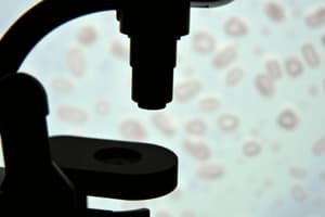
Light Microscopy and the Discovery of Microorganisms
- Robert Hooke, in 1665, was the first to describe microbes using a microscope.
- Hooke's book, Micrographia, illustrated the fruiting structures of molds.
- Antoni van Leeuwenhoek was the first to describe bacteria using a microscope.
Improving Contrast in Light Microscopy
- Magnification is the ability to make an object appear larger.
- Resolution is the ability to distinguish two adjacent objects as distinct and separate.
- The limit of resolution for a light microscope is approximately 0.2 micrometers, which is half the wavelength of visible light.
- Several types of light microscopy exist:
- Bright-field
- Phase-contrast
- Differential interference contrast
- Dark-field
- Fluorescence
- Compound microscopes use two sets of lenses to form an image: an objective lens (magnifies 10-100x) and an ocular lens (magnifies 10-30x).
- The total magnification of a compound microscope is the product of the objective lens magnification and the ocular lens magnification.
- A magnification of 1,000x is needed to reach the 0.2-micrometer resolution limit of most light microscopes.
- Bright-field microscopy uses normal light and visualization depends on differences in contrast or density between the specimen and its surroundings.
Microscopy & Staining
- Staining can improve contrast by using dyes that bind to specific cellular materials.
- Basic dyes, like methylene blue, crystal violet, and safranin, are positively charged and bind to negatively charged cell components, such as nucleic acids, acidic polysaccharides, and cell surfaces.
- Simple staining uses dried cells to highlight the overall shape, size, and arrangement of cells.
- Differential stains differentiate between cell types by staining them different colors.
- Gram staining is a differential stain widely used for identifying bacteria, which differentiates them into two main groups: gram-positive and gram-negative.
- Gram-positive bacteria stain purple-violet due to a thick peptidoglycan layer in their cell walls.
- Gram-negative bacteria stain pink because they have a thinner peptidoglycan layer and an outer membrane.
Phase-Contrast Microscopy
- Phase-contrast microscopy enhances image contrast of unstained, live cells.
- A phase ring amplifies differences in refractive index between the cell and its surroundings.
- An image with dark cells on a light background is produced.
Dark-Field Microscopy
- Light reaches the specimen from the sides.
- Only scattered light reaches the lens, resulting in an image that appears light on a dark background.
- Better resolution than bright-field microscopy.
- Excellent for observing motility, such as flagella.
Fluorescence Microscopy
- Used to visualize specimens that fluoresce, meaning they emit light after being illuminated at a different wavelength.
- Cells appear to glow on a black background due to filters.
- Some cells fluoresce naturally (autofluorescence), while others can be stained with fluorescent dyes like DAPI.
- Widely used in microbial ecology to enumerate bacteria in natural samples.
Differential Interference Contrast (DIC) Microscopy
- Uses a polarizer to generate two polarized light beams
- Provides a three-dimensional appearance to structures like nuclei, endospores, vacuoles, and inclusions
Confocal Scanning Laser Microscopy (CSLM)
- Uses a computerized microscope with a laser source to create a 3D image
- Computer focuses the laser on individual layers of the specimen
- Different layers can be compiled for a complete 3D image
Electron Microscopy
- Uses electrons instead of visible light (photons)
- Electromagnets act as lenses
- Operates in a vacuum
- Images are captured using a camera, resulting in an electron micrograph
- Two primary types: Transmission Electron Microscopy (TEM) and Scanning Electron Microscopy (SEM)
Transmission Electron Microscopy (TEM)
- Offers significantly higher resolving power (0.2 nm) than light microscopes (200 nm)
- Enables visualization of structures at the molecular level
- Requires thin specimens (20–60 nm) stained with high atomic weight substances for contrast
Scanning Electron Microscopy (SEM)
- Provides detailed, three-dimensional images of surfaces
Cryo-Electron Microscopy (Cryo-EM)
- Used to dissect protein structure
- Involves rapidly freezing a specimen in liquid ethane
- Cryo-samples are then visualized at -170 °C
- 3D structures are determined by analyzing thousands of individual protein particle images
- Applicable for very large protein complexes
Enriched Media
- Contain complex media plus highly nutritious materials, for example serum or blood
- Used to culture fastidious microbes, which are nutritionally demanding
Selective Media
- Contain compounds selectively inhibiting the growth of some microbes but not others
Differential Media
- Contain an indicator, typically a dye, that detects particular metabolic reactions during growth
Considerations for Growth Media
- For successful microbial cultivation, it is crucial to know the nutritional requirements and supply them in the proper form and proportions in a culture medium.
- Cells can be cultivated in liquid or solid culture media.
- Solid media are prepared by adding the gelling agent agar to liquid media.
- When grown on solid media, cells form isolated masses called colonies.
Properties of Bacteria Utilized for Differential Media
- Example: Lactose utilization and anaerobic fermentation in E. coli
- Involves the conversion of lactose to glucose by β-galactosidase
- Under anaerobic fermentative conditions, a mixture of succinate, formate, acetate, lactate, and ethanol is produced
- The products, including e.g., acetate and lactate, are acidic
MacConkey Agar
- A selective and differential medium
- Selective: Bile salts inhibit most Gram-positive bacteria, and crystal violet inhibits certain Gram-positive bacteria.
- Differential: Detects lactose fermentation
- Lactose-fermenting bacteria: Consumes lactose or other six-carbon sugars and metabolizes them.
- Acid production: Reduces the pH, turning the neutral red dye red.
- Composition: Peptone, proteose peptone, lactose, bile salts, sodium chloride, neutral red, crystal violet, agar.
CHROMagar™
- Enzyme-based chromogenic detection system
- Invented by Dr. Alain Rambach in 1979
- Pioneered chromogenic differentiation culture media for detecting E.coli
- Patented a chromogenic system for detecting Salmonella in 1989, known as Rambach Agar
- Established CHROMagar Microbiology in 1993
Typical Appearance of Microorganisms on CHROMagar™
- VRE (Vancomycin-Resistant Enterococci) faecalis/faecium: Pink to mauve
- E. gallinarum/E. casseliflavus: Blue or inhibited
- Other Bacteria: Inhibited
CHROMagar™ E. coli and CHROMagar™ VRE
- CHROMagar™ E. coli: β-galactosidase-based
- Most chromogenic mixes are proprietary
Growth Media and Laboratory Culture
- Microbes are ubiquitous.
- Sterilization of media is crucial.
- Aseptic technique should be followed to prevent contamination.
Viable Plate Counts
- Measures living, reproducing microbial populations
- Two main methods:
- Spread-plate method
- Pour-plate method
- Serial dilution is key for accurate counts, which are typically between 30 and 300 colonies
Sources of Error
- Inoculum size, viability, culture medium, and incubation conditions affect accuracy
- Mixed cultures grow at different rates, leading to inaccuracies
- Colony-forming units (CFUs) are reported instead of viable cell numbers to address clumping
Applications of Plate Counts
- Quick and easy
- Used in food, dairy, medical, and aquatic microbiology, as well as water analyses
- High sensitivity
- Can target specific species in mixed samples
The Great Plate Count Anomaly
- Direct microscopic counts often reveal far more organisms than plate counts
- Dead cells are counted microscopically, but not by viable methods
- Different organisms have vastly different growth requirements, biasing culture methods
Turbidimetric Measures of Microbial Cell Numbers
- Cell suspensions are turbid (cloudy) due to cells scattering light.
- Turbidity is measured with a spectrophotometer, and the measurement is called optical density (OD)
- OD is proportional to cell number for unicellular organisms within a certain range.
- A standard curve must be established to relate OD to cell count.
Bacterial Growth Phases
- E. coli can grow quickly, doubling every 30 minutes under optimal conditions.
- Growth typically goes through three phases:
- Lag phase
- Exponential phase
- Stationary phase
E. Coli Culture and Turbidity
- E. coli is typically cultured at 37°C with shaking for aeration.
- Absorbance at 600 nm is commonly used to measure bacterial density, particularly for E. coli.
- Absorbance is correlated to bacterial cell concentration.
Mechanism of a Spectrophotometer
- A beam of light is passed through the sample, and the intensity of the transmitted light (I) is measured, compared to the initial light intensity (Io).
- Transmittance (T) = I/Io
- Optical density (OD) or Absorbance (A) = -log10T
How to Calculate Concentration
- The Beer-Lambert Law relates absorbance (A) to concentration, path length (ℓ) and absorptivity (ɛ):
- A = ɛℓC*
- Absorbance is linearly correlated with concentration within a certain range, typically absorbance < 1.0
Advantages of Turbidimetric Methods
- Quick and easy to perform
- Does not require sample destruction or significant disturbance
- Allows repeated measurements on the same sample
Disadvantages of Turbidimetric Methods
- Can be problematic for microbes that form clumps or biofilms in liquid medium.
Studying That Suits You
Use AI to generate personalized quizzes and flashcards to suit your learning preferences.