Podcast
Questions and Answers
What functional consequence would be most likely to arise from a lesion affecting the subarachnoid space?
What functional consequence would be most likely to arise from a lesion affecting the subarachnoid space?
- Impaired motor control due to disrupted Purkinje cell function.
- Compromised blood-brain barrier integrity, leading to widespread neuronal damage.
- Increased intracranial pressure and hydrocephalus due to impaired CSF flow. (correct)
- Loss of sensory perception in the dermatome served by the affected spinal nerve.
Which statement accurately links a structural component of the spinal cord to its primary functional role?
Which statement accurately links a structural component of the spinal cord to its primary functional role?
- Anterior horns consist mainly of sensory neuron cell bodies, receiving afferent signals from the periphery.
- Posterior horns process primarily efferent signals, sending motor commands to the periphery.
- White matter tracts transmit sensory and motor information over long distances. (correct)
- Myelinated axons in gray matter facilitate rapid integration of sensory and motor information.
How does the unique cytoarchitecture of the cerebral cortex support its complex functions?
How does the unique cytoarchitecture of the cerebral cortex support its complex functions?
- A single, uniform layer of neurons allows for basic, reflexive responses.
- The distribution of white matter throughout the cortex enhances local processing.
- The six distinct layers each contain specific cell types and connections for specialized information processing. (correct)
- A predominantly glial cell composition provides structural support but limits information processing capabilities.
In what ways does the arrangement of meningeal layers serve to protect the central nervous system (CNS)?
In what ways does the arrangement of meningeal layers serve to protect the central nervous system (CNS)?
What distinguishes the organization of neurons in the cerebral cortex from that in the cerebellar cortex, and how does this relate to their respective functions?
What distinguishes the organization of neurons in the cerebral cortex from that in the cerebellar cortex, and how does this relate to their respective functions?
How might a compromised blood-brain barrier within the choroid plexus impact the function of the central nervous system?
How might a compromised blood-brain barrier within the choroid plexus impact the function of the central nervous system?
Consider a patient diagnosed with meningitis. How would inflammation of the meninges most directly contribute to the neurological symptoms observed?
Consider a patient diagnosed with meningitis. How would inflammation of the meninges most directly contribute to the neurological symptoms observed?
How do the distinct structural features of the choroid plexus support its primary function?
How do the distinct structural features of the choroid plexus support its primary function?
How does the cytoarchitecture of the cerebellum contribute to its role in motor coordination and learning?
How does the cytoarchitecture of the cerebellum contribute to its role in motor coordination and learning?
What distinguishes the organization of gray and white matter in the spinal cord, and how does this arrangement contribute to spinal cord function?
What distinguishes the organization of gray and white matter in the spinal cord, and how does this arrangement contribute to spinal cord function?
If a patient is diagnosed with hydrocephalus, resulting from impaired absorption of cerebrospinal fluid (CSF), which structure is most likely malfunctioning?
If a patient is diagnosed with hydrocephalus, resulting from impaired absorption of cerebrospinal fluid (CSF), which structure is most likely malfunctioning?
A neuroanatomist is examining a cross-section of neural tissue and observes a high concentration of neuron cell bodies, dendrites, and synapses. Based on this observation, which region of the central nervous system is the neuroanatomist most likely examining?
A neuroanatomist is examining a cross-section of neural tissue and observes a high concentration of neuron cell bodies, dendrites, and synapses. Based on this observation, which region of the central nervous system is the neuroanatomist most likely examining?
In the context of the central nervous system (CNS), what role do the meninges play in maintaining the health and functionality of neural tissue?
In the context of the central nervous system (CNS), what role do the meninges play in maintaining the health and functionality of neural tissue?
How does the structural organization of the cerebral cortex support its role in higher-order cognitive functions?
How does the structural organization of the cerebral cortex support its role in higher-order cognitive functions?
How does damage specifically to the anterior horns of the spinal cord manifest differently from damage to the posterior horns?
How does damage specifically to the anterior horns of the spinal cord manifest differently from damage to the posterior horns?
What role does the myelin sheath play in facilitating neural communication, and what specific cell types are responsible for its formation in the central nervous system (CNS)?
What role does the myelin sheath play in facilitating neural communication, and what specific cell types are responsible for its formation in the central nervous system (CNS)?
In a scenario involving traumatic brain injury, what specific role does cerebrospinal fluid (CSF) play in mitigating damage to the central nervous system (CNS)?
In a scenario involving traumatic brain injury, what specific role does cerebrospinal fluid (CSF) play in mitigating damage to the central nervous system (CNS)?
How might a blockage in the central canal of the spinal cord impact the function of the nervous system?
How might a blockage in the central canal of the spinal cord impact the function of the nervous system?
A researcher is investigating the effects of a neurotoxin that selectively targets and impairs the function of Purkinje cells in the cerebellar cortex. What specific motor deficits would be most likely to result from this neurotoxin?
A researcher is investigating the effects of a neurotoxin that selectively targets and impairs the function of Purkinje cells in the cerebellar cortex. What specific motor deficits would be most likely to result from this neurotoxin?
What is the key function of the choroid plexus, and where is it primarily located within the central nervous system (CNS)?
What is the key function of the choroid plexus, and where is it primarily located within the central nervous system (CNS)?
How do the unique structural characteristics of the capillary endothelial cells within the blood-brain barrier contribute to its function?
How do the unique structural characteristics of the capillary endothelial cells within the blood-brain barrier contribute to its function?
Considering a patient presenting with sensory deficits, what would indicate damage to the posterior horns over damage to white matter tracts of the spinal cord?
Considering a patient presenting with sensory deficits, what would indicate damage to the posterior horns over damage to white matter tracts of the spinal cord?
What is the primary function of the cerebrospinal fluid (CSF) within the central nervous system (CNS)?
What is the primary function of the cerebrospinal fluid (CSF) within the central nervous system (CNS)?
A neurologist is evaluating a patient who presents with progressive motor deficits, including impaired coordination, tremors, and difficulty maintaining balance. Based on these clinical findings, which region of the central nervous system is most likely affected?
A neurologist is evaluating a patient who presents with progressive motor deficits, including impaired coordination, tremors, and difficulty maintaining balance. Based on these clinical findings, which region of the central nervous system is most likely affected?
What statement best describes the relationship between the cerebral cortex and its underlying white matter?
What statement best describes the relationship between the cerebral cortex and its underlying white matter?
What are the implications of damage to the choroid plexus on the central nervous system's function?
What are the implications of damage to the choroid plexus on the central nervous system's function?
In what ways do the meninges act as a physical barrier to protect the central nervous system (CNS)?
In what ways do the meninges act as a physical barrier to protect the central nervous system (CNS)?
What role does the ependymal cell layer lining the ventricles of the brain play in regulating the internal environment of the central nervous system (CNS)?
What role does the ependymal cell layer lining the ventricles of the brain play in regulating the internal environment of the central nervous system (CNS)?
How does the arrangement of meningeal layers contribute to the unique functional properties of the subarachnoid space?
How does the arrangement of meningeal layers contribute to the unique functional properties of the subarachnoid space?
What specific neurological deficits are associated with damage to the anterior horns of the spinal cord?
What specific neurological deficits are associated with damage to the anterior horns of the spinal cord?
What are the functional consequences of disrupting the blood-brain barrier, considering its critical role in maintaining the delicate environment of the central nervous system?
What are the functional consequences of disrupting the blood-brain barrier, considering its critical role in maintaining the delicate environment of the central nervous system?
How does the unique composition of cerebrospinal fluid (CSF) contribute to the optimal functioning of the central nervous system (CNS)?
How does the unique composition of cerebrospinal fluid (CSF) contribute to the optimal functioning of the central nervous system (CNS)?
How does the organization of the cerebral cortex into distinct layers contribute to its complex functions?
How does the organization of the cerebral cortex into distinct layers contribute to its complex functions?
Why is the precise regulation of cerebrospinal fluid (CSF) pressure essential, and what mechanisms does the body employ to maintain this delicate balance?
Why is the precise regulation of cerebrospinal fluid (CSF) pressure essential, and what mechanisms does the body employ to maintain this delicate balance?
A researcher discovers a novel neurotoxin that selectively disrupts the function of oligodendrocytes. How would the researcher's discovery impact signal transmission?
A researcher discovers a novel neurotoxin that selectively disrupts the function of oligodendrocytes. How would the researcher's discovery impact signal transmission?
If a patient were to experience a traumatic injury resulting in damage to the anterior horns of the spinal cord, which clinical findings would be consistent with the injury?
If a patient were to experience a traumatic injury resulting in damage to the anterior horns of the spinal cord, which clinical findings would be consistent with the injury?
Flashcards
What are meninges?
What are meninges?
Membranes of connective tissue surrounding the brain and spinal cord.
What is the dura mater?
What is the dura mater?
Tough, outermost connective tissue layer around the CNS.
What is the arachnoid mater?
What is the arachnoid mater?
Delicate layer located below the dura mater.
What is the pia mater?
What is the pia mater?
Signup and view all the flashcards
What is the subarachnoid space?
What is the subarachnoid space?
Signup and view all the flashcards
What is Meningitis?
What is Meningitis?
Signup and view all the flashcards
What are the major regions of the CNS?
What are the major regions of the CNS?
Signup and view all the flashcards
What is white matter?
What is white matter?
Signup and view all the flashcards
What is gray matter?
What is gray matter?
Signup and view all the flashcards
What are posterior horns?
What are posterior horns?
Signup and view all the flashcards
What is cerebrospinal fluid (CSF)?
What is cerebrospinal fluid (CSF)?
Signup and view all the flashcards
What is hydrocephalus?
What is hydrocephalus?
Signup and view all the flashcards
What is the cerebral cortex?
What is the cerebral cortex?
Signup and view all the flashcards
What is the Molecular layer (I)?
What is the Molecular layer (I)?
Signup and view all the flashcards
What is the External granular layer (II)?
What is the External granular layer (II)?
Signup and view all the flashcards
What is the External pyramidal layer (III)?
What is the External pyramidal layer (III)?
Signup and view all the flashcards
What is the Internal granular layer (IV)?
What is the Internal granular layer (IV)?
Signup and view all the flashcards
What is the Internal pyramidal layer (V)?
What is the Internal pyramidal layer (V)?
Signup and view all the flashcards
What is the Multiform layer (VI)?
What is the Multiform layer (VI)?
Signup and view all the flashcards
What is the Cerebellar cortex?
What is the Cerebellar cortex?
Signup and view all the flashcards
What is the Outer molecular layer?
What is the Outer molecular layer?
Signup and view all the flashcards
What is the Middle Purkinje layer?
What is the Middle Purkinje layer?
Signup and view all the flashcards
What is the Granule cell layer?
What is the Granule cell layer?
Signup and view all the flashcards
What is the choroid plexus?
What is the choroid plexus?
Signup and view all the flashcards
What are ganglions?
What are ganglions?
Signup and view all the flashcards
Study Notes
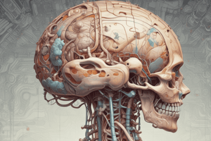
Meninges
- Meninges are membranes of connective tissue between bone and nervous tissue that protect the CNS
- The three meningeal layers are the dura mater, arachnoid mater, and pia mater
Protective Layers of the CNS
- Bones, connective tissue, and cerebrospinal fluid (CSF) surround and protect the brain and spinal cord
Dura Mater
- The tough outermost connective tissue layer around the CNS
Arachnoid Mater
- Located below the dura mater
Pia Mater
- The innermost layer and adheres directly to the surface of the brain and spinal cord
Subarachnoid Space
- Located between the pia mater and arachnoid mater
- Filled with CSF
Meningitis
- Meningitis is an inflammation (swelling) of the protective membranes covering the brain and spinal cord
- It is usually caused by a bacterial or viral infection of the fluid surrounding the brain and spinal cord
- Injuries, cancer, certain drugs, and other types of infections can also cause meningitis
Central Nervous System (CNS)
- The major regions of the CNS are the cerebrum, cerebellum, and spinal cord
- It is covered by three connective tissue layers, the meninges
- The CNS is considered relatively soft and easily damaged by injuries affecting its protective cranium or vertebral bones
Spinal Cord
- The entire CNS displays organized areas of white and gray matter
White Matter
- The differences in white and gray matter is caused by the differential distribution of myelin
- The main components of white matter are myelinated axons commonly grouped together as tracts and myelin-producing oligodendrocytes
Gray Matter
- Contains neurons, dendrites, and neuroglia
- Posterior horns of the spinal cord are associated with axons of posterior roots
- Anterior horns of the spinal cord are associated with axons of anterior roots
Cerebrospinal Fluid (CSF)
- A clear, colorless fluid that cushions and protects the brain and spinal cord
- Continuously produced by choroid plexuses in brain ventricles, with most in the lateral ventricles
- CSF is important for homeostasis, brain metabolism, and optimal neuronal environments
Clinical Significance of CSF
- A decrease in the absorption of CSF or a blockage of outflow from the ventricles during fetal or postnatal development results in hydrocephalus
- Hydrocephalus causes a progressive enlargement of the head followed by mental impairment.
Cerebral Cortex Structure
- Six layers of neurons with different sizes and shapes located in the cerebral cortex (cerebrum)
- Efferent pyramidal neurons are the most noticeable of these cells and come in many sizes
- Neurons of the cerebral cortex function in the integration of sensory information and the initiation of voluntary motor responses
Gray Matter & Synapses
- Gray matter is where most synapses occur
- It occupies the thick surface or cortex of both the cerebrum and the cerebellum
- Most white matter is found in deeper regions of the CNS
Cerebrum Layers
- Molecular layer (I) is most superficial and covered by pia mater, which contains neuroglial cells and horizontal cells of Cajal
- External granular layer (II) contains neuroglial cells and small pyramidal cells
- External pyramidal layer (III) contains medium-sized pyramidal cells and is the predominant type
- Internal granular layer (IV) is a thin layer with small granules, pyramidal cells, and neuroglia
- Internal pyramidal layer (V) contains neuroglial cells and the largest pyramidal cells
- Multiform layer (VI) is the deepest layer, adjacent to white matter and contains various cell types
Cerebellar Cortex
- The cerebellar cortex is highly folded with folds or folia, separated by closely set parallel transverse fissure
- Deep folds in the cortex are called cerebellar folia which are separated by sulci
- Outer molecular layer contains small neurons and fibers
- Middle Purkinje layer contains large Purkinje cells
- Purkinje cells have dendrites that branch in the molecular layer
- Granule cell layer contains small granule cells, Golgi type II cells, and empty spaces called glomeruli
Choroid Plexus
- Consists of highly specialized tissue with elaborate folds and many villi that project into the four large ventricles of the brain
- Removes water from blood and releases it as CSF
- CSF is clear and contains Na+, K+, and Cl- ions with very little protein
- Its only cells are normally very sparse lymphocytes
- It is produced continuously and completely fills the ventricles, the central canal of the spinal cord, and the subarachnoid and perivascular spaces
- Provides the ions required for CNS neuronal activity
- Serves to help absorb mechanical shocks in the arachnoid
Peripheral Nervous System
Ganglia
- Accumulations of neurons covered by connective tissue
- Contain both sensory and motor nerves
- Neurons of peripheral nerves can be located in the CNS or in ganglia
Studying That Suits You
Use AI to generate personalized quizzes and flashcards to suit your learning preferences.