Podcast
Questions and Answers
What is the primary function of the basal nuclei?
What is the primary function of the basal nuclei?
- Regulation of involuntary motor control
- Control of skeletal muscles to maintain balance
- Regulation of voluntary motor control (correct)
- Processing of sensory information
Which part of the brain attaches to the spinal cord?
Which part of the brain attaches to the spinal cord?
- Medulla oblongata (correct)
- Cerebrum
- Hypothalamus
- Thalamus
What is the function of the cerebellum in relation to motor control?
What is the function of the cerebellum in relation to motor control?
- Comparing motor commands of the cerebrum with proprioceptor information (correct)
- Processing sensory information from the eyes
- Regulating involuntary motor control
- Sending motor commands to muscles
What is the function of the thalamus in relation to sensations?
What is the function of the thalamus in relation to sensations?
What is the term for the dominance of one cerebral hemisphere over the other?
What is the term for the dominance of one cerebral hemisphere over the other?
What is the location of the pons in relation to the medulla oblongata and midbrain?
What is the location of the pons in relation to the medulla oblongata and midbrain?
What is the main purpose of the falx cerebri?
What is the main purpose of the falx cerebri?
What is the function of the cerebrospinal fluid (CSF)?
What is the function of the cerebrospinal fluid (CSF)?
What is the function of the cerebral tracts?
What is the function of the cerebral tracts?
What is the subarachnoid space located between?
What is the subarachnoid space located between?
What is the epidural space located immediately outside of?
What is the epidural space located immediately outside of?
What is the main function of the tentorium cerebelli?
What is the main function of the tentorium cerebelli?
What is the medulla oblongata?
What is the medulla oblongata?
What is the subdural space normally?
What is the subdural space normally?
Which of the following structures is responsible for associating sensory impulses with feelings of pleasantness and unpleasantness?
Which of the following structures is responsible for associating sensory impulses with feelings of pleasantness and unpleasantness?
Which of the following tracts originates in the motor cortex and crosses at the level of the medulla?
Which of the following tracts originates in the motor cortex and crosses at the level of the medulla?
Which of the following systems consists of centers in the brainstem's reticular formation that receive impulses from the spinal cord and relay them to the thalamus and from the thalamus to all parts of the cerebral cortex?
Which of the following systems consists of centers in the brainstem's reticular formation that receive impulses from the spinal cord and relay them to the thalamus and from the thalamus to all parts of the cerebral cortex?
Which of the following structures is responsible for providing conduction routes to and from the brain and serves as the integrator, or reflex centre, for all spinal reflexes?
Which of the following structures is responsible for providing conduction routes to and from the brain and serves as the integrator, or reflex centre, for all spinal reflexes?
Which of the following tracts is responsible for transmitting impulses that coordinate body movements and maintenance of posture?
Which of the following tracts is responsible for transmitting impulses that coordinate body movements and maintenance of posture?
Which of the following structures is often referred to as the 'link between the psyche and the soma'?
Which of the following structures is often referred to as the 'link between the psyche and the soma'?
Which of the following tracts is responsible for head and neck movement related to visual reflexes?
Which of the following tracts is responsible for head and neck movement related to visual reflexes?
What is the primary function of the motor tracts mentioned in the content?
What is the primary function of the motor tracts mentioned in the content?
Where is cerebrospinal fluid primarily formed?
Where is cerebrospinal fluid primarily formed?
What is the approximate volume of CSF in the ventricles of an average adult?
What is the approximate volume of CSF in the ventricles of an average adult?
What is the primary function of cerebrospinal fluid?
What is the primary function of cerebrospinal fluid?
What is the composition of cerebrospinal fluid similar to?
What is the composition of cerebrospinal fluid similar to?
What is the process by which CSF is reabsorbed into the bloodstream?
What is the process by which CSF is reabsorbed into the bloodstream?
What is the approximate total volume of CSF in an average adult?
What is the approximate total volume of CSF in an average adult?
What is the location of cerebrospinal fluid in the CNS?
What is the location of cerebrospinal fluid in the CNS?
What are the major solutes in cerebrospinal fluid?
What are the major solutes in cerebrospinal fluid?
Flashcards are hidden until you start studying
Study Notes
Structures that Protect the Brain and Spinal Cord
- The brain and spinal cord are protected by outer and inner coverings.
- The outer covering consists of bone: cranial bones encase the brain, and vertebrae encase the spinal cord.
- The inner covering consists of three distinct layers of membranes known as meninges: dura mater, arachnoid mater, and pia mater.
Meninges
- Dura mater has three extra protective and supportive inward extensions or dura septa:
- Falx cerebri: separates the two cerebral hemispheres.
- Falx cerebelli: separates the two halves of the cerebellum.
- Tentorium cerebelli: separates the cerebellum from the cerebrum.
- The meninges provide a cushion of fluid, cerebrospinal fluid (CSF), both around the organs and within them.
Cerebrospinal Fluid (CSF)
- CSF is found in the subarachnoid space around the brain and spinal cord and within the cavities and canals of the brain and spinal cord.
- Formation of CSF occurs mainly by separation of fluid from blood in the choroid plexuses.
- The CSF circulates in the subarachnoid space and then is reabsorbed into venous blood through the arachnoid villi.
- The amount of CSF in the average adult is about 140 mL (about 23 mL in the ventricles and 117 mL in the subarachnoid space of the brain and cord).
- CSF is a watery broth with a composition similar to blood plasma, and its major solutes include glucose, proteins, and NaCl.
Subarachnoid, Subdural, and Epidural Spaces
- Epidural space: immediately outside the dura mater but inside the bony coverings of the spinal cord, containing a supporting cushion of fat and other connective tissues.
- Subdural space: between the dura mater and arachnoid mater, typically a potential space but can become a real space if blood leaks into it, forming a subdural hematoma.
- Subarachnoid space: under the arachnoid and outside the pia mater, containing a significant amount of CSF.
Brain Divisions
- The brain is divided into three main parts: medulla oblongata, midbrain, and pons.
- Medulla oblongata: forms the lowest part of the brainstem, attaching to the spinal cord.
- Midbrain: forms the uppermost part, lying above the pons and below the cerebrum.
- Pons: lies between the medulla and midbrain.
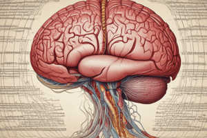
Cerebrum
- The cerebrum is the largest and uppermost division of the brain, consisting of two halves: the right and left cerebral hemispheres.
- The surface of the cerebrum, the cerebral cortex, is made up of gray matter only 2-4 mm thick.
- The cerebral hemisphere that dominates motor and language function is usually the left hemisphere, responsible for language, mathematical abilities, and logic.
- The right hemisphere is more involved in visual-spatial skills, intuition, emotion, and art, music appreciation.
Basal Nuclei
- Structure: islands of gray matter located deep inside the white matter of each hemisphere.
- Function: regulation of voluntary (conscious) motor control related to posture, walking, and other repetitive movements; possible roles in thinking and learning.
Cerebellum
- Structure: second largest part of the brain, containing more neurons than the rest of the nervous system, located just below the posterior portion of the cerebrum.
- Function: compares the motor commands of the cerebrum with the information coming from proprioceptors in the muscle, coordinating movements to produce the intended action.
Brainstem
- Located beneath the cerebrum and anterior to the cerebellum, composed of three divisions: medulla oblongata, midbrain, and pons.
Thalamus, Hypothalamus, and Medulla Oblongata
- Thalamus:
- Plays a part in the mechanism responsible for sensations, emotions, and arousal.
- Relays sensory impulses to the cerebrum.
- Hypothalamus:
- Consists of several structures that lie beneath the thalamus and form the floor of the third ventricle and the lower part of its lateral walls.
- Links the nervous system to the endocrine system and is known as a link between the psyche (mind) and the soma (body).
- Medulla oblongata:
- Part of the brain that attaches to the spinal cord.
- Located just above the foramen magnum.
Reticular Activating System (RAS)
- Consists of centers in the brainstem's reticular formation that receive impulses from the spinal cord and relay them to the thalamus and from the thalamus to all parts of the cerebral cortex.
- A complex network of nerve fibers and gray matter scattered throughout the brain.
- Activates the cerebral cortex into a state of wakefulness, and a decrease in activity results in sleep.
Limbic System
- A complex network of fiber tracts and gray matter found in interconnecting parts of the cerebral cortex, hypothalamus, thalamus, and basal nuclei.
- Monitors our emotional experiences and expression.
Spinal Cord
- Structure: elongated cylinder extending from the brainstem through the foramen magnum of the skull, gray matter interior surrounded by white matter, with 31 pairs of spinal nerves attached by dorsal and ventral nerve roots.
- Function: provides conduction routes to and from the brain and serves as the integrator, or reflex center, for all spinal reflexes.
Tracts
- Ascending tracts: conduct sensory impulses up the cord to the brain.
- Descending tracts: conduct motor impulses down the cord from the brain.
- Specific tracts:
- Pyramidal tracts: corticospinal tract impulses, stimulating individual muscle groups.
- Extrapyramidal tracts: movements that express emotion, including large automatic movements and automatic expressions.
Studying That Suits You
Use AI to generate personalized quizzes and flashcards to suit your learning preferences.