Podcast
Questions and Answers
Define pneumonia.
Define pneumonia.
Pneumonia is defined as the collection of inflammatory exudate in lung parenchyma distal to terminal bronchioles, mostly resulting in consolidation of lung parts.
Which aetiological agent is associated with bronchopneumonia?
Which aetiological agent is associated with bronchopneumonia?
- Influenza
- Cryptococcus
- Pneumocystis jiroveci
- Streptococcus pneumoniae (correct)
Lobar pneumonia affects mainly females.
Lobar pneumonia affects mainly females.
False (B)
_____ pneumonia typically affects otherwise healthy adults between 20 and 50 years of age.
_____ pneumonia typically affects otherwise healthy adults between 20 and 50 years of age.
Match the morphological changes with the stages of Lobar Pneumonia:
Match the morphological changes with the stages of Lobar Pneumonia:
What is the main difference in the distribution of lesions between bronchopneumonia and lobar pneumonia?
What is the main difference in the distribution of lesions between bronchopneumonia and lobar pneumonia?
What is the definition of lobar pneumonia?
What is the definition of lobar pneumonia?
Is bronchopneumonia more severe than lobar pneumonia?
Is bronchopneumonia more severe than lobar pneumonia?
Study Notes
Pneumonia
- Pneumonia is defined as a collection of inflammatory exudate in lung parenchyma distal to terminal bronchioles, resulting in consolidation (solidification) of lung parts.
Classifications of Pneumonia
- Anatomical pattern:
- Bronchopneumonia
- Lobar pneumonia
- Clinical circumstances:
- Primary (in an otherwise healthy person)
- Secondary (with local or systemic defects in defense)
- Aetiological agents:
- Bacterial (Streptococcus pneumoniae, Staphylococcus aureus, Mycobacterium tuberculosis, etc.)
- Viral (Influenza, measles, etc.)
- Fungal (Cryptococcus, Candida, Aspergillus, etc.)
- Other (Pneumocystis jiroveci, Mycoplasma, Aspiration, lipid, eosinophilic)
- Host reaction:
- Fibrinous
- Suppurative
Pathogenesis
- Pneumonia occurs when defense mechanisms of the respiratory system are impaired or immunity of the host is low.
- Factors that compromise pulmonary defense mechanisms:
- Loss or suppression of the cough reflex
- Dysfunction of the mucociliary apparatus
- Interference with phagocytic and bactericidal activities of alveolar macrophages
- Pulmonary congestion and edema
Lobar Pneumonia
- Affects anatomically delineated segment(s) or the entirety of a lobe or lung
- Relatively uncommon in infancy and old age
- Affects males more than females
- 90% due to Streptococcus pneumoniae (pneumococcus)
- Pneumococcal pneumonia typically affects otherwise healthy adults between 20 and 50 years of age
Clinical Features of Lobar Pneumonia
- Symptoms:
- Cough, fever, and production of sputum
- The sputum appears purulent and may contain flecks of blood (rusty sputum)
- Occasional patients may have hemoptysis
- Fever can be very high (over 40°C), with rigors (shaking chills)
- Acute pleuritic chest pain on deep inspiration reflects inflammation of the pleura (pleurisy)
- Signs:
- Dullness to percussion with bronchial breathing
- Pleural friction rub
Morphological Changes in Lobar Pneumonia
- Congestion:
- Outpouring of a protein-rich exudate into alveolar spaces, with venous congestion
- The lung is heavy, oedematous, and red
- Red hepatization:
- Massive accumulation of polymorphs, together with some lymphocytes and macrophages
- Many red cells are also extravasated from the distended capillaries
- The overlying pleura bears a fibrinous exudate
- The lung is red, solid, and airless, with a consistency resembling fresh liver
- Grey hepatization:
- Further accumulation of fibrin, with destruction of white cells and red cells
- The lung is now grey-brown and solid
- Resolution:
- Resorption of exudate and enzymatic digestion of inflammatory debris, with preservation of the underlying alveolar wall architecture
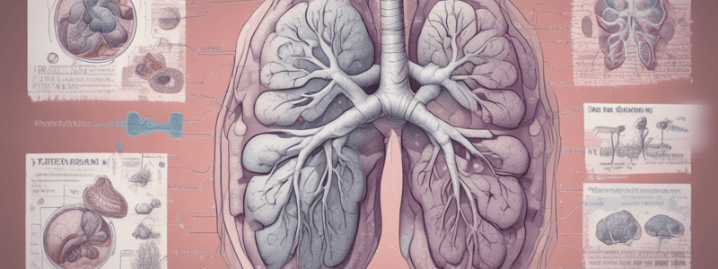
Bronchopneumonia
- Has a characteristic patchy distribution, centred on inflamed bronchioles and bronchi with subsequent spread to surrounding alveoli
- Often several lobes or bilateral
- It occurs most commonly in old age, in infancy, and in patients with debilitating diseases
- Typical organisms include staphylococci, streptococci, and Haemophilus influenzae
Complications of Pneumonia
- Abscess formation:
- Results from tissue destruction (more in case of Klebsiella or type III Pneumococcal infections)
- Empyema:
- Virulent bacterial strains induce suppuration in the pleural cavity, causing the intra-pleural fibrosuppurative reaction
- Fibrosis:
- Organization of intra-alveolar exudate may convert affected lung into solid fibrous tissue
- Bacterial dissemination:
- Dissemination of bacteria may lead to endocarditis, pericarditis, meningitis, suppurative arthritis, and formation of metastatic abscesses in various organs
Atypical Pneumonia
- Pneumonias caused by organisms other than traditional bacteria are often referred to as ‘atypical pneumonias’
- Causative organisms:
- Mycoplasma pneumoniae
- Influenza virus type A and B
- Respiratory syncytial viruses, adenovirus, rhinovirus, rubeola, and varicella virus
- Chlamydia
- Coxiella burnetii
Non-Infective Pneumonias
- Cryptogenic organising pneumonia
- Aspiration pneumonia:
- When fluid or food is aspirated into the lung
- Lipid pneumonia:
- Endogenous: associated with airway obstruction causing distal collections of foamy macrophages and giant cells
- Exogenous: due to aspiration of material containing a high concentration of lipid
Differences between Bronchopneumonia and Lobar Pneumonia
- Definition:
- Bronchopneumonia: Patchy consolidation of multiple lobes (bilateral)
- Lobar pneumonia: Involvement of a large part of a lobe or entire lobe, diffuse consolidation
- Predisposing illness:
- Bronchopneumonia: Bronchitis/bronchiolitis, chronic debility
- Lobar pneumonia: Affects healthy individuals
- Immune status:
- Bronchopneumonia: Usually affects immunosuppressed individuals
- Lobar pneumonia: Affects previously healthy individuals
Studying That Suits You
Use AI to generate personalized quizzes and flashcards to suit your learning preferences.
Related Documents
Description
Classify and describe different types of pneumonia, including lobar pneumonia and bronchopneumonia, and compare their clinicopathological features.