Podcast
Questions and Answers
What do the initials MRI stand for?
What do the initials MRI stand for?
Magnetic Resonance Imaging
What is the primary focus of MRI imaging?
What is the primary focus of MRI imaging?
- Mapping the distribution of carbon nuclei within the body
- Mapping the distribution of helium nuclei within the body
- Mapping the distribution of hydrogen nuclei within the body (correct)
- Mapping the distribution of oxygen nuclei within the body
MRI stands for Magnetic Resonance Imaging?
MRI stands for Magnetic Resonance Imaging?
True (A)
What do hydrogen atoms primarily contribute to in MRI imaging?
What do hydrogen atoms primarily contribute to in MRI imaging?
In MRI, the nucleus of a hydrogen atom contains a single positively-charged ____________.
In MRI, the nucleus of a hydrogen atom contains a single positively-charged ____________.
What is needed to generate a visible image in Magnetic Resonance Imaging (MRI)?
What is needed to generate a visible image in Magnetic Resonance Imaging (MRI)?
Which of the following are types of MRI pulse sequences? (Select all that apply)
Which of the following are types of MRI pulse sequences? (Select all that apply)
Spin-spin relaxation refers to the rate at which Mxy increases back to its initial value.
Spin-spin relaxation refers to the rate at which Mxy increases back to its initial value.
The time for Mz to grow back to 63% of M0 is defined as __________.
The time for Mz to grow back to 63% of M0 is defined as __________.
Match the biological tissue with its relaxation time:
Match the biological tissue with its relaxation time:
Flashcards are hidden until you start studying
Study Notes
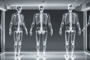
Magnetic Resonance Imaging (MRI)
- MRI is a medical imaging modality that uses magnetic fields and resonance to produce images of the body.
Unit Overview
- The unit will be delivered onsite over 4 sessions with additional learning resources available on CANVAS.
- A Question and Answer session will be held on the 31st October, and a revision session will be facilitated on the 30th January 2024.
Learning Outcomes
- Differentiate between MRI and Computed Tomography (CT).
- Describe the basics of image generation in MRI.
- Correctly identify, view, and orientate an MR image.
- Identify common pulse sequences used in MRI.
Physical Principles of MRI
- MRI sounds different from CT sounds.
- MRI measures different things than CT.
- MRI developed into an important imaging modality from 1978.
- MRI utilizes the fact that magnetic nuclei in a static magnetic field exhibit a characteristic resonance frequency proportional to the field strength.
Generating the MRI Signal
- MRI primarily maps the distribution of hydrogen nuclei within the body.
- The medical imaging community has recognized MRI's unique ability to image soft tissue, differentiate between benign and malignant tissue, and provide multi-planar capabilities.
MRI Terminology
- MRI stands for Magnetic Resonance Imaging.
- MRA stands for MR Angiography.
- MRS stands for MR Spectroscopy.
- fMRI stands for Functional MRI.
MRI Hardware
- Gradient coils have evolved over time.
- Inside an MRI scanner, there are strong magnetic fields, gradient coils, and RF coils.
MRI Field Strengths
- High field systems: 1.0 to 3.0 Tesla (standard clinical systems).
- Low field systems: 0.2 – 0.5 Tesla (some clinical sites, but mainly research or spectroscopy).
- Up to 7 Tesla (high field systems).
Principles of Magnetism and Resonance
- MRI relies on the principles of magnetism and resonance to produce an image.
- The hydrogen proton is targeted because of its abundance in the body.
- A spinning proton produces a magnetic charge.
Atomic Structure
- Atoms are made up of electrons, protons, and neutrons.
- Protons and neutrons have a property of spin.
Nuclear Spin
- Protons and neutrons have a net spin that can be aligned with an external magnetic field.
- In most nuclei, they cancel each other out, leaving no net spin.
Hydrogen Atoms
- Hydrogen atoms are abundant in the human body.
- They have a strong magnetic moment.
Magnetic Moment
- A moving (or rotating) particle with electric charge has an associated magnetic field.
- Protons can be thought of as a tiny bar magnet.
Magnetic Distribution
- Magnetic moment in an external magnetic field becomes aligned with the field.
- Alignment is not exact because of the proton's spin.
Energy States
- Hydrogen atoms have two stable orientations: spin up (lower energy) and spin down (higher energy).
- There is always a small excess of spins in the lower energy state, producing a net magnetization M0 parallel to the direction of B0.
Net Magnetization Vector
- M0 represents the average behavior of all protons.
- M0 can be separated into components: longitudinal (z-axis) and transverse (x and y axes).
Vector Components
- In MRI, the z-axis is defined by the direction of B0.
- There is no difference between x and y axes, which are both perpendicular to B0.
Review
- MRI is based on the interaction between hydrogen atoms and a magnetic field.
- The magnetic moment of the protons precesses around the magnetic field at a particular frequency.
Generating the MR Signal
- Magnetic Resonance Imaging (MRI) requires a scanner to generate a strong magnetic field (B0) and radiofrequency (RF) pulses to excite the signals.
- Gradients are used to localize the signals by changing the magnetic field strength in a defined way, causing protons to change their resonant frequencies to match.
- The signal is optimized by exciting it multiple times and adjusting the signal-to-noise ratio (SNR).
Generating B0
- The main magnet is a cylindrical bore with a diameter of approximately 60cm, and a field strength of 1.5-3 Tesla.
- The magnet is superconducting, and it is surrounded by gradients and body RF coils.
- The gradients and body coil are used to localize the signal and improve the signal-to-noise ratio.
Localizing MR Signal
- The Larmor frequency is used to localize the signal, which depends on the strength of the magnetic field and the gyromagnetic ratio.
- The frequency of the signal changes depending on the location within the magnetic field.
- The isocentre is the point where the magnetic field strength is at its maximum.
Slice Selection
- Slice selection is achieved by applying a gradient to the magnetic field, which causes the resonant frequency to change.
- The frequency of the signal changes depending on the location within the slice.
- The slice selection gradient is used to select the desired slice.
Signals
- The signal is composed of many frequencies, which are added together to form a complete signal.
- The signal is a mixture of frequencies, which are used to create an image.
Total Signal and Fourier Transform
- The total signal is composed of all the frequencies, which are added together.
- The Fourier transform is used to convert the time-domain signal into a frequency-domain signal.
- The frequency-domain signal is used to create an image.
Displaying an Image
- The image is created by taking the Fourier transform of the signal and displaying it as a function of frequency.
- The image is a representation of the spatial distribution of the signal.
Image Matrix
- The image matrix is a 2D array of pixels, which represents the spatial distribution of the signal.
- The image matrix is used to create a visible image.
Review
- Gradients are used to modify the frequency of the signals in a controlled way.
- The frequency and amplitude of the signals are used to localize the signals within the magnet.
- Pulse sequences are used to create an image with different contrast.
Pulse Sequences
- A pulse sequence is a set of changing magnetic gradients and RF pulses.
- Each pulse sequence has a number of parameters, and multiple sequences are grouped together into an MRI protocol.
- Pulse sequences are used to produce images with different contrast.
Spin Echo Pulse Sequence
- The spin echo pulse sequence is a basic sequence that produces a high signal-to-noise ratio (SNR) and resolution.
- The sequence consists of a 90° RF pulse, followed by a 180° RF pulse, and a data collection period.
Spin Echo Pulse Sequence Parameters
- The time to echo (TE) is the time between the 90° RF pulse and the data collection period.
- The time to repeat (TR) is the time between successive 90° RF pulses.
Rephasing
- Rephasing is the process of re-aligning the magnetic moments of the protons.
- Rephasing is used to refocus the signal and improve the signal-to-noise ratio.
Image Contrast
- The contrast in the image depends on the TR and TE.
- The TR controls the amount of T1 relaxation, which affects the contrast.
- The TE controls the amount of T2 relaxation, which affects the contrast.
T1 and T2 Relaxation
- T1 relaxation is the process of longitudinal magnetization recovery.
- T2 relaxation is the process of transverse magnetization decay.
- T1 and T2 relaxation times are tissue-dependent and affect the contrast in the image.
T1 Relaxation Times
- Typical T1 relaxation times for different tissues:
- Muscle: 630 ms
- Fat: 190 ms
- Grey matter: 825 ms
- White matter: 685 ms
- CSF: 1200 ms
T2 Relaxation Times
- Typical T2 relaxation times for different tissues:
- Muscle: 40 ms
- Fat: 110 ms
- Grey matter: 110 ms
- White matter: 105 ms
- CSF: 800 ms
Studying That Suits You
Use AI to generate personalized quizzes and flashcards to suit your learning preferences.