Podcast
Questions and Answers
What is the primary function of the lower limbs?
What is the primary function of the lower limbs?
- To maintain balance and posture
- To facilitate movement of the upper limb
- To regulate blood pressure and circulation
- To support the weight of the body and produce locomotion (correct)
What is the term for the joint that connects the two hip bones anteriorly?
What is the term for the joint that connects the two hip bones anteriorly?
- Pelvis
- Coccyx
- Sacrum
- Symphysis pubis (correct)
Which of the following bones is NOT part of the lower limb?
Which of the following bones is NOT part of the lower limb?
- Fibula
- Scapula (correct)
- Os coxae
- Tibia
What is the term for the arches of the foot that are oriented longitudinally?
What is the term for the arches of the foot that are oriented longitudinally?
What is the purpose of the gluteal region in the lower limb?
What is the purpose of the gluteal region in the lower limb?
What is the term for the joint that connects the hip bones posteriorly with the trunk?
What is the term for the joint that connects the hip bones posteriorly with the trunk?
What is the purpose of the tarsal bones in the lower limb?
What is the purpose of the tarsal bones in the lower limb?
What is the general arrangement of the bones in the lower limb compared to the upper limb?
What is the general arrangement of the bones in the lower limb compared to the upper limb?
What is the function of the lower limb girdle?
What is the function of the lower limb girdle?
Which of the following bones is not part of the os coxae?
Which of the following bones is not part of the os coxae?
What is the name of the surface where the three bones of the os coxae fuse?
What is the name of the surface where the three bones of the os coxae fuse?
Which muscle is located near the inguinal ligament?
Which muscle is located near the inguinal ligament?
What is the point of attachment for the rectus femoris muscle?
What is the point of attachment for the rectus femoris muscle?
What is the topographical and functional equivalent of the os coxae in the upper limb?
What is the topographical and functional equivalent of the os coxae in the upper limb?
What is the function of the gluteus minimus muscle?
What is the function of the gluteus minimus muscle?
Which of the following muscles is not a part of the gluteal muscles?
Which of the following muscles is not a part of the gluteal muscles?
What is the name of the bony structure that forms the lower limb girdle?
What is the name of the bony structure that forms the lower limb girdle?
What is the landmarks of the tensor fasciae latae muscle?
What is the landmarks of the tensor fasciae latae muscle?
How many skeletal elements form the os coxae?
How many skeletal elements form the os coxae?
What is the purpose of the compartmentalization of the thigh and leg?
What is the purpose of the compartmentalization of the thigh and leg?
What is the anatomical structure that separates the femoral artery from the saphenous opening?
What is the anatomical structure that separates the femoral artery from the saphenous opening?
Where is the membranous layer of superficial fascia attached?
Where is the membranous layer of superficial fascia attached?
What is the name of the group of lymph nodes located horizontally near the inguinal ligament?
What is the name of the group of lymph nodes located horizontally near the inguinal ligament?
What is the name of the structure that surrounds the femoral artery?
What is the name of the structure that surrounds the femoral artery?
Which structure is located near the saphenous opening?
Which structure is located near the saphenous opening?
What is the path of the sciatic nerve in the gluteal region?
What is the path of the sciatic nerve in the gluteal region?
What is the main supply of the trochanteric anastomosis?
What is the main supply of the trochanteric anastomosis?
Which nerve distributes branches throughout the gluteal region?
Which nerve distributes branches throughout the gluteal region?
Where does the superior gluteal nerve leave the pelvis?
Where does the superior gluteal nerve leave the pelvis?
What is the origin of the posterior cutaneous nerve of the thigh?
What is the origin of the posterior cutaneous nerve of the thigh?
What is the main branch of the tibial nerve in the lower limb?
What is the main branch of the tibial nerve in the lower limb?
Which muscle is supplied by the posterior cutaneous nerve of the thigh?
Which muscle is supplied by the posterior cutaneous nerve of the thigh?
Which artery ends by supplying the tensor fasciae latae?
Which artery ends by supplying the tensor fasciae latae?
What is the main function of the trochanteric anastomosis?
What is the main function of the trochanteric anastomosis?
Through which foramen do the Inferior gluteal nerve and Nerve to Quadratus Femoris leave the pelvis?
Through which foramen do the Inferior gluteal nerve and Nerve to Quadratus Femoris leave the pelvis?
What is the name of the anastomosis situated at the level of the lesser trochanter of the femur?
What is the name of the anastomosis situated at the level of the lesser trochanter of the femur?
What receives lymph from the lower half of the anal canal?
What receives lymph from the lower half of the anal canal?
Which muscle is supplied by the Inferior gluteal Nerve?
Which muscle is supplied by the Inferior gluteal Nerve?
What is the function of the anastomosis formed by the inferior gluteal artery, medial femoral circumflex artery, and lateral femoral circumflex artery?
What is the function of the anastomosis formed by the inferior gluteal artery, medial femoral circumflex artery, and lateral femoral circumflex artery?
What is the route by which all lymph from the superficial and deep inguinal lymph nodes ultimately drains?
What is the route by which all lymph from the superficial and deep inguinal lymph nodes ultimately drains?
Which nerves leave the pelvis through the lower part of the greater sciatic foramen?
Which nerves leave the pelvis through the lower part of the greater sciatic foramen?
What forms the greater sciatic foramen?
What forms the greater sciatic foramen?
What is located near the saphenous opening in the deep fascia?
What is located near the saphenous opening in the deep fascia?
What is the name of the proximal segment of the lower limb?
What is the name of the proximal segment of the lower limb?
What is the bony core of the thigh?
What is the bony core of the thigh?
What is the anatomical region that includes the umbilicus?
What is the anatomical region that includes the umbilicus?
What is the relationship between the superficial and deep inguinal lymph nodes?
What is the relationship between the superficial and deep inguinal lymph nodes?
What is the term for the foramen formed by the greater sciatic notch of the hip bone and the sacrotuberous and sacrospinous ligaments?
What is the term for the foramen formed by the greater sciatic notch of the hip bone and the sacrotuberous and sacrospinous ligaments?
What receives lymph from the abdominal walls below the level of the umbilicus?
What receives lymph from the abdominal walls below the level of the umbilicus?
What is the function of the perforating veins in preventing high-pressure venous blood from flowing outward into superficial veins?
What is the function of the perforating veins in preventing high-pressure venous blood from flowing outward into superficial veins?
What is the result of the muscles within the closed fascial compartments relaxing?
What is the result of the muscles within the closed fascial compartments relaxing?
What is the characteristic of a varicosed vein?
What is the characteristic of a varicosed vein?
What is the nerve that supplies the skin immediately in front of the medial malleolus of the tibia?
What is the nerve that supplies the skin immediately in front of the medial malleolus of the tibia?
What is the purpose of blocking the saphenous nerve branches with local anesthetic?
What is the purpose of blocking the saphenous nerve branches with local anesthetic?
What is the location of the transverse incision made through the skin and subcutaneous tissue?
What is the location of the transverse incision made through the skin and subcutaneous tissue?
What is the consequence of incompetent valves in the veins?
What is the consequence of incompetent valves in the veins?
What is the common site for varicose veins?
What is the common site for varicose veins?
What is the location of the great saphenous vein in relation to the pubic tubercle?
What is the location of the great saphenous vein in relation to the pubic tubercle?
Which nerve lies just anterior to the great saphenous vein?
Which nerve lies just anterior to the great saphenous vein?
What is the purpose of reversing the great saphenous vein in coronary bypass surgery?
What is the purpose of reversing the great saphenous vein in coronary bypass surgery?
What is the location of the incision made for the great saphenous vein cutdown?
What is the location of the incision made for the great saphenous vein cutdown?
What is the significance of the saphenous opening in the femoral vein?
What is the significance of the saphenous opening in the femoral vein?
What is the purpose of blocking the branches of the ilioinguinal nerve and the intermediate cutaneous nerve of the thigh?
What is the purpose of blocking the branches of the ilioinguinal nerve and the intermediate cutaneous nerve of the thigh?
What is the anatomical structure that separates the femoral artery from the saphenous opening?
What is the anatomical structure that separates the femoral artery from the saphenous opening?
What is the name of the nerve that supplies the area of skin below and lateral to the scrotum or labium majus?
What is the name of the nerve that supplies the area of skin below and lateral to the scrotum or labium majus?
Which artery is located near the femoral canal?
Which artery is located near the femoral canal?
What is the name of the bony structure that forms the lower limb girdle in the right lower limb?
What is the name of the bony structure that forms the lower limb girdle in the right lower limb?
Which muscle is located near the pubic tubercle?
Which muscle is located near the pubic tubercle?
What is the name of the canal that contains the femoral artery and vein?
What is the name of the canal that contains the femoral artery and vein?
Which structure is located near the inguinal ligament?
Which structure is located near the inguinal ligament?
What is the name of the artery that supplies the tensor fasciae latae muscle?
What is the name of the artery that supplies the tensor fasciae latae muscle?
What is the primary function of the perforating veins in the lower limb?
What is the primary function of the perforating veins in the lower limb?
What is the route by which lymph from the superficial and deep inguinal lymph nodes ultimately drains?
What is the route by which lymph from the superficial and deep inguinal lymph nodes ultimately drains?
Which of the following structures is not located near the inguinal ligament?
Which of the following structures is not located near the inguinal ligament?
What is the main function of the great saphenous vein?
What is the main function of the great saphenous vein?
What receives lymph from the skin and superficial fascia of the anterior and posterior abdominal walls?
What receives lymph from the skin and superficial fascia of the anterior and posterior abdominal walls?
What is the anatomical structure that separates the femoral artery from the saphenous opening?
What is the anatomical structure that separates the femoral artery from the saphenous opening?
Which of the following structures is not a part of the superficial inguinal lymph nodes?
Which of the following structures is not a part of the superficial inguinal lymph nodes?
What is the relationship between the superficial and deep inguinal lymph nodes?
What is the relationship between the superficial and deep inguinal lymph nodes?
What is the common location of the deep inguinal lymph nodes?
What is the common location of the deep inguinal lymph nodes?
What is the main action of the quadriceps femoris muscle?
What is the main action of the quadriceps femoris muscle?
Which muscles form the iliopsoas muscle in the thigh?
Which muscles form the iliopsoas muscle in the thigh?
What is the path of the efferent lymph vessels from the deep inguinal lymph nodes?
What is the path of the efferent lymph vessels from the deep inguinal lymph nodes?
What is the function of the retinacula formed by the tendinous fibers of the vastus lateralis and vastus medialis?
What is the function of the retinacula formed by the tendinous fibers of the vastus lateralis and vastus medialis?
What is the origin of the lymph that drains into the deep inguinal lymph nodes?
What is the origin of the lymph that drains into the deep inguinal lymph nodes?
What is the insertion point of the quadriceps femoris muscle?
What is the insertion point of the quadriceps femoris muscle?
What is the location of the deep inguinal lymph nodes in relation to the femoral vein?
What is the location of the deep inguinal lymph nodes in relation to the femoral vein?
What is the relationship between the obturator nerve and the adductor muscles in the right lower limb?
What is the relationship between the obturator nerve and the adductor muscles in the right lower limb?
What is the function of the sartorius muscle?
What is the function of the sartorius muscle?
What is the point of attachment of the iliacus muscle?
What is the point of attachment of the iliacus muscle?
Which nerve supplies the sartorius muscle?
Which nerve supplies the sartorius muscle?
What is the relationship between the profunda femoris artery and the adductor magnus muscle?
What is the relationship between the profunda femoris artery and the adductor magnus muscle?
What is the segment of the lower limb that includes the thigh?
What is the segment of the lower limb that includes the thigh?
Which nerve descends in the midline of the posterior compartment of the thigh?
Which nerve descends in the midline of the posterior compartment of the thigh?
Which muscle is located near the inguinal ligament?
Which muscle is located near the inguinal ligament?
What is the name of the vein that drains the greater part of the blood from the compartment?
What is the name of the vein that drains the greater part of the blood from the compartment?
Which bone forms the bony core of the thigh?
Which bone forms the bony core of the thigh?
Which nerve supplies the gluteus maximus muscle?
Which nerve supplies the gluteus maximus muscle?
What is the name of the artery that ends by supplying the tensor fasciae latae muscle?
What is the name of the artery that ends by supplying the tensor fasciae latae muscle?
Which structure is located near the saphenous opening?
Which structure is located near the saphenous opening?
What is the name of the anastomosis situated at the level of the lesser trochanter of the femur?
What is the name of the anastomosis situated at the level of the lesser trochanter of the femur?
Which muscle does the tibial nerve supply in the posterior compartment of the thigh?
Which muscle does the tibial nerve supply in the posterior compartment of the thigh?
At what level does the sciatic nerve occasionally divide into its two terminal parts?
At what level does the sciatic nerve occasionally divide into its two terminal parts?
Which nerve innervates the short head of the biceps?
Which nerve innervates the short head of the biceps?
What is the relationship between the two terminal parts of the sciatic nerve?
What is the relationship between the two terminal parts of the sciatic nerve?
What is the location of the tibial nerve and the common fibular nerve in the lower limb?
What is the location of the tibial nerve and the common fibular nerve in the lower limb?
What muscles overlap the sciatic nerve posteriorly?
What muscles overlap the sciatic nerve posteriorly?
What is the relation of the popliteal surface of the femur with the knee joint?
What is the relation of the popliteal surface of the femur with the knee joint?
Which structures are located posterior to the popliteal artery?
Which structures are located posterior to the popliteal artery?
What is the cause of popliteal aneurysms?
What is the cause of popliteal aneurysms?
What is the characteristic of a semimembranosus bursa swelling?
What is the characteristic of a semimembranosus bursa swelling?
What is the purpose of the anastomosis of small branches of the femoral artery?
What is the purpose of the anastomosis of small branches of the femoral artery?
What is the characteristic of the popliteal artery?
What is the characteristic of the popliteal artery?
What is the significance of the profuse anastomosis of small branches of the femoral artery?
What is the significance of the profuse anastomosis of small branches of the femoral artery?
What is the difference between a semimembranosus bursa swelling and a Baker's cyst?
What is the difference between a semimembranosus bursa swelling and a Baker's cyst?
Which ligament contributes to the popliteus fascia?
Which ligament contributes to the popliteus fascia?
What is the name of the artery that branches into the sural artery and the lateral calcaneal artery?
What is the name of the artery that branches into the sural artery and the lateral calcaneal artery?
Which muscle is located near the tibial nerve?
Which muscle is located near the tibial nerve?
What is the name of the structure that separates the tibia and fibula?
What is the name of the structure that separates the tibia and fibula?
Which nerve is a branch of the common fibular nerve?
Which nerve is a branch of the common fibular nerve?
What is the name of the muscle that is located near the saphenous opening?
What is the name of the muscle that is located near the saphenous opening?
Which artery is a branch of the popliteal artery?
Which artery is a branch of the popliteal artery?
What is the name of the ligament that connects the tibia and fibula?
What is the name of the ligament that connects the tibia and fibula?
What is the name of the fibrous expansion that reinforces the capsule on the back of the knee joint?
What is the name of the fibrous expansion that reinforces the capsule on the back of the knee joint?
Which nerve supplies the short head of the biceps femoris muscle?
Which nerve supplies the short head of the biceps femoris muscle?
What is the name of the muscle that has two parts, an upper adductor part and a lower hamstring part, and receives a dual innervation from the tibial nerve and obturator nerve?
What is the name of the muscle that has two parts, an upper adductor part and a lower hamstring part, and receives a dual innervation from the tibial nerve and obturator nerve?
Which artery gives rise to the four perforating branches that supply the posterior compartment of the thigh?
Which artery gives rise to the four perforating branches that supply the posterior compartment of the thigh?
What is the name of the ligament that sends a fibrous expansion upward and laterally, which reinforces the capsule on the back of the knee joint?
What is the name of the ligament that sends a fibrous expansion upward and laterally, which reinforces the capsule on the back of the knee joint?
Which muscle has two heads, a long head (hamstring portion) and a short head (gluteal portion), and receives a dual nerve supply from the sciatic nerve?
Which muscle has two heads, a long head (hamstring portion) and a short head (gluteal portion), and receives a dual nerve supply from the sciatic nerve?
What is the name of the nerve that supplies the long head of the biceps femoris muscle?
What is the name of the nerve that supplies the long head of the biceps femoris muscle?
What is the term for the muscles of the posterior fascial compartment of the thigh?
What is the term for the muscles of the posterior fascial compartment of the thigh?
What is the function of the synovial sheath surrounding the tendons that pass beneath the flexor retinaculum?
What is the function of the synovial sheath surrounding the tendons that pass beneath the flexor retinaculum?
Which of the following structures is NOT located near the medial malleolus of the tibia?
Which of the following structures is NOT located near the medial malleolus of the tibia?
What is the term for the top of the foot?
What is the term for the top of the foot?
Which of the following tendons passes behind the medial malleolus?
Which of the following tendons passes behind the medial malleolus?
What is the function of the arches of the foot?
What is the function of the arches of the foot?
Which of the following structures is responsible for supplying the skin over the medial surface of the heel?
Which of the following structures is responsible for supplying the skin over the medial surface of the heel?
What is the term for the joint that connects the tibia and fibula?
What is the term for the joint that connects the tibia and fibula?
Which of the following muscles has a tendon that passes behind the medial malleolus?
Which of the following muscles has a tendon that passes behind the medial malleolus?
Which structure pierces the interosseous membrane to reach the lower part of the front of the leg?
Which structure pierces the interosseous membrane to reach the lower part of the front of the leg?
What type of embolism is often fatal if the thrombus becomes dislodged?
What type of embolism is often fatal if the thrombus becomes dislodged?
Which artery forms the popliteal vein in the popliteal fossa?
Which artery forms the popliteal vein in the popliteal fossa?
What is the primary action of the tibialis posterior muscle?
What is the primary action of the tibialis posterior muscle?
What is the purpose of stretching the legs every hour?
What is the purpose of stretching the legs every hour?
Which nerve is located on the front of the medial malleolus?
Which nerve is located on the front of the medial malleolus?
What is the location of the tibialis posterior muscle in the lower leg?
What is the location of the tibialis posterior muscle in the lower leg?
What is the anatomical structure that connects the medial and lateral plantar arteries to the sole of the foot?
What is the anatomical structure that connects the medial and lateral plantar arteries to the sole of the foot?
What is the relationship between the popliteus muscle and the lateral meniscus of the knee joint?
What is the relationship between the popliteus muscle and the lateral meniscus of the knee joint?
What is the action of the popliteus muscle on the knee joint?
What is the action of the popliteus muscle on the knee joint?
Which artery is responsible for supplying the tibia?
Which artery is responsible for supplying the tibia?
What is the relationship between the venae comitantes of the posterior tibial artery and the anterior tibial artery?
What is the relationship between the venae comitantes of the posterior tibial artery and the anterior tibial artery?
What is the course of the posterior tibial artery in the lower leg?
What is the course of the posterior tibial artery in the lower leg?
What is the relationship between the posterior tibial artery and the deep transverse fascia of the leg?
What is the relationship between the posterior tibial artery and the deep transverse fascia of the leg?
What is the nerve root supply of the tibialis posterior muscle?
What is the nerve root supply of the tibialis posterior muscle?
What is the function of the interosseous membrane between the tibia and fibula?
What is the function of the interosseous membrane between the tibia and fibula?
Which nerve branch innervates the medial side of the heel?
Which nerve branch innervates the medial side of the heel?
What is the purpose of the fibrous bands in the skin of the sole of the foot?
What is the purpose of the fibrous bands in the skin of the sole of the foot?
What structures lie behind the ankle?
What structures lie behind the ankle?
What is the function of the foot?
What is the function of the foot?
What is the term for the bottom of the foot?
What is the term for the bottom of the foot?
What is the location of the skin of the sole of the foot?
What is the location of the skin of the sole of the foot?
What is the function of the plantar aponeurosis?
What is the function of the plantar aponeurosis?
What is the point of attachment of the apex of the plantar aponeurosis?
What is the point of attachment of the apex of the plantar aponeurosis?
Which nerve innervates the lateral third of the sole?
Which nerve innervates the lateral third of the sole?
What is the condition that causes pain and tenderness of the sole of the foot?
What is the condition that causes pain and tenderness of the sole of the foot?
What is the result of repeated attacks of plantar fasciitis?
What is the result of repeated attacks of plantar fasciitis?
How many layers are the muscles of the sole conveniently described in?
How many layers are the muscles of the sole conveniently described in?
What is the name of the muscle that is located near the inguinal ligament?
What is the name of the muscle that is located near the inguinal ligament?
What is the course of the medial plantar nerve?
What is the course of the medial plantar nerve?
What is the distribution of the lateral plantar nerve?
What is the distribution of the lateral plantar nerve?
What is the termination of the tibial nerve?
What is the termination of the tibial nerve?
What is the accompanied artery of the medial plantar vein?
What is the accompanied artery of the medial plantar vein?
Where does the lateral plantar nerve arise?
Where does the lateral plantar nerve arise?
What is the course of the plantar arch?
What is the course of the plantar arch?
What is the relationship between the medial and lateral plantar veins?
What is the relationship between the medial and lateral plantar veins?
What is the distribution of the medial plantar nerve?
What is the distribution of the medial plantar nerve?
What is the characteristic of the skin on the dorsum of the foot?
What is the characteristic of the skin on the dorsum of the foot?
How does the tendon divide on the dorsum of the foot?
How does the tendon divide on the dorsum of the foot?
What is the origin of the superficial fibular (peroneal) nerve?
What is the origin of the superficial fibular (peroneal) nerve?
What is the function of the extensor expansion near the proximal interphalangeal joint?
What is the function of the extensor expansion near the proximal interphalangeal joint?
What is the nerve supply to the skin on the dorsum of the foot?
What is the nerve supply to the skin on the dorsum of the foot?
What is the structure that joins the extensor tendon on its lateral side?
What is the structure that joins the extensor tendon on its lateral side?
What is the insertion of the central part of the extensor expansion?
What is the insertion of the central part of the extensor expansion?
What is the characteristic of the dorsum of the foot similar to?
What is the characteristic of the dorsum of the foot similar to?
What is the name of the artery that completes the plantar arterial arch?
What is the name of the artery that completes the plantar arterial arch?
Which nerve supplies the skin of the adjacent sides of the big and second toes?
Which nerve supplies the skin of the adjacent sides of the big and second toes?
What is the name of the muscle that sends a long tendon to the big toe?
What is the name of the muscle that sends a long tendon to the big toe?
Which structure does the tendon of extensor digitorum longus pass through?
Which structure does the tendon of extensor digitorum longus pass through?
What is the name of the branch of the deep fibular nerve that supplies the skin of the adjacent sides of the big and second toes?
What is the name of the branch of the deep fibular nerve that supplies the skin of the adjacent sides of the big and second toes?
Which muscle is related to the fourth dorsal interosseous muscle?
Which muscle is related to the fourth dorsal interosseous muscle?
What is the name of the artery that passes deep to the extensor retinacula on the lateral side of the dorsalis pedis artery?
What is the name of the artery that passes deep to the extensor retinacula on the lateral side of the dorsalis pedis artery?
Which nerve enters the dorsum of the foot by passing deep to the extensor retinacula on the lateral side of the dorsalis pedis artery?
Which nerve enters the dorsum of the foot by passing deep to the extensor retinacula on the lateral side of the dorsalis pedis artery?
What is the main purpose of the synovial sheath that surrounds the extensor digitorum longus and fibularis tertius tendons?
What is the main purpose of the synovial sheath that surrounds the extensor digitorum longus and fibularis tertius tendons?
Which nerve supplies the skin along the lateral margin of the foot and the lateral side of the little toe?
Which nerve supplies the skin along the lateral margin of the foot and the lateral side of the little toe?
What is the continuation of the anterior tibial artery that supplies the dorsum of the foot?
What is the continuation of the anterior tibial artery that supplies the dorsum of the foot?
What is the relationship between the medial and lateral cutaneous branches of the deep fibular nerve?
What is the relationship between the medial and lateral cutaneous branches of the deep fibular nerve?
What is the common synovial sheath that surrounds the extensor digitorum longus and fibularis tertius tendons proximal to?
What is the common synovial sheath that surrounds the extensor digitorum longus and fibularis tertius tendons proximal to?
What is the nerve that supplies the nail beds and the skin covering the dorsal surfaces of the terminal phalanges?
What is the nerve that supplies the nail beds and the skin covering the dorsal surfaces of the terminal phalanges?
What is the dorsal venous arch also known as?
What is the dorsal venous arch also known as?
What is the landmark where the dorsal artery of the foot begins?
What is the landmark where the dorsal artery of the foot begins?
What is the function of the ligament of the head of the femur?
What is the function of the ligament of the head of the femur?
What is the significance of the shape of the bones in the hip joint?
What is the significance of the shape of the bones in the hip joint?
What is the effect of flexion of the knee on the hip joint?
What is the effect of flexion of the knee on the hip joint?
What is the function of the synovial membrane in the hip joint?
What is the function of the synovial membrane in the hip joint?
What is the significance of the tension of the hamstring group of muscles on the hip joint?
What is the significance of the tension of the hamstring group of muscles on the hip joint?
What is the attachment point of the ligament of the head of the femur?
What is the attachment point of the ligament of the head of the femur?
What is the purpose of the transverse ligament in the hip joint?
What is the purpose of the transverse ligament in the hip joint?
What is the relationship between the hip joint and the surrounding muscles?
What is the relationship between the hip joint and the surrounding muscles?
What is the primary factor contributing to the stability of the knee joint?
What is the primary factor contributing to the stability of the knee joint?
Which muscle group is most important for stabilizing the knee joint?
Which muscle group is most important for stabilizing the knee joint?
What is the result of forced abduction of the tibia on the femur?
What is the result of forced abduction of the tibia on the femur?
What is the name of the bursa located anteriorly to the knee joint?
What is the name of the bursa located anteriorly to the knee joint?
What is the role of the physiotherapist in relation to the knee joint?
What is the role of the physiotherapist in relation to the knee joint?
Which muscle forms the boundary of the popliteal fossa?
Which muscle forms the boundary of the popliteal fossa?
What is the location of tenderness when there is a tear in the menisci?
What is the location of tenderness when there is a tear in the menisci?
What is the effect of damage to the articular surfaces, menisci, or ligaments of the joint?
What is the effect of damage to the articular surfaces, menisci, or ligaments of the joint?
What is the result of forced adduction of the tibia on the femur?
What is the result of forced adduction of the tibia on the femur?
Which ligament is the most frequently injured in the body?
Which ligament is the most frequently injured in the body?
What is the result of injury to the cruciate ligaments?
What is the result of injury to the cruciate ligaments?
What is the extent of swelling in the knee joint when the synovial membrane is distended with fluid?
What is the extent of swelling in the knee joint when the synovial membrane is distended with fluid?
What is the relationship between the suprapatellar bursa and the joint cavity?
What is the relationship between the suprapatellar bursa and the joint cavity?
Which gender is more commonly affected by injury to the cruciate ligaments?
Which gender is more commonly affected by injury to the cruciate ligaments?
What occurs to the femur when the foot is off the ground?
What occurs to the femur when the foot is off the ground?
What is the role of the popliteus muscle in the knee joint?
What is the role of the popliteus muscle in the knee joint?
What happens to the menisci when the knee joint is in full extension?
What happens to the menisci when the knee joint is in full extension?
What is the result of the major ligaments being twisted and tightened in the knee joint?
What is the result of the major ligaments being twisted and tightened in the knee joint?
What muscles produce medial rotation of the knee joint?
What muscles produce medial rotation of the knee joint?
What is the limitation of flexion in the knee joint?
What is the limitation of flexion in the knee joint?
What is the role of the quadriceps femoris in the knee joint?
What is the role of the quadriceps femoris in the knee joint?
What happens to the menisci when the knee joint is in flexion?
What happens to the menisci when the knee joint is in flexion?
What is the function of the menisci in the knee joint?
What is the function of the menisci in the knee joint?
Where is the suprapatellar bursa located?
Where is the suprapatellar bursa located?
What is the purpose of the posterior cruciate ligament (PCL)?
What is the purpose of the posterior cruciate ligament (PCL)?
What is the function of the prepatellar bursa?
What is the function of the prepatellar bursa?
Where is the deep infrapatellar bursa located?
Where is the deep infrapatellar bursa located?
What is the shape of the menisci?
What is the shape of the menisci?
Where is the popliteal bursa located?
Where is the popliteal bursa located?
What is the function of the superficial infrapatellar bursa?
What is the function of the superficial infrapatellar bursa?
What is the likely reason for the increased risk of injury to the collateral ligament in women?
What is the likely reason for the increased risk of injury to the collateral ligament in women?
Which meniscus is more prone to damage?
Which meniscus is more prone to damage?
What is the characteristic of the joint cavity after an injury to the cruciate ligaments?
What is the characteristic of the joint cavity after an injury to the cruciate ligaments?
What is the movement of the tibia in relation to the femur when the posterior cruciate ligament is ruptured?
What is the movement of the tibia in relation to the femur when the posterior cruciate ligament is ruptured?
What is the position of the medial meniscus during injury?
What is the position of the medial meniscus during injury?
What is the influence of the female sex hormones on the risk of injury to the cruciate ligaments?
What is the influence of the female sex hormones on the risk of injury to the cruciate ligaments?
What is the characteristic of the capsule during an injury to the cruciate ligaments?
What is the characteristic of the capsule during an injury to the cruciate ligaments?
What is the movement of the femur and tibia during an injury to the menisci?
What is the movement of the femur and tibia during an injury to the menisci?
What is the name of the bony structure visible in the lateral surface of the thigh that can be felt and moves beneath the examining finger as the hip joint is flexed and extended?
What is the name of the bony structure visible in the lateral surface of the thigh that can be felt and moves beneath the examining finger as the hip joint is flexed and extended?
Which nerve is visible in the posterior aspect of the knee in the provided magnetic resonance image?
Which nerve is visible in the posterior aspect of the knee in the provided magnetic resonance image?
What is the name of the ligament that connects the patella to the lateral condyle of the femur?
What is the name of the ligament that connects the patella to the lateral condyle of the femur?
What is the term for the solution used in the magnetic resonance image to enhance the visibility of the soft tissues?
What is the term for the solution used in the magnetic resonance image to enhance the visibility of the soft tissues?
Which muscle is located near the lateral meniscus of the knee joint?
Which muscle is located near the lateral meniscus of the knee joint?
What is the name of the artery that accompanies the tibial nerve in the posterior aspect of the knee?
What is the name of the artery that accompanies the tibial nerve in the posterior aspect of the knee?
Which muscle is responsible for flexing the hip joint?
Which muscle is responsible for flexing the hip joint?
Where is the anterosuperior iliac spine located in relation to the greater trochanter?
Where is the anterosuperior iliac spine located in relation to the greater trochanter?
What is the significance of the fold of the buttocks?
What is the significance of the fold of the buttocks?
What is the name of the bony structure that forms the lower limb girdle?
What is the name of the bony structure that forms the lower limb girdle?
What is the function of the sacroiliac joint?
What is the function of the sacroiliac joint?
Where can the tip of the coccyx be palpated?
Where can the tip of the coccyx be palpated?
What is the significance of the spinous processes of the sacrum?
What is the significance of the spinous processes of the sacrum?
What is the relation of the iliac crest to the greater trochanter?
What is the relation of the iliac crest to the greater trochanter?
What is the location of the gluteal region?
What is the location of the gluteal region?
What is the significance of the ischial tuberosity?
What is the significance of the ischial tuberosity?
What is the location of the medial and lateral menisci in the knee joint?
What is the location of the medial and lateral menisci in the knee joint?
What muscle bounds the lower part of the popliteal fossa?
What muscle bounds the lower part of the popliteal fossa?
What is the name of the ligament that connects the patella to the tibia?
What is the name of the ligament that connects the patella to the tibia?
What is the location of the popliteal artery in the knee joint?
What is the location of the popliteal artery in the knee joint?
What is the name of the muscle that lies lateral to the patella?
What is the name of the muscle that lies lateral to the patella?
What is the name of the structure that forms the joint line between the ligamentum patellae and the medial and lateral collateral ligaments?
What is the name of the structure that forms the joint line between the ligamentum patellae and the medial and lateral collateral ligaments?
What is the name of the anterior border of the tibia?
What is the name of the anterior border of the tibia?
What is the location of the patella in the knee joint?
What is the location of the patella in the knee joint?
Which muscle is located near the adductor magnus tendon?
Which muscle is located near the adductor magnus tendon?
Which nerve is located near the head of the fibula?
Which nerve is located near the head of the fibula?
What is the anatomical structure that separates the femoral artery from the saphenous opening?
What is the anatomical structure that separates the femoral artery from the saphenous opening?
Which muscle is supplied by the posterior cutaneous nerve of the thigh?
Which muscle is supplied by the posterior cutaneous nerve of the thigh?
What is the name of the bony structure that forms the lower limb girdle?
What is the name of the bony structure that forms the lower limb girdle?
Which of the following is NOT a landmark of the tensor fasciae latae muscle?
Which of the following is NOT a landmark of the tensor fasciae latae muscle?
What is the name of the tendon connected to the lateral malleolus?
What is the name of the tendon connected to the lateral malleolus?
Which bone is connected to the medial malleolus?
Which bone is connected to the medial malleolus?
What is the name of the tendon connected to the head of the talus?
What is the name of the tendon connected to the head of the talus?
Which tendon is located near the medial malleolus?
Which tendon is located near the medial malleolus?
What is the name of the muscle connected to the extensor digitorum longus tendon?
What is the name of the muscle connected to the extensor digitorum longus tendon?
Which tendon is connected to the talus bone?
Which tendon is connected to the talus bone?
Flashcards
What are the segments of the lower limb?
What are the segments of the lower limb?
The lower limbs are divided into the gluteal region, thigh, popliteal region, leg, ankle, and foot.
What are the primary functions of the lower limbs?
What are the primary functions of the lower limbs?
The lower limbs are responsible for supporting the body's weight and locomotion.
What is the role of the os coxae?
What is the role of the os coxae?
The os coxae, also known as the hip bone, forms the lower limb girdle, connecting the limb to the vertebral column.
What are the three skeletal elements that make up the os coxae?
What are the three skeletal elements that make up the os coxae?
Signup and view all the flashcards
What is the iliac crest and its function?
What is the iliac crest and its function?
Signup and view all the flashcards
What is the ischial tuberosity and its location?
What is the ischial tuberosity and its location?
Signup and view all the flashcards
What is the femoral triangle and its contents?
What is the femoral triangle and its contents?
Signup and view all the flashcards
What is the fascia lata and its attachment?
What is the fascia lata and its attachment?
Signup and view all the flashcards
What is the saphenous opening and its relation to the femoral sheath?
What is the saphenous opening and its relation to the femoral sheath?
Signup and view all the flashcards
Where does lymph from the lower limb and abdomen drain?
Where does lymph from the lower limb and abdomen drain?
Signup and view all the flashcards
What is the greater sciatic foramen and its role?
What is the greater sciatic foramen and its role?
Signup and view all the flashcards
Where does the sciatic nerve pass?
Where does the sciatic nerve pass?
Signup and view all the flashcards
What muscles does the superior gluteal nerve supply?
What muscles does the superior gluteal nerve supply?
Signup and view all the flashcards
What muscle does the inferior gluteal nerve supply?
What muscle does the inferior gluteal nerve supply?
Signup and view all the flashcards
What muscles does the nerve to the quadratus femoris supply?
What muscles does the nerve to the quadratus femoris supply?
Signup and view all the flashcards
What is the function of the perforating veins in the thigh?
What is the function of the perforating veins in the thigh?
Signup and view all the flashcards
What is involved in the ankle vein cutdown procedure?
What is involved in the ankle vein cutdown procedure?
Signup and view all the flashcards
What are varicose veins and their consequences?
What are varicose veins and their consequences?
Signup and view all the flashcards
Where does the great saphenous vein join the femoral vein?
Where does the great saphenous vein join the femoral vein?
Signup and view all the flashcards
Where is the saphenous nerve located in relation to the great saphenous vein?
Where is the saphenous nerve located in relation to the great saphenous vein?
Signup and view all the flashcards
What is the main action of the quadriceps femoris muscle group?
What is the main action of the quadriceps femoris muscle group?
Signup and view all the flashcards
What is the main action of the hamstring muscle group?
What is the main action of the hamstring muscle group?
Signup and view all the flashcards
What is the popliteal fossa and its contents?
What is the popliteal fossa and its contents?
Signup and view all the flashcards
What is a common cause of popliteal aneurysms?
What is a common cause of popliteal aneurysms?
Signup and view all the flashcards
What is the role of the popliteus muscle?
What is the role of the popliteus muscle?
Signup and view all the flashcards
What is the plantar aponeurosis and its function?
What is the plantar aponeurosis and its function?
Signup and view all the flashcards
What are the sensory innervation patterns of the medial and lateral plantar nerves?
What are the sensory innervation patterns of the medial and lateral plantar nerves?
Signup and view all the flashcards
What is the function of the posterior cruciate ligament (PCL) in the knee joint?
What is the function of the posterior cruciate ligament (PCL) in the knee joint?
Signup and view all the flashcards
What are the menisci and their role in the knee joint?
What are the menisci and their role in the knee joint?
Signup and view all the flashcards
What is the greater trochanter and its movement?
What is the greater trochanter and its movement?
Signup and view all the flashcards
What is the tibial tuberosity and its function?
What is the tibial tuberosity and its function?
Signup and view all the flashcards
Study Notes
Lower Limb Overview
- The primary functions of the lower limbs are to support the body's weight and produce locomotion.
- The lower limbs are stable and can bear the body's weight due to the strong sacroiliac joints and symphysis pubis.
Organization of the Lower Limb
- Each lower limb is organized into the gluteal region, the thigh, the popliteal region, the leg, the ankle, and the foot.
- The thigh and leg are compartmentalized, with each compartment having its own muscles, nerve, and blood supply.
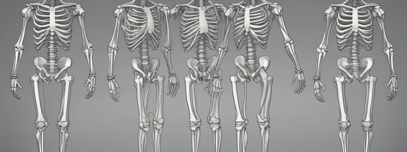
Osteology of the Lower Limb
- The bones of the lower limb are the os coxae (hip bone), femur, patella, tibia, fibula, metatarsal bones, tarsal bones, and phalanges.
- The os coxae is topographically and functionally equivalent to the upper limb clavicle and scapula.
- The os coxae forms the lower limb girdle that attaches the limb to the vertebral column.
- The os coxae is composed of three skeletal elements: the ilium, ischium, and pubis.
Os Coxae (Hip Bone)
- The os coxae has a rough surface for attachment of muscles and ligaments.
- The iliac crest is a site for interosseous ligament attachment.
- The posterior superior iliac spine is a prominent landmark.
- The ischial tuberosity is a characteristic feature of the ischium.
- The pubis forms the anterior part of the os coxae.
Lower Limb Problems
- Lower limb problems are common and dealt with by health professionals in various fields.
- Some common conditions encountered by physicians include arthritis, varicose veins, vascular deficiencies, fractures, dislocations, sprains, lacerations, knee effusions, leg pain, ankle injuries, and peripheral nerve injuries.
Lower Limb
- The superficial fascia of the thigh has a membranous layer that attaches to the deep fascia (fascia lata) about a fingerbreadth below the inguinal ligament.
- The femoral triangle contains the femoral artery, femoral vein, and femoral canal.
- The saphenous opening in the deep fascia is related to the femoral sheath.
Lymph Drainage
- Lymph from the superficial tissues of the lower limb and abdominal walls below the umbilicus drains into the external iliac nodes via the femoral canal.
- The horizontal group of superficial inguinal lymph nodes is located above the inguinal ligament.
- The vertical group of superficial inguinal lymph nodes is located below the inguinal ligament.
Gluteal Region
- The greater sciatic foramen is formed by the greater sciatic notch of the hip bone and the sacrotuberous and sacrospinous ligaments.
- The sciatic nerve usually gives no branches in the gluteal region.
- The posterior cutaneous nerve of the thigh enters the gluteal region through the lower part of the greater sciatic foramen.
Nerves of the Gluteal Region
- The superior gluteal nerve leaves the pelvis through the upper part of the greater sciatic foramen and supplies the gluteus medius and minimus muscles.
- The inferior gluteal nerve leaves the pelvis through the lower part of the greater sciatic foramen and supplies the gluteus maximus muscle.
- The nerve to the quadratus femoris leaves the pelvis through the lower part of the greater sciatic foramen and supplies the quadratus femoris and inferior gemellus muscles.
Thigh
- The thigh is the proximal segment of the lower limb, from the hip to the knee.
- The femur is the bony core of the thigh.
- The perforating veins prevent high-pressure venous blood from being forced outward into the low-pressure superficial veins.
Anatomy of Ankle Vein Cutdown
- The procedure involves blocking the sensory nerve supply to the skin in front of the medial malleolus of the tibia with local anesthetic.
- A transverse incision is made through the skin and subcutaneous tissue across the long axis of the vein just anterior and superior to the medial malleolus.
Varicose Veins
- Varicose veins are superficial veins that have a larger diameter than normal and are elongated and tortuous.
- Varicose veins commonly occur in the superficial veins of the lower limb and can cause discomfort and pain.
- Causes of varicose veins include hereditary weakness of the vein walls, incompetent valves, and elevated pressure.
Anatomy of Groin Vein Cutdown
- The great saphenous vein is constantly found at a site 1.5 inches (4 cm) below and lateral to the pubic tubercle, where it joins the femoral vein.
- The saphenous nerve usually lies just anterior to the vein.
- The great saphenous vein passes through the saphenous opening to gain entrance to the femoral vein.
Incision and Dissection
- A transverse incision is made through the skin and subcutaneous tissue centered on a point about 1.5 inches (4 cm) below and lateral to the pubic tubercle.
- The incision is carried medially just medial to the pulse if the femoral pulse can be felt.
Superficial Inguinal Lymph Nodes
- The superficial inguinal lymph nodes lie in the superficial fascia below the inguinal ligament.
- They are divided into a horizontal and a vertical group.
- The horizontal group lies just below and parallel to the inguinal ligament.
- The medial members of the group receive superficial lymph vessels from the anterior abdominal wall below the level of the umbilicus and from the perineum.
Deep Inguinal Lymph Nodes
- The deep inguinal lymph nodes are variable in number, but there are commonly three.
- They lie along the medial side of the terminal part of the femoral vein.
- They receive all the lymph from the superficial inguinal nodes via lymph vessels that pass through the cribriform fascia of the saphenous opening.
Action of Quadriceps Femoris Muscle
- The quadriceps femoris muscle (consisting of the rectus femoris, the vastus intermedius, the vastus lateralis, and the vastus medialis) inserts into the patella and, via the ligamentum patellae (patellar ligament), attaches to the tibial tuberosity.
- Together, they provide a powerful extensor of the knee joint.
Posterior Fascial Compartment Muscles
- The muscles of the posterior fascial compartment are collectively called the hamstrings.
- The biceps femoris muscle has two heads: a long head (hamstring portion) and a short head (gluteal portion).
- The biceps femoris muscle receives a dual nerve supply from the sciatic nerve.
- The tibial nerve component innervates the long head, and the common fibular (peroneal) component supplies the short head.
- The adductor magnus muscle also has two parts (an upper adductor part and a lower hamstring part) and a dual innervation.
- The tibial nerve component of the sciatic nerve supplies the hamstring portion, and the obturator nerve supplies the adductor part.
- The semimembranosus insertion sends a fibrous expansion upward and laterally, which reinforces the capsule on the back of the knee joint.
Posterior Compartment Blood Supply
- The four perforating branches of the profunda femoris artery provide a rich blood supply to this compartment.
- The profunda femoris vein drains the greater part of the blood from the compartment.
Posterior Compartment Nerve Supply
- The sciatic nerve leaves the gluteal region and descends in the midline of the posterior compartment of the thigh.
- The sciatic nerve is overlapped posteriorly by the adjacent margins of the biceps femoris and semimembranosus muscles and lies on the posterior aspect of the adductor magnus muscle.
- The sciatic nerve ends in the lower third of the thigh by dividing into the separate tibial and common fibular (peroneal) nerves.
- The tibial nerve supplies most of the posterior compartment of the thigh.
- The common fibular nerve innervates only the short head of the biceps femoris.
Popliteal Fossa
- The popliteal fossa is bounded by the posterior aspect of the femur, the knee joint, and the popliteus muscle.
- The contents of the popliteal fossa include the popliteal artery, popliteal vein, tibial nerve, and fascia.
- The proximal end of the soleus muscle is shown in outline only.
Clinical Notes
- Popliteal aneurysms are thought to be caused by the pulsations of the wall of the femoral artery against the tendon of the adductor magnus at the opening of the adductor hiatus.
- Semimembranosus bursa swelling is the most common swelling found in the popliteal space.
- It can be distinguished from a Baker's cyst, which is centrally located and arises as a pathologic (osteoarthritis) diverticulum of the synovial membrane through a hole in the back of the capsule of the knee joint.
Popliteus Muscle
- Arises inside the capsule of the knee joint and inserts into the upper part of the posterior surface of the tibia
- Separates the lateral ligament of the knee joint from the lateral meniscus
- Responsible for "unlocking" the knee joint
Posterior Tibial Artery
- Lies on the posterior surface of the tibia below the tibialis posterior muscle
- Covered only by skin and fascia in the lower part of the leg
- Passes behind the medial malleolus deep to the flexor retinaculum
- Terminate by dividing into medial and lateral plantar arteries
Popliteal Fossa
- Contains the popliteal artery and vein, as well as the tibial nerve
- Formed by the popliteus muscle above and the posterior surface of the tibia below
Structures That Pass Superficial to Extensor Retinaculum
- Saphenous nerve and great saphenous vein
- Superficial fibular (peroneal) nerve
- Anterior tibial artery with venae comitantes
- Extensor hallucis longus tendon
- Tibialis anterior tendon
Structures That Pass Deep to or through Extensor Retinaculum
- Tibialis posterior tendon
- Flexor digitorum longus tendon
- Flexor hallucis longus tendon
- Posterior tibial artery with venae comitantes
- Tibial nerve
Foot
- Constructed in the form of arches, enabling it to adapt to uneven surfaces and absorb shocks
- The skin of the sole of the foot is thick and hairless, firmly bound down to the underlying deep fascia
- Sole muscles are described in four layers, from superficial to deep
Plantar Aponeurosis
- A triangular thickening of the deep fascia that protects the underlying nerves, blood vessels, and muscles
- Attaches to the medial and lateral tubercles of the calcaneum
- Divides into five slips that pass into the toes
Plantar Arteries and Veins
- Plantar arch gives off plantar metatarsal arteries to the toes
- Medial and lateral plantar veins accompany the corresponding arteries and unite behind the medial malleolus to form the posterior tibial venae comitantes
Nerves of Sole
- Tibial nerve passes behind the medial malleolus and terminates by dividing into the medial and lateral plantar nerves
- Medial plantar nerve supplies the skin of the medial three and a half toes and the nail beds
- Lateral plantar nerve supplies the skin of the lateral one and a half toes and the nail beds
Dorsal Surface of the Foot
- Skin is thin, hairy, and freely mobile on the underlying tendons and bones
- Sensory nerve supply is derived from the superficial fibular (peroneal) nerve, assisted by the deep fibular (peroneal), saphenous, and sural nerves
Dorsal Nerve Supply
- Superficial fibular (peroneal) nerve supplies the skin on the dorsum of the foot, the medial side of the big toe, and the adjacent sides of the second, third, fourth, and fifth toes
- Deep fibular (peroneal) nerve supplies the skin of the adjacent sides of the big and second toes
- Saphenous nerve supplies the skin along the medial side of the foot as far forward as the head of the first metatarsal bone
- Sural nerve supplies the skin along the lateral margin of the foot and the lateral side of the little toe
Dorsum Arterial Supply
- Dorsalis pedis artery is the continuation of the anterior tibial artery and supplies the dorsum of the foot
- It is superficial in position and is crossed by the inferior extensor retinaculum and the first tendon of extensor digitorum brevis
Dorsal Venous Arch (or Network)
- Formed by the union of the end of the lateral plantar artery and the dorsalis pedis artery to complete the plantar arterial arch
Long Extensor Tendon Insertion
- Tendon of extensor digitorum longus passes deep to the superior extensor retinaculum and through the extensor expansion
- The extensor expansion, as in the fingers, receives the tendons of insertion of the interosseous and lumbrical muscles
Knee Joint
- The posterior intercondylar area of the tibia passes upward, forward, and medially to attach to the anterior part of the lateral surface of the medial femoral condyle.
- The posterior cruciate ligament (PCL) prevents anterior displacement of the femur on the tibia.
Anterior Bursae
- The suprapatellar bursa lies beneath the quadriceps muscle and communicates with the joint cavity.
- The prepatellar bursa lies in the subcutaneous tissue between the skin and the front of the lower half of the patella and the upper part of the ligamentum patellae.
- The superficial infrapatellar bursa lies in the subcutaneous tissue between the skin and the front of the lower part of the ligamentum patellae.
- The deep infrapatellar bursa lies between the ligamentum patellae and the tibia.
Menisci
- The menisci are C-shaped sheets of fibrocartilage.
- The peripheral border is thick and attached to the capsule, and the inner border is thin and concave and forms a free edge.
- The upper surfaces are in contact with the femoral condyles, and the lower surfaces are in contact with the tibial condyles.
- The menisci deepen the articular surfaces of the tibial condyles to receive the convex femoral condyles and serve as cushions between the two bones and distribute forces transmitted through the joint.
Posterior Bursae
- The popliteal bursa is found in association with the tendon of the popliteus and communicates with the joint cavity.
Movement and Stability
- In the position of full extension, the femur medially rotates on the tibia, resulting in a twisting and tightening of all the major ligaments of the joint, and the knee becomes a mechanically rigid structure.
- The cartilaginous menisci are compressed like rubber cushions between the femoral and tibial condyles.
- The fully extended knee is said to be in the locked and stabilized position.
- Before flexion of the knee joint can occur, it is essential that the major ligaments be untwisted and slackened to permit movements between the joint surfaces.
Flexion
- The biceps femoris, semitendinosus, and semimembranosus muscles, assisted by the gracilis, sartorius, and popliteus muscles, produce flexion.
- Flexion is limited by the contact of the back of the leg with the thigh.
Extension
- The quadriceps femoris produces extension.
- Extension is limited by the tension of all the major ligaments of the joint.
Medial Rotation
- The sartorius, gracilis, and semitendinosus muscles produce medial rotation.
Lateral Rotation
- The biceps femoris produces lateral rotation.
Important Relations
- Anteriorly: The prepatellar bursa.
- Posteriorly: The popliteal vessels; tibial and common fibular nerves; lymph nodes; and the muscles that form the boundaries of the popliteal fossa.
- Medially: Sartorius, gracilis, and semitendinosus muscles.
- Laterally: Biceps femoris and common fibular nerve.
Clinical Notes
- Knee Joint Strength: The strength of the knee joint depends on the tone of the muscles acting on the joint and the strength of the ligaments that bind the femur to the tibia.
- The quadriceps femoris is the most important muscle group.
- Knee Injury and Synovial Membrane: Damage to the articular surfaces, menisci, or ligaments of the joint results in distension of the synovial cavity with fluid.
Ligamentous Injury of Knee Joint
- The four ligaments (medial collateral ligament, lateral collateral ligament, ACL, and PCL) are commonly injured.
- Tears of the ACL are common, especially in women, and may be explained by the different alignment of the thigh on the leg.
- Tears of the PCL are less common.
Meniscal Injury
- Injuries of the menisci are common, with the medial meniscus damaged more frequently than the lateral.
- The injury occurs when the femur is rotated on the tibia or the tibia is rotated on the femur, with the knee joint partially flexed and taking the weight of the body.
Surface Anatomy of the Knee
- The patella, patellar surface of the femur, and vastus medialis can be identified in a transverse (axial) proton density magnetic resonance image of the right knee.
- The lateral collateral ligament, vastus lateralis, edge of the lateral meniscus, and biceps femoris are also visible in the image.
- The posterior cruciate ligament, popliteal artery and vein, and tibial nerve are located in the knee joint.
Surface Anatomy of the Hip and Gluteal Region
- The greater trochanter of the femur can be felt on the lateral surface of the thigh and moves beneath the examining finger as the hip joint is flexed and extended.
- The upper border of the greater trochanter lies on a line connecting the anterosuperior iliac spine to the ischial tuberosity.
- The spinous process of the sacrum is fused with each other to form the median sacral crest, which can be felt beneath the skin in the upper part of the cleft between the buttocks.
- The tip of the coccyx can be palpated beneath the skin in the cleft between the buttocks about 1 inch (2.5 cm) behind the anus.
Surface Anatomy of the Thigh
- The iliac crest is easily palpable along its entire length, ending in front at the anterosuperior iliac spine and behind at the posterior iliac spine.
- The ischial tuberosity can be palpated in the lower part of the buttock.
- The sacral nerve in the buttock lies under the cover of the gluteus maximus muscle.
- The popliteal artery can be felt by gentle palpation in the depths of the popliteal fossa, provided that the deep fascia is fully relaxed by passively flexing the knee joint.
Surface Anatomy of the Lower Leg
- The medial and lateral condyles of the femur, patella, and medial and lateral collateral ligaments can be identified in the anterior aspect of the right knee.
- The tibial tuberosity, anterior border of the tibia, and subcutaneous surface of the tibia can be palpated.
- The head of the fibula, biceps femoris, and common fibular nerve can be identified in the lateral aspect of the knee.
- The medial malleolus, lateral malleolus, and tendo calcaneus can be palpated in the lower leg.
- The extensor hallucis longus tendon, extensor digitorum longus tendons, and tibialis anterior tendon can be identified in the lower leg.
Studying That Suits You
Use AI to generate personalized quizzes and flashcards to suit your learning preferences.


