Podcast
Questions and Answers
Which of the following muscles is responsible for adducting and flexing the thigh?
Which of the following muscles is responsible for adducting and flexing the thigh?
- Adductor brevis, adductor magnus, and adductor longus (correct)
- Gluteus maximus
- Biceps femoris
- Semitendinosus
What is the primary nerve that innervates the muscles of the medial thigh?
What is the primary nerve that innervates the muscles of the medial thigh?
- Sciatic nerve
- Lumbar vessels
- Femoral nerve
- Obturator nerve (correct)
Where does the iliacus muscle originate from?
Where does the iliacus muscle originate from?
- Upper femoral shaft
- Upper part of the femur
- Iliac fossa within the bony pelvis (correct)
- Lower border of the T12 vertebra to the L5 vertebra
Which muscle is responsible for extending the thigh and rotating the tibia medially?
Which muscle is responsible for extending the thigh and rotating the tibia medially?
Where does the biceps femoris muscle insert?
Where does the biceps femoris muscle insert?
What is the primary action of the gluteus maximus muscle?
What is the primary action of the gluteus maximus muscle?
Which muscle in the anterior compartment of the thigh acts as a hip flexor and knee extensor?
Which muscle in the anterior compartment of the thigh acts as a hip flexor and knee extensor?
The sartorius muscle originates from the:
The sartorius muscle originates from the:
Which muscle in the medial compartment of the thigh is responsible for adduction?
Which muscle in the medial compartment of the thigh is responsible for adduction?
Which of the following muscles is innervated by the femoral nerve?
Which of the following muscles is innervated by the femoral nerve?
The action of the pectineus muscle includes:
The action of the pectineus muscle includes:
Which muscle in the anterior compartment of the thigh is responsible for abduction and lateral rotation of the leg?
Which muscle in the anterior compartment of the thigh is responsible for abduction and lateral rotation of the leg?
Flashcards are hidden until you start studying
Study Notes
Lower Body Extremity Anatomy of Muscles
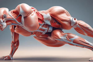
Our legs play crucial roles in supporting us while we're standing, facilitate movement like running, jumping, and cycling. The muscles of the lower extremities can be categorized into the anterior, medial, and posterior compartments of the thigh, providing various functions to the lower limb. Let's dive into the fascinating world of muscle structure and function.
Thigh Muscles
The thigh muscles include three primary groups: the anterior, medial, and posterior compartments. Each compartment contains muscles with similar functional actions, innervation, and arterial supplies.
Anterior Compartment
This compartment consists of muscles responsible for hip flexion and knee extension. Notable muscles here include the iliacus, psoas major, sartorius, and pectineus. The iliacus and psoas major originate from the bony pelvis and attach to the upper part of the femur, serving as hip flexors. The sartorius runs from the anterior superior iliac spine to the medial surface of the tibia, flexing the thigh and knee while also abducting the leg. The pectineus arises from the pubis and attaches to the upper femoral shaft, contributing to hip flexion.
Medial Compartment
The medial compartment includes muscles responsible for adduction, which means moving the leg toward the center of the body. Some key players here are the adductor brevis, adductor magnus, and adductor longus, which all arise from different origins but insert onto the linea aspera of the femur, adducting and flexing the thigh.
Posterior Compartment
The posterior compartment houses muscles like the hamstrings and gluteals, which play a significant role in extending the thigh and rotating the tibia medially. Notable muscles include the biceps femoris, semitendinosus, and semimembranosus. The biceps femoris consists of long and short heads, contributing to knee extension, hip extension, and tibial rotation. The semitendinosus and semimembranosus both extend the thigh and rotate the tibia medially, especially when the knee is flexed.
Origin of Muscles
Muscle origins refer to the specific locations where muscles attach or originate from. For example, the iliacus arises from the iliac fossa within the bony pelvis, while the psoas major arises from the lower border of the T12 vertebra to the L5 vertebra.
Innervation of Muscles
Muscle innervation concerns the nerves that supply and control muscle function. The muscles of the anterior thigh receive blood supply from the lumbar vessels, with the iliacus being supplied by branches of the femoral nerve, while the sartorius receives innervation from the muscular branch of the femoral nerve. The medial and posterior thigh muscles are innervated by the obturator nerve for the medial muscles and the sciatic nerve for the posterior muscles.
Insertion of Muscles
Muscle insertions refer to the specific locations where muscles attach or insert into bones or other structures. For example, the iliacus inserts at the upper part of the femur, while the pectineus attaches to the upper femoral shaft. The biceps femoris inserts at the head of the fibula and the lateral condyle of the tibia.
Action of Muscles
Muscle actions describe the movements a muscle can create or facilitate. For instance, the iliacus and psoas major are hip flexors, while the quadriceps femoris group consists of four muscles located in the anterior thigh, allowing you to extend your leg and lift your foot forward. The gluteus maximus, a key muscle of the posterior thigh, facilitates hip extension, helping you to walk uphill or rise from a seated position.
Understanding the anatomy of thigh muscles is essential for maintaining optimal physical performance and preventing injuries. By acknowledging these variations in muscle structure and function, individuals can tailor exercise routines and rehabilitation programs to address specific deficiencies or imbalances within their lower body extremities.
Studying That Suits You
Use AI to generate personalized quizzes and flashcards to suit your learning preferences.


