Podcast
Questions and Answers
Светнлиният микроскоп се състои от две основни системи
Светнлиният микроскоп се състои от две основни системи
- Механична и осветителна
- Механична и оптична (correct)
- Механична и увеличителна
Лофотрихите се характеризират с
Лофотрихите се характеризират с
- Снопче ресни на единият край (correct)
- Една полярно разположена ресна
- Една или повече ресни на двата полюса
За повишаване на оцветителните качества на разтворите се прибавят
За повишаване на оцветителните качества на разтворите се прибавят
- Основи (correct)
- Фенол
- Основи или фенол
Най бързо бактериите се движат с
Най бързо бактериите се движат с
Максималното увеличение което де достига с електронен микроскоп е
Максималното увеличение което де достига с електронен микроскоп е
По техника на Shaeffer & Fulton спорите се оцветяват в
По техника на Shaeffer & Fulton спорите се оцветяват в
Морфологични параметри на бактериите са
Морфологични параметри на бактериите са
Фиксинът по Пфайфер оцветява натривките най-добре за
Фиксинът по Пфайфер оцветява натривките най-добре за
Кой от изброените методи е за оцветяване на капсули
Кой от изброените методи е за оцветяване на капсули
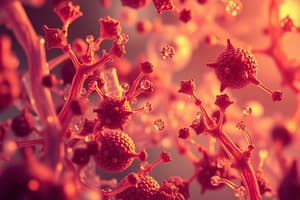
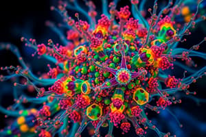
Без клетъчни живи системи са
Без клетъчни живи системи са
Flashcards are hidden until you start studying
Study Notes
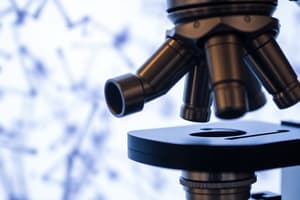
Light Microscopes
Light microscopes are powerful tools used extensively across various scientific disciplines. They allow us to magnify objects beyond what our eyes can see alone. In this article, we'll explore how light microscopes work, their history, applications, features, and limitations.
How Does A Light Microscope Work?
A basic light microscope consists of three main parts: a base with a stage, a tube system, and an eyepiece with lenses. When you look through the eyepiece, the image appears reversed because it travels backwards from your eye through the lens. Here is a more detailed description:
- An object is placed on the stage.
- Light enters the objective lens (the first lens closest to the sample) through the bottom port.
- The objective bends the light passing through it, creating a larger, brighter image.
- This enlarged image moves up through the tube system towards the ocular lens (the second lens close to your eye).
- As the light passes back through the ocular lens, another set of lenses fixes any imperfections and creates yet one last bigger viewable image.
- Finally, when looking into the eyepiece, everything seems smaller due to its focusing ability.
This process repeats numerous times until the final viewing angle allows you to see details many times larger than your naked eye would ordinarily pick out.
History Of Light Microscopy
Microscopes have been around since ancient Greece, but the development of practical optical instruments began during Renaissance Italy. By 1670s, Robert Hooke invented the compound microscope by combining several small lenses together to achieve higher levels of magnification.
In the early stages of development, scientists were limited to observing only opaque samples under high magnifications. However, thanks to new techniques such as phase contrast illumination—which brings out even faint differences between transparent regions of living cells—and differential interference contrast, where thin slices of specimens reveal subcellular structures clearly, much more accurate observations became possible.
Applications And Features
The primary function of a light microscope is visualization, which makes it useful in many fields including biology, medicine, forensic science, environmental studies, archaeology, geology, agriculture, materials science, and engineering among others. It offers advantages like affordability compared to other forms of high-resolution imagery; they also come in portable versions suitable for fieldwork.
Some advanced models feature computer interfaces allowing digital images to be taken and stored electronically, which eliminates the need for photographic film. Many modern models now include specialized attachments designed for specific purposes such as fluorescence, polarized light, or darkfield observation.
Limitations Of Light Microscopes
Despite being widely used, there are some drawbacks associated with light microscopes:
-
Resolving Power: The smallest detail visible under normal conditions using visible light before diffraction effects limit resolution is approximately 0.2 micrometers. For finer details, transmission electron microscopy might be needed.
-
Depth Perception: Unlike binocular vision with two separate images coming to each eye allowing for depth perception, most light microscopes display a single flat image making it difficult to determine exact distance relationships within the viewed scene.
-
Sample Preparation And Staining: Samples must often be chemically treated, stained, or fixed so that fine structural elements become apparent enough to observe. Unfortunately, these treatments may alter the natural state of the cells or tissues.
As technology advances, researchers continue exploring ways to push the boundaries of light microscopy further, leading to innovations such as super-resolution microscopy methods capable of achieving nanometer resolutions, thereby revealing previously hidden cellular complexity and dynamics.
Studying That Suits You
Use AI to generate personalized quizzes and flashcards to suit your learning preferences.