Podcast
Questions and Answers
Which of the following self-report measures is specifically designed for individuals diagnosed with Achilles tendinopathy?
Which of the following self-report measures is specifically designed for individuals diagnosed with Achilles tendinopathy?
- Foot and Ankle Ability Measure (FAAM)
- Foot and Ankle Disability Index (FADI)
- Achilles Tendon Total Rupture Scale (ATRS)
- Victorian Institute of Sport Assessment – Achilles (VISA-A) (correct)
The Tampa Scale of Kinesiophobia (TSK) is primarily used to assess a patient's fear of movement and potential psychological barriers to recovery.
The Tampa Scale of Kinesiophobia (TSK) is primarily used to assess a patient's fear of movement and potential psychological barriers to recovery.
True (A)
What does a higher score on the Foot and Ankle Ability Measure (FAAM) indicate regarding a patient's functional ability?
What does a higher score on the Foot and Ankle Ability Measure (FAAM) indicate regarding a patient's functional ability?
higher ability
A patient with plantar heel pain that is worse in the morning is indicative of ______.
A patient with plantar heel pain that is worse in the morning is indicative of ______.
Match the following outcome measures with their primary focus or application:
Match the following outcome measures with their primary focus or application:
A patient reports ankle stiffness that is at its peak in the morning. This symptom is most indicative of what condition?
A patient reports ankle stiffness that is at its peak in the morning. This symptom is most indicative of what condition?
Pain-producing structures located outside the symptomatic area should not be considered as potential sources of the patient's symptoms.
Pain-producing structures located outside the symptomatic area should not be considered as potential sources of the patient's symptoms.
What historical information is crucial to obtain regarding a patient's injury to help determine the potential cause?
What historical information is crucial to obtain regarding a patient's injury to help determine the potential cause?
The acronym S.I.N.S.S. is useful in physical exam planning to determine exam vigor and stands for Severity, Irritability, ______, Stage, and Stability.
The acronym S.I.N.S.S. is useful in physical exam planning to determine exam vigor and stands for Severity, Irritability, ______, Stage, and Stability.
Match the following components to what a social history and patient goals assessment would include:
Match the following components to what a social history and patient goals assessment would include:
According to the Ottawa Ankle Rule, radiographs are indicated after acute ankle injury if there is tenderness upon palpation along which of the following?
According to the Ottawa Ankle Rule, radiographs are indicated after acute ankle injury if there is tenderness upon palpation along which of the following?
The Ottawa Foot Rule suggests radiographs are unnecessary if a patient is unable to bear weight for four steps but has no tenderness at the base of the 5th metatarsal or navicular bone.
The Ottawa Foot Rule suggests radiographs are unnecessary if a patient is unable to bear weight for four steps but has no tenderness at the base of the 5th metatarsal or navicular bone.
What is a key early symptom of acute compartment syndrome that should prompt immediate medical attention?
What is a key early symptom of acute compartment syndrome that should prompt immediate medical attention?
In cases of suspected acute compartment syndrome, a ______ is performed to relieve pressure within the affected compartment.
In cases of suspected acute compartment syndrome, a ______ is performed to relieve pressure within the affected compartment.
Match the following signs and symptoms with the '6 P's' of acute compartment syndrome:
Match the following signs and symptoms with the '6 P's' of acute compartment syndrome:
Which joint is most commonly affected by gout?
Which joint is most commonly affected by gout?
The posterior talofibular ligament (PTFL) is the most commonly injured ligament in a lateral ankle sprain.
The posterior talofibular ligament (PTFL) is the most commonly injured ligament in a lateral ankle sprain.
For a patient with a Grade 2 (moderate) ankle sprain, approximately how long would it take for a patient to recover?
For a patient with a Grade 2 (moderate) ankle sprain, approximately how long would it take for a patient to recover?
If a patient who sustained an ankle sprain has pain at the calcaneocuboid joint and reports a 'rock' feeling under the cuboid, they may have injured their ______.
If a patient who sustained an ankle sprain has pain at the calcaneocuboid joint and reports a 'rock' feeling under the cuboid, they may have injured their ______.
Match the grades of ankle sprains with their pathology:
Match the grades of ankle sprains with their pathology:
Which of the following is characteristic of a 'high' ankle sprain?
Which of the following is characteristic of a 'high' ankle sprain?
Medial ankle sprains are more common than lateral ankle sprains.
Medial ankle sprains are more common than lateral ankle sprains.
In a 'high' ankle sprain, what ligament is typically injured?
In a 'high' ankle sprain, what ligament is typically injured?
A positive ______ test where there is pain when the foot is externally rotated is indicative of high ankle sprain.
A positive ______ test where there is pain when the foot is externally rotated is indicative of high ankle sprain.
Match each grade of high ankle sprain with its appropriate symptom:
Match each grade of high ankle sprain with its appropriate symptom:
What defines chronic ankle instability (CAI)?
What defines chronic ankle instability (CAI)?
Functional ankle instability always presents with mechanical laxity.
Functional ankle instability always presents with mechanical laxity.
What are 2 primary components of chronic ankle instability?
What are 2 primary components of chronic ankle instability?
Anterior ankle pain with dorsiflexion is indicative of ______.
Anterior ankle pain with dorsiflexion is indicative of ______.
Match the operative management techniques used for addressing chronic ankle instability with their description:
Match the operative management techniques used for addressing chronic ankle instability with their description:
What percentage of ankle osteoarthritis cases are post-traumatic in nature?
What percentage of ankle osteoarthritis cases are post-traumatic in nature?
A patient must always experience pain if they have radiographic evidence of ankle osteoarthritis.
A patient must always experience pain if they have radiographic evidence of ankle osteoarthritis.
A patient with ankle OA might report an increase in what in after being at prolonged rest?
A patient with ankle OA might report an increase in what in after being at prolonged rest?
A patient with medial ankle pain being considered for surgery is likely to undergo a ______ (FDL).
A patient with medial ankle pain being considered for surgery is likely to undergo a ______ (FDL).
Match the following interventions with rationale:
Match the following interventions with rationale:
What is another name given to posterior tibial tendinopathy (PTTD)?
What is another name given to posterior tibial tendinopathy (PTTD)?
Caucasian, women, age > 40 years old, obesity, diabetes, and hypertension are all risk factors for posterior tibial tendinopathy.
Caucasian, women, age > 40 years old, obesity, diabetes, and hypertension are all risk factors for posterior tibial tendinopathy.
What are some early clinical findings of posterior tibial tendinopathy?
What are some early clinical findings of posterior tibial tendinopathy?
Tarsal tunnel syndrome is caused by tibial nerve irritation or entrapment neuropathy in the tarsal ______.
Tarsal tunnel syndrome is caused by tibial nerve irritation or entrapment neuropathy in the tarsal ______.
Match the following interventions to the appropriate pathology:
Match the following interventions to the appropriate pathology:
During a bone stress injury, there is more damage occurring at a faster rate and what is not laying down new bone fast enough?
During a bone stress injury, there is more damage occurring at a faster rate and what is not laying down new bone fast enough?
Which of the following is NOT a key objective when taking a patient history for leg, ankle, and foot pathologies?
Which of the following is NOT a key objective when taking a patient history for leg, ankle, and foot pathologies?
The Ottawa Ankle Rule is designed to increase the use of radiographs in the diagnosis of acute foot and ankle injuries.
The Ottawa Ankle Rule is designed to increase the use of radiographs in the diagnosis of acute foot and ankle injuries.
What is the most commonly injured ligament in a lateral ankle sprain?
What is the most commonly injured ligament in a lateral ankle sprain?
The VISA-A is specific for __________ or ___________ .
The VISA-A is specific for __________ or ___________ .
Match each outcome measure with its specific application:
Match each outcome measure with its specific application:
Which of the following findings in a patient's history would be MOST indicative of potential ankle osteoarthritis?
Which of the following findings in a patient's history would be MOST indicative of potential ankle osteoarthritis?
In acute compartment syndrome, pulselessness is an early finding, critical for initial diagnosis.
In acute compartment syndrome, pulselessness is an early finding, critical for initial diagnosis.
What foot position during injury is MOST commonly associated with a lateral ankle sprain?
What foot position during injury is MOST commonly associated with a lateral ankle sprain?
In the FAAM, items marked __________ are not scored, and total possible points are deducted.
In the FAAM, items marked __________ are not scored, and total possible points are deducted.
Match each structure with the contents of the anterior compartment of the lower leg with their corresponding function or characteristic:
Match each structure with the contents of the anterior compartment of the lower leg with their corresponding function or characteristic:
A patient presents with exquisite joint pain, red, shiny, tense, warm skin at the joint, especially at the 1st MTP. Which of the following conditions is MOST likely?
A patient presents with exquisite joint pain, red, shiny, tense, warm skin at the joint, especially at the 1st MTP. Which of the following conditions is MOST likely?
In a high ankle sprain, what is the MOST common mechanism of injury?
In a high ankle sprain, what is the MOST common mechanism of injury?
In patients with chronic ankle instability (CAI), therapeutic exercise and activity are Grade _______ recommendations for improved outcomes.
In patients with chronic ankle instability (CAI), therapeutic exercise and activity are Grade _______ recommendations for improved outcomes.
Match the following descriptions with the corresponding stage of posterior tibial tendon dysfunction:
Match the following descriptions with the corresponding stage of posterior tibial tendon dysfunction:
Which of the following is NOT typically associated with tarsal tunnel syndrome?
Which of the following is NOT typically associated with tarsal tunnel syndrome?
In the management of bone stress injuries, some pain is acceptable during exercise within a safe and acceptable range.
In the management of bone stress injuries, some pain is acceptable during exercise within a safe and acceptable range.
What is the MAIN difference in acceptable pain levels during exercise between bone stress injury (BSI) and medial tibial stress syndrome (MTSS)?
What is the MAIN difference in acceptable pain levels during exercise between bone stress injury (BSI) and medial tibial stress syndrome (MTSS)?
A fracture of the 5th metatarsal metadiaphyseal junction is known as a __________ fracture.
A fracture of the 5th metatarsal metadiaphyseal junction is known as a __________ fracture.
Match the following ankle fractures with their descriptions:
Match the following ankle fractures with their descriptions:
Which of the following is a characteristic of insertional Achilles tendinopathy that differentiates it from mid-portion Achilles tendinopathy?
Which of the following is a characteristic of insertional Achilles tendinopathy that differentiates it from mid-portion Achilles tendinopathy?
Previous Achilles tendinopathy is a significant risk factor for Achilles tendon rupture.
Previous Achilles tendinopathy is a significant risk factor for Achilles tendon rupture.
What specific structure is affected in Sever's disease?
What specific structure is affected in Sever's disease?
Plantar fasciopathy is characterized by an absence of inflammatory __________ .
Plantar fasciopathy is characterized by an absence of inflammatory __________ .
Match the Plantar Fasciopathy phase management with the appropriate intervention:
Match the Plantar Fasciopathy phase management with the appropriate intervention:
A patient reports sharp, burning pain in the third web space of their foot, feeling like a pebble in their shoe. Which condition is MOST likely responsible for these symptoms?
A patient reports sharp, burning pain in the third web space of their foot, feeling like a pebble in their shoe. Which condition is MOST likely responsible for these symptoms?
Hallux rigidus primarily involves inflammation of the 1st MTP joint.
Hallux rigidus primarily involves inflammation of the 1st MTP joint.
What is the primary mechanism of injury for “turf toe”?
What is the primary mechanism of injury for “turf toe”?
Tampa Scale of Kinesiophobia scores greater than __________ may indicate kinesiophobia.
Tampa Scale of Kinesiophobia scores greater than __________ may indicate kinesiophobia.
Match the following aspects of medial ankle sprains:
Match the following aspects of medial ankle sprains:
According to the LEFS, what does a higher total score indicate?
According to the LEFS, what does a higher total score indicate?
A positive anterior drawer test always indicates a Grade 3 (severe) ankle sprain.
A positive anterior drawer test always indicates a Grade 3 (severe) ankle sprain.
Briefly describe a Positive syndesmosis squeeze test.
Briefly describe a Positive syndesmosis squeeze test.
A decreased ankle dorsiflexion range of motion could lead to overload of which structures in the forefoot?
A decreased ankle dorsiflexion range of motion could lead to overload of which structures in the forefoot?
Match the following BSI risks with the population:
Match the following BSI risks with the population:
Compared to lateral ankle sprains, about how much longer will high ankle sprains take to recover and return to sport?
Compared to lateral ankle sprains, about how much longer will high ankle sprains take to recover and return to sport?
The MCID for the ATRS is 14 points.
The MCID for the ATRS is 14 points.
What is the cutoff number for a kinesiophobia indication on the TSK score?
What is the cutoff number for a kinesiophobia indication on the TSK score?
The ATFL is most commonly injured during ________/__________.
The ATFL is most commonly injured during ________/__________.
Match the correct finding with the appropriate diagnosis
Match the correct finding with the appropriate diagnosis
Which of the following outcome measures is specifically designed for patients diagnosed with Achilles tendinopathy?
Which of the following outcome measures is specifically designed for patients diagnosed with Achilles tendinopathy?
A higher score on the Foot and Ankle Ability Measure (FAAM) indicates lower functional ability of the foot and ankle.
A higher score on the Foot and Ankle Ability Measure (FAAM) indicates lower functional ability of the foot and ankle.
What does a score greater than 37 on the Tampa Scale of Kinesiophobia (TSK) potentially indicate about a patient?
What does a score greater than 37 on the Tampa Scale of Kinesiophobia (TSK) potentially indicate about a patient?
According to the Ottawa Ankle Rule, radiographs are indicated after an acute ankle injury if there is tenderness to palpation along the distal 6 cm of the posterior tibia or tip of the ______ malleolus.
According to the Ottawa Ankle Rule, radiographs are indicated after an acute ankle injury if there is tenderness to palpation along the distal 6 cm of the posterior tibia or tip of the ______ malleolus.
Match each compartment of the lower leg with its corresponding nerve supply:
Match each compartment of the lower leg with its corresponding nerve supply:
Which of the following is a late finding associated with acute compartment syndrome?
Which of the following is a late finding associated with acute compartment syndrome?
Gout is caused by an overproduction of collagen in the synovial fluid.
Gout is caused by an overproduction of collagen in the synovial fluid.
According to the grading system for lateral ankle sprains, what specific pathology defines a Grade 3 (severe) sprain?
According to the grading system for lateral ankle sprains, what specific pathology defines a Grade 3 (severe) sprain?
In a 'high' ankle sprain, injury occurs to the tibiofibular ligaments and ______ ligament.
In a 'high' ankle sprain, injury occurs to the tibiofibular ligaments and ______ ligament.
Match each surgical procedure with its description for chronic ankle instability:
Match each surgical procedure with its description for chronic ankle instability:
Which of the following is the most common cause of ankle osteoarthritis (OA)?
Which of the following is the most common cause of ankle osteoarthritis (OA)?
In the later stages of posterior tibial tendinopathy, pain always remains localized to the medial ankle due to the tendon's dysfunction.
In the later stages of posterior tibial tendinopathy, pain always remains localized to the medial ankle due to the tendon's dysfunction.
Describe the primary location of pain and discomfort associated with Tarsal Tunnel Syndrome.
Describe the primary location of pain and discomfort associated with Tarsal Tunnel Syndrome.
For bone stress injuries (BSI), the only safe and acceptable pain level during, after, and following loading is ______ on a 0-10 pain scale.
For bone stress injuries (BSI), the only safe and acceptable pain level during, after, and following loading is ______ on a 0-10 pain scale.
Match each fracture type with its corresponding description in ankle fractures:
Match each fracture type with its corresponding description in ankle fractures:
Which of the following Achilles tendinopathy characteristics is NOT typically managed by early dorsiflexion with strengthening exercises?
Which of the following Achilles tendinopathy characteristics is NOT typically managed by early dorsiflexion with strengthening exercises?
Previous Achilles tendinopathy is considered a significant risk factor for Achilles tendon rupture.
Previous Achilles tendinopathy is considered a significant risk factor for Achilles tendon rupture.
What is the primary age range typically associated with Sever's disease?
What is the primary age range typically associated with Sever's disease?
Plantar fasciopathy is a condition characterized by an absence of ______ cells.
Plantar fasciopathy is a condition characterized by an absence of ______ cells.
Match each forefoot and great toe pathology with relevant features:
Match each forefoot and great toe pathology with relevant features:
Flashcards
VISA-A
VISA-A
A self-report measure specific to Achilles tendinopathy or tendinosis.
LEFS
LEFS
A global lower extremity outcome measure useful for patients with hip, knee, and foot/ankle pain.
TSK
TSK
An instrument used for a patient who is fearful of movement.
PSFS
PSFS
Signup and view all the flashcards
Ottawa Rules
Ottawa Rules
Signup and view all the flashcards
Lateral Ankle Sprain
Lateral Ankle Sprain
Signup and view all the flashcards
Cuboid Syndrome
Cuboid Syndrome
Signup and view all the flashcards
Medial Ankle Sprains
Medial Ankle Sprains
Signup and view all the flashcards
“High” Ankle Sprains
“High” Ankle Sprains
Signup and view all the flashcards
Chronic Ankle Instability
Chronic Ankle Instability
Signup and view all the flashcards
Anterior Ankle Impingement
Anterior Ankle Impingement
Signup and view all the flashcards
Ankle Osteoarthritis
Ankle Osteoarthritis
Signup and view all the flashcards
Posterior Tibial Tendinopathy
Posterior Tibial Tendinopathy
Signup and view all the flashcards
Tarsal Tunnel Syndrome
Tarsal Tunnel Syndrome
Signup and view all the flashcards
Bone Stress Injury Pathophysiology
Bone Stress Injury Pathophysiology
Signup and view all the flashcards
Medial Tibial Stress Syndrome
Medial Tibial Stress Syndrome
Signup and view all the flashcards
Chronic Exertional Compartment Syndrome
Chronic Exertional Compartment Syndrome
Signup and view all the flashcards
Traumatic Fractures
Traumatic Fractures
Signup and view all the flashcards
Ankle Fractures
Ankle Fractures
Signup and view all the flashcards
Achilles Tendon Rupture
Achilles Tendon Rupture
Signup and view all the flashcards
Sever’s Disease
Sever’s Disease
Signup and view all the flashcards
Plantar Fasciopathy
Plantar Fasciopathy
Signup and view all the flashcards
Lateral Plantar Neuropathy
Lateral Plantar Neuropathy
Signup and view all the flashcards
Metatarsalgia
Metatarsalgia
Signup and view all the flashcards
MTP Fractures
MTP Fractures
Signup and view all the flashcards
Interdigital Neuralgia
Interdigital Neuralgia
Signup and view all the flashcards
Turf Toe
Turf Toe
Signup and view all the flashcards
Hallux Rigidus
Hallux Rigidus
Signup and view all the flashcards
Hallux Valgus
Hallux Valgus
Signup and view all the flashcards
Study Notes
- The following study notes cover leg, ankle, and foot pathologies, patient self-reports, patient history, screening for medical referral, acute ankle sprains, chronic ankle instability, anterior ankle impingement, ankle osteoarthritis, medial ankle pain, leg pain (BSI, MTSS, CECS), traumatic fracture, Achilles pain, plantar heel pain, and forefoot pain.
Patient Self-Report
- Using the ICF framework and Movement System helps choose the right outcome measures for musculoskeletal disorders
- Choose reliable and valid tools to assess biological, physical, and psychosocial factors affecting pain, impairment, and disability
- Key self-report measure features include MCID and MDC
Self-Report Measures
- Foot and Ankle Ability Measure (FAAM) and Foot and Ankle Disability Index (FADI) assess the foot and ankle region, but are not pathology specific
- Victorian Institute of Sport Assessment – Achilles (VISA-A) specifically assesses Achilles tendinopathy or tendinosis
- Achilles Tendon Total Rupture Scale assesses ruptured or torn Achilles tendons
- Lower Extremity Functional Scale (LEFS) is a global lower extremity outcome measure useful for hip, knee, and foot/ankle pain
- Tampa Scale of Kinesiophobia (TSK) assesses a patient's fear of movement
- Numeric Pain Rating Scale (NPRS) rates pain intensity
- Patient-Specific Functional Scale (PSFS) assesses the patient’s functional ability to complete specific activities
Foot and Ankle Ability Measure (FAAM)
- Used for general leg, foot, and ankle disorders
- Items scored from 4 (no difficulty) to 0 (unable to do); mark "N/A" if necessary
- Total score is divided by the total possible score to get a percentage, with higher scores indicating higher ability
- Contains a 21-item ADL subscale (0-84) and an 8-item sport subscale (0-32)
- ADL MDC = 5.7, Sport MDC = 12.3
- ADL MCID = 8 points, Sport MCID = 9 points
Foot and Ankle Disability Index (FADI)
- Used for general foot and ankle conditions
- The 26-item instrument has items scored from 4 (no difficulty) to 0 (unable to do)
- Pain items score from 4 (none) to 0 (unbearable)
- Raw scoring ranges from 0-104, with 100% representing no dysfunction
- MDC = 3-4.48
Victorian Institute of Sport Assessment – Achilles (VISA-A)
- Specific to Achilles tendinopathy and only given to those diagnosed with it
- An 8-item instrument in 3 domains: pain (questions 1-3), function (questions 4-6), and activity (questions 7 and 8)
- Scoring ranges from 0-100; 70 points is the maximum score if not participating in sports
- At 12 weeks in patients with Achilles tendinopathy, MCID = 14 points
- At 24 weeks in patients with Achilles tendinopathy, MCID = 7 points
Achilles Tendon Total Rupture Score (ATRS)
- Specific to Achilles tendon rupture
- A 10-item instrument with a range of 0-10 for each item
- 0 = worse symptoms and greater limitations, 10 = no symptoms or limitations
- MCID = 8 points
Lower Extremity Functional Scale (LEFS)
- A 20-item instrument with a scoring scale of 0-80
- A higher score represents higher functional ability, with a maximum score of 4 for each item
- MDC = 9 points, MCID = 9 points
Tampa Scale of Kinesiophobia (TSK)
- Assesses kinesiophobia, the fear of movement
- The 17-item instrument has items scored from 1-4, giving a score scale of 17-68
- Higher scores indicate kinesiophobia, with scores > 37 possibly indicating kinesiophobia
Numeric Pain Rating Scale (NPRS)
- Patients rate pain on a scale of 0-10, with 0 indicating no pain and 10 representing the worst possible pain
- Demonstrates adequate construct validity as a measure of pain intensity
- MCID = 1.3 points in a non-specific patient population
Patient-Specific Functional Scale (PSFS)
- Assesses a patient’s functional ability to complete specific activities
- Patients rate their ability to complete an activity on a scale from 0 to 10
- "0" represents "unable to perform", and "10" represents "able to perform at prior level"
- Total score = sum of activity scores/number of activities
- MDC for average score = 2 points, MDC for single activity score = 3 points
Using Self-Report Information
- Guides physical examinations, interventions, and the plan of care
- Assists in determining prognosis
- Used in re-evaluation and discharge to determine progress toward goals
Patient History
- Objectives include recognizing key patient history findings associated with leg, ankle, and foot pathologies
- The goal is to create hypotheses based on the patient history
- Data from the patient history is used to make clinical judgments, establish a prognosis, and determine a movement system diagnosis
Chief Complaint and Symptom Behavior
- Location of the chief complaint helps identify if symptoms may be coming from a more proximal region (back, buttock, thigh, or knee)
- Determine if there are radiating symptoms, and whether they are localized or generalized
- Bone stress injury or stress fracture in the tibia can be very localized
- Medial tibia stress syndrome may have more vague and generalized symptoms
- For Achilles tendinopathy, it is important to differentiate whether it is mid-portion or insertional
- Aggravating and easing factors, along with altered symptoms with movements of the back, hip, and knee, help determine the source of symptoms
- Ankle stiffness in the morning points to ankle OA
- Plantar heel pain in the morning points to plantar fasciosis
- Achilles pain in the morning points to Achilles tendinopathy
Areas and Structures to Consider as a Possible Source of Symptoms
- Joint and bony structures under the area of symptoms
- Muscles, tendons, and other soft tissue under and in the area of symptoms
- Pain-producing structures that may refer into the area of symptoms
- Other structures or conditions that must be considered or ruled out
Symptom History
- Mechanism of injury (traumatic vs. atraumatic)
- Foot position during injury, and whether it was a contact or non-contact injury
- Preceding change in activity, such as footwear or increase in running/walking
- Timeline since onset
- Prior episodes, and increasing frequency of painful episodes
- Previous tests/treatments, including imaging or diagnostic testing
Medical History
- Additional conditions or related injuries, like RED-S in suspected Bone Stress Injury
- Related surgical history of the lower extremity or lumbar region
- Medications that could contribute to the development of Achilles tendinopathy or preclude manual therapy/exercise (e.g., fluoroquinolones)
Social History and Patient Goals
- The goal is to learn about the patient's living environment, occupation, and activities/participation
- Understand what they are experiencing, their thoughts on care, and their goals
Physical Exam Planning
- Prioritize based on the most likely hypotheses and the S.I.N.S.S (Severity, Irritability, Nature, Stage, Stability)
- Determine the vigor of the exam, from first onset to end of passive range of motion, with or without overpressure
- Use sustained, repeated, or combined movements
Screening for Medical Referral
- Symptoms are evaluated to determine if a patient is appropriate for physical therapy or requires a referral
- The most appropriate healthcare practitioner and initial imaging (if needed) are determined
- Red flag pathologies include traumatic fracture, acute compartment syndrome, gout, cellulitis, DVT/PE, septic arthritis, and PAOD/PAD
Ottawa Ankle Rule and Ottawa Foot Rule
- Reduce the use of unnecessary radiographs in the diagnosis of acute foot and ankle injuries in emergency departments
Ottawa Ankle Rule
- After acute ankle injury or ankle trauma, radiographs are indicated if:
- There is tenderness to palpation along distal 6 cm of posterior fibula or tip of lateral malleolus
- There is tenderness to palpation along distal 6 cm of posterior tibia or tip of medial malleolus
- There is an inability to bear weight both immediately and in the emergency department for 4 steps
- The rule is sensitive, so a negative result ensures a fracture is unlikely
Ottawa Foot Rule
- After acute foot injury (trauma), radiographs are indicated if:
- There is tenderness to palpation at the base of the 5th metatarsal
- There is tenderness to palpation at the navicular bone
- There is an inability to bear weight both immediately and in the emergency department for 4 steps
Standard Radiograph Views
- Ankle: AP, mortise, and lateral views
- Foot: AP, oblique, and lateral views
Acute Compartment Syndrome
- Increased pressure within a compartment of the lower leg, diminishing blood flow to nerves and muscles
- Can be acute (direct trauma- emergency situation) or chronic exertional (repetitive overuse or recent rigorous exercise)
Lower-Leg Compartment Contents
- Anterior: extensor hallicus longus, extensor digitorum communis, tibialis anterior, peroneous tertius; deep peroneal (fibular) nerve; anterior tibial artery
- Lateral: peroneous brevis and longus; superficial peroneal (fibular) nerve, proximal portion of deep peroneal nerve; peroneal (fibular) artery
- Posterior superficial: gastrocnemius, soleus, plantaris; tibial nerve branches; posterior tibial artery, popliteal artery, peroneal (fibular) artery, sural arteries
- Posterior deep: popliteus; tibialis posterior, flexor hallicus longus, flexor digitorum longus, popliteus; tibial nerve; posterior tibial artery, peroneal artery
Acute Compartment Syndrome Presentation (6 P’s)
- Pain: Characteristically out of proportion to the injury and increases with passive stretching
- Pulselessness: A late finding distal to the compartment in the posterior tibial or dorsalis pedis pulse
- Pallor: Loss of distal color in the foot; less common finding
- Paresthesia: Nerve tissues become ischemic, contributing to dysfunction and paresthesia/paralysis; loss of light touch precedes weakness
- Paralysis
- Poikilothermia: Change of temperature/coolness in the extremity
- Monitor those with increased risk (trauma, fracture, bleeding disorders, anticoagulation)
Acute Compartment Syndrome Diagnosis and Treatment
- Diagnosis: Intracompartmental pressure measurement via needle or MRI
- Treatment: Medical emergency; irreversible damage after >8 hours
- Emergent fasciotomy: Incision to relieve the pressure in the compartment
Gout
- Inflammatory metabolic joint disease marked by hyperuricemia and formation of monosodium urate crystals in the synovial fluid of joints and other tissues
- Risk factors: Family history, alcohol use, chronic kidney disease, obesity, renal insufficiency, hypertension
- Symptomatic in 40-50 year olds, but may be asymptomatic for years
- Causes exquisite joint pain, and red, shiny, tense, warm skin at the joint
- The 1st MTP is most common site
Acute Ankle Sprains
- Objectives include identifying risks factors/clinical findings associated with an acute lateral ankle sprain and understanding appropriate treatment considerations
- Can affect lateral ankle, medial ankle, or be a "high" ankle sprain
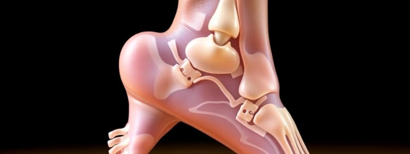
Lateral Ankle Sprain
- An acute injury to the lateral ankle ligaments
- The anterior talofibular ligament (ATFL) is the most commonly injured ligament
- The calcaneofibular ligament is the second most common, and the posterior talofibular ligament is least common
- Mechanism of injury: Forced plantarflexion/inversion
Lateral Ankle Sprain Prevalence and Risk Factors
- Most prevalent musculoskeletal injury in active populations
- About 50% of individuals seek medical attention
- Prevalence of 11.88% in the general population
- Risk decreases with increased age
- Most common in indoor court sports
- Risk factors: Previous history of ankle sprain, female, decreased ankle dorsiflexion ROM, hip abductor/extensor weakness, poor performance on balance/hopping tests, participating in court sports
Classification of Ankle Sprain Severity
- Grade 1 (mild) stable: Mild stretch, no instability, single ligament involved (usually ATFL), no hemorrhage, minimal swelling, point tenderness, no anterior drawer/varus laxity, no or little limp, minimal functional loss, difficulty hopping, recovery 8 days (range 2-10)
- Grade 2 (moderate) stable: Large injury spectrum, mild to moderate instability, complete ATFL tear, or partial ATFL/CFL tear, some hemorrhage, localized swelling (margins of Achilles tendon less defined), anterior drawer sign may be present, no varus laxity, walking with limp, unable to toe raise/hop/run, recovery 20 days (range 10-30)
- Grade 3 (severe) two-ligament, unstable: Significant instability, complete tear of anterior capsule, ATFL and CFL, diffuse swelling on both sides of Achilles tendon, early hemorrhage, possible tenderness medially and laterally, positive anterior drawer/varus laxity, unable to bear weight fully, significant pain inhibition, initially almost complete loss of ROM, recovery 40 days (range 30-90)
Clinical Findings; Patient History
- Sudden onset of symptoms with acute ankle inversion-related injury
- Bruising
- Swelling
- Difficulty weightbearing
Clinical Findings; Physical Examination
- Negative Ottawa Ankle and Foot Rules
- Pain and/or reported instability with weight-bearing
- Decreased ankle range of motion
- Decreased postural control and strength
- Positive Anterior Drawer Test
- Positive Tilt Test
- Tenderness to palpation over ATFL and possibly also CFL
Associated Injuries; Cuboid Syndrome
- Pain at the calcaneocuboid joint
- Clinical presentation: Plantar lateral midfoot pain and feeling of a "rock" under cuboid when walking, pain with palpation and joint mobility assessment of cuboid, and pain with resisted eversion
- Management: Cuboid mobilization/manipulation and cuboid sling taping technique
Associated Injuries
- Lisfranc injuries
- Talar dome lesion (osteochondral defect, Fracture)
- Fractures of the 5th metatarsal, fibula, tibia, talus
- Fibular/sural nerve injury: Manifests as a positive slump or straight leg raise; managed via nerve mobilizations
- Fibularis dysfunction (subluxation, tendinopathy)
- Ruptured retinaculum leading to tendon subluxation
- Clinical presentation: Posterlolateral ankle pain and potential swelling, pain to palpation of fibularii tendons, supinated foot position during gait, pain with AROM and resisted eversion, and positive peroneal (fibularis) subluxation test
- Management: Impairment-based interventions; acute subluxation managed with a boot for up to 6 weeks; tendinopathy managed with isometric loading for symptom modulation followed by progressive loading
Imaging for Lateral Ankle Sprain
- Acute trauma, negative Ottawa Ankle Rules: Typically, no imaging indicated
- Positive Ottawa ankle or foot rules: Ankle radiographs
- Positive radiographs demonstrate potential osetochondral injury or negative radiographs with persistent symptoms (1-3 weeks): MRI or CT scan without contrast
- Negative ankle radiographs, suspected syndesmotic injury: Leg radiographs, ankle stress radiographs, MRI without contrast and CT scan without contrast
Management of Lateral Ankle Sprain
- Protected Motion Phase: Protect injured structures, manage pain/inflammation/effusion, and provide early, low grade ROM/mobilizations
- Controlled Motion Phase: Normalize ROM/joint mobility, progressive strengthening, progressive neuromuscular control exercises, and normalize gait
- Return to Function Phase: Progress strength/neuromuscular re-education exercises, agility/plyometric training, and return to functional/sport-related skills
Protected Motion Phase
- Physical agents: Cryotherapy, diathermy, and low-level laser therapy
- Therapeutic exercise: Protected (pain-free) active range of motion, stretching exercises, and strengthening
- Manual therapy: Lymphatic drainage and joint mobilizations; anterior-to-posterior talar glides
- Loading: Use braces or taping, and progressive weightbearing
- Use semi-rigid bracing to casting for up to 10 days in more severe sprains
- Initially sagittal plane motion, protecting frontal and transverse planes
- Progress from non-weightbearing to weightbearing
- Open chain strengthening is in the sagittal plane, while isometrics in the frontal plane
Controlled Motion Phase
- Manual Therapy: Graded mobilizations/manipulations and mobilization with movement (MWM)
- Therapeutic Exercise: Range of motion in all planes, open chain strengthening in all planes, closed chain strengthening in sagittal plane- progressing to all planes, weightbearing functional exercises, and single limb balance activities progressive from stable to unstable surfaces
Return to Function Phase
- Ankle should be pain-free and feel stable with no giving way episodes
- Practice and game-specific loads should not be a novel experience at return to sport and functional activity
- Prophylactic bracing reduces the risk of a first-time lateral ankle sprain
- Prophylactic bracing and proprioceptive/balance-focused therapeutic exercise training programs reduce the risk of subsequent injury
Tertiary Prevention
- Acute: Early weight bearing with support, therapeutic exercise, and manual therapy procedures; do not administer therapeutic ultrasound
- Chronic: Therapeutic exercise (proprioceptive and neuromuscular exercise) and manual therapy procedures; combined treatment programs
Lateral Ankle Sprain Prognosis
- Full return to participation ranges from 1 day to ~3 weeks depending on sport demands
- Full, asymptomatic recovery may take months to years
- About 40% develop Chronic Ankle Instability (CAI)
- Variables with increased risk of developing CAI include inability to perform single-leg drop landing/drop vertical jump 2 weeks post sprain, increased BMI, high level sport participation, not participating in an exercise-balance program, and not using prophylactic bracing
Medial Ankle Sprains
- Objectives include identifying key clinical findings associated with medial ankle sprains and "high" ankle sprains, and understanding appropriate treatment considerations
- An acute injury to the medial ankle ligaments, specifically the deltoid ligaments
- The deltoid ligaments are a strong fan-shaped ligament that runs form the distal aspect of the tibia down to the navicular, calcaneus, and talus
- Mechanism of injury: Forced eversion
Medial Ankle Sprains Epidemiology and Risk Factors
- Incidence in athletes is 0.06 per 1000 athlete exposures (3-5% of all ankle sprains)
- Males are more affected then females
- Higher injury rates in women's gymnastics, men's rugby, and men's and women’s soccer and men's American football
- Clinical Findings
- Patient History: Acute or recent traumatic injury, medial ankle swelling/bruising, and may have difficulty weightbearing
- Physical Examination: Tenderness to palpation of deltoid ligament, pain/apprehension with passive eversion, and fractures
Medial Ankle Sprains Management
- Limited evidence specifically for medial ankle sprain
- Initially, protect medial ankle ligaments
- Similar to lateral ankle sprain after acute phase
"High" Ankle Sprains
- An acute injury to the tibiofibular and interosseous ligaments
- Mechanism of injury: Forced external rotation of the foot/ankle and/or hyper-dorsiflexion
- This injures the anteroinferior tibiofibular ligament (AITFL) as the talus moves forcefully into a closed pack position and into the mortise
Grading of Syndesmosis Injuries Based on Clinical Presentation
- Grade 1: Sprain without diastasis: Mild point tenderness over tibiofibular ligaments, mild laxity with stable endpoint to stress, stable with stress on plain radiographs, weight bearing to tolerance, and immobilization for 0-3 days
- Grade 2: Sprain with latent diastasis: Point tenderness extends proximally over the IOM, moderate laxity with soft endpoint to stress, and mild laxity present with stress but absent with plain radiographs
- Progress to full weight bearing after 1-2 weeks, and immobilize for 3-7 days
- Grade 3: Sprain with frank diastasis: Significant tenderness/inability to weight bear, notable laxity with absence of endpoint, instability and/or fracture evident on plain radiograph, minimum 2-3 weeks non-weight bearing, and 7+ days of immobilization
"High" Ankle Sprains Epidemiology and Clinical Findings
- Epidemiology: About 10% of ankle sprains, often occur with medial ankle sprains, with males > females
- Clinical Findings: Anterior/posterior talocrural region pain, pain with weightbearing (dorsiflexion), pain and limited ROM in dorsiflexion, pain with AROM eversion, positive syndesmosis squeeze test and Kleiger’s test, and tenderness to palpation over AITFL
Diagnostic Imaging
- Anteroposterior, lateral, and mortise views
- Greater space/less overlap between the fibula and tibia can indicate rupture of the syndesmosis
Non-Operative Management; Acute, Stable Sprains
- Protect the patient and control the inflammatory response
- Non-weightbearing (up to 2 weeks) then partial weightbearing
- Proximal hip/knee strengthening and foot intrinsic strengthening
- Limit dorsiflexion ROM
- Restore strength, mobility, and neuromuscular control
- Gradually increase weightbearing and normalize gait pattern
- Progress pain-free ankle strengthening and proprioceptive exercises
"High" Ankle Sprains: Conscientious Progression
- Emphasis: restore activity specific skills
- Think about how much forced dorsiflexion they are in, and then gradually progressing that amount
- Limit dorsiflexion in the early phases of rehab
- Operative Management
- Screw fixation or knotless suture button fixation
"High" Ankle Sprain Prognosis
- Takes 2-4x as long to recover/return to sport compared to a lateral ankle sprain
- Mean return time to sport is 46.4 days (non-operative) and 55.2 days (operative)
- 15% of college athletes will miss >21 days of sport and 47% will miss >7 days of sport
Chronic Ankle Instability and Anterior Ankle Impingement
- Objectives include identifying key clinical findings and understanding treatment considerations
- Chronic Ankle Instability: Patient with history of at least 1 ankle sprain, who has perceived of episodic “giving way” of the ankle that persists >1 year after the initial sprain, and causes resultant activity limitation
Two Primary Components
- Functional Ankle Instability: Subjective feelings of instability, that can occur without mechanical laxity
- Mechanical Ankle Instability: Mechanical laxity= injury to the ATFL or CFL, results in a positive special test for anterior drawer and talar tilt
- Both components are often present
Epidemiology of Chronic Ankle Instability
- A 20% prevalence in adolescent athletes
- A 23.4% prevalence in high school and collegiate athletes
- A 40% prevalence in patients who sought care for a first-time lateral ankle sprain
- Not using prophylactic bracing, not participating in exercise programs, poor functional performance, participating in sports, and high BMI increase risk
Chronic Ankle Instability Clinical Findings
- Recurrent ankle sprains and/or feelings of instability
- Impaired postural control (static and dynamic)
- Muscle weakness of the hip abductors/extensors/external rotators and ankle invertors/evertors/PF's
- Limited ankle DF ROM (open and closed chain)
- Decreased talocrural joint mobility (anterior-to-posterior glide)
- Deficits with hopping and jumping along with altered movement patterns during functional activates
Chronic Ankle Instability Pathways and Management
- After an acute lateral ankle sprain, there are two pathways for a patient: Chronic ankle instability or "coper"
- Management:
- Grade B: Do not use external support (braces/taping) as a stand-alone intervention to improve balance and postural stability
- Grade A: Proprioceptive and neuromuscular therapeutic exercise should used to improve dynamic postural stability
- Grade A: Manual therapy can improve weight-bearing ankle dorsiflexion and dynamic balance
Chronic Ankle Instability Operative Management
- Rehabilitation Precautions: Initial period of Non-weight bearing, and avoid Active/passive inversion for 6 weeks
- Outcomes: Most patients report "good" or "excellent" ankle function ~2 years post surgery
- Techniques: Brostrom/Carlson procedure or Brostrom-Gould procedure for extra reinforcement
Chronic Ankle Instability Prognosis
- Increased risk of ankle osteoarthritis
- Diminished quality of life and physical activity lead to compromised health and wellness
- Increased levels of fear and kinesiophobia
- The importance of early intervention
Anterior Ankle Impingement
- Anterior ankle pain with dorsiflexion
- May result in bony exostoses on the anterior rim of the tibia and talus
- As high as 25% of individuals with the condition also possess chronic ankle instability
Anterior Ankle Impingement Clinical Findings
- Patient History: History of recurrent ankle sprains and pain/limited mobility with squat/single leg squat and descending stairs/ascending hills
- Physical Examination: Limited and painful ankle dorsiflexion ROM, decreased talocrural joint mobility; and pain/limited mobility with single leg squat
Anterior Ankle Impingement Management
- Depends on severity and bony exostoses
- Nonoperative: Relative rest, activity modification, heel lift, manal therapy, and NSAIDs/corticosteroid injection
- Operative: Arthroscopic debridement
Ankle Osteoarthritis (OA)
- Progressive gradual loss of cartilage, resulting in joint related changes, stiffness, and pain
- Patient can have radiographic findings of OA without pain
- Radiographic prevalence =13.9% in patients > 50 years old
- Symptomatic prevalence = 1.2-3.4% in patients > 50 years old
- Approximately 70% were post-traumatic, 12% were rheumatoid disease, and 7% were idiopathic
Clinical Presentation
- Typically, post traumatic onset especially rotational injuries
- Gradual worsening over time
- Stiffness after prolonged rest, especially in the morning
- Pain with weight bearing activities, like walking and stairs
- Limited ankle ROM and stiffness with passive ROM
- Hypomobility with joint passive accessory examination
Ankle Osteoarthritis Management
- Patient education: Activity modification and assistive devices to offload weight
- Strengthening/proprioception exercises and talocrural joint mobilizations
- Rocker bottom footwear and orthoses/taping
Ankle Osteoarthritis Pharmacological Management
- NSAIDs, low dose paracetamol, intra-articular corticosteriods, hyaluronic acid injection, and platelet-rich plasma PRP
- Intra-articular corticosteriods should be used no more than 3-4 times per year, each spaced 3-4 months apart
Ankle Osteoarthritis Operative Management
- Typically for patients with mild to early moderate ankle arthritis, ankle arthrodesis (fusion), and total ankle arthroplasty
Medial Ankle Pain
- Objectives include identifying key clinical findings associated with medial ankle and foot pain, and understanding appropriate treatment considerations
- The notes cover Posterior Tibial Tendinopathy and Tarsal Tunnel Syndrome
Posterior Tibial Tendinopathy
- Progressive microtrauma and insufficiency of the posterior tibialis tendon
- Can be classified into 4 stages
- Occurs in 5-15% of the general population
- Risk factors: Caucasian, women, age >40 years old, obesity, diabetes, and hypertension
Posterior Tibial Tendinopathy Body Chart
- Localized, achy pain that is superficial
Posterior Tibial Tendon Dysfunction Stages
- Stage 1: No deformity, mild pain/swelling at medial ankle, normal heel raise test, normal tendon length. with tenosynovitis
- Stage 2: Flexible deformity, moderate medial ankle and foot pain, pain/weakness with heel raise test and increased tendon length
- Stage 3: Rigid deformity, sever pain both medially and laterally at the ankle and foot, abnormal heel raise test, and tendon tearing
- Stage 4: Talus is titled laterally: Stage IVA contains a flexible ankle valgus and Stage IVB has a rigid ankle valgus. Arthritic changes may or may not be present
Clinical Presentation; Early Stages
- Pain increased with waling and standing
- Medial foot and ankle pain and/or swelling
- Standing on toes may be apinful and difficult
- Inability to invert heel with single limb heel raise and pain/weakness with resisted plantarflexion and inversion
- Forefoot may be abducted “too many toes sign”, pronatory foot type, and positive navicular drop test
Management – Early Stages
- Patient education for activity and footwear modifications
- Relative rest, anti-inflammatory medications, orthotics/taping to support medial longitudinal arch, and foot intrinsic strengthening
- Progressive loading of posterior tibialis and Talocrural joint mobilizations if needed
- Gastrocnemius, soleus flexibility
Clinical Presentation; Later Stages
- Pain may shift laterally because of calcaneofibular or lateral subtalar impingement
- Progressive collapse of the medial longitudinal arch and development of severe hindfoot valgus
Management – Later Stages
- Orthotics/ankle-foot orthosis to support medial longitudinal arch
- Foot intrinsic strengthening and progressive loading of posterior tibialis if intact
- Talocrural joint mobilizations and Gastrocnemius, soleus flexibility
- Operative Management may be warranted if continued pain and disability; Tendon transfer (FDL), triple arthrodesis or Total ankle arthroplasty
Tarsal Tunnel Syndrome
- Tibial nerve irritation/entrapment neuropathy in tarsal tunnel
- Made up of the flexor retinaculum that runs from the medial malleolus posteriorly to the calcaneus
- The tibial nerve runs underneath the roof of the flexor retinaculum
Tarsal Tunnel Syndrome Body Chart
- Generalized, achy, numbness pain that is superficial
Tarsal Tunnel Syndrome Clinical Presentation
- Patient History: Pain and paresthesias in medial ankle, heel, and/or foot, aggravated by walking/running/standing, worse after activity and at the end of the day, insidious or post-traumatic onset, and burning sensation
- Possible numbness and/or weakness in ankle or foot, presenting as a positive straight leg raise with dorsiflexion/eversion
Tarsal Tunnel Syndrome Clinical Presentation; Physical Examination
- Patient symptoms with passive eversion and a positive Tinel’s sign, dorsiflexion-eversion test, triple compression test, and straight leg raise with dorsiflexion/eversion
Tarsal Tunnel Syndrome Management
- Orthoses/taping to limit excessive pronation
- Gastrocnemius and soleus flexibility and Talocrural mobilizations
- Nerve mobilizations
- Posterior tibialis and foot intrinsic strengthening and patient education for activity modification
- Operative management if failed non-operative management; Decompression of the tarsal tunnel
Leg Pain
- Focuses on Bone Stress Injury BSI, Medial Tibial Stress Syndrome MTSS and Chronic Exertional Compartment Syndrome CECS
- Objectives include identifying key clinical findings and understanding treatment considerations
Bone Stress Injury; Pathophysiology
- Progression from Bone loading, causing bone strain leading to no damage or strain-related remodeling
- Bone damage and damage-related remodeling create a damage repair and altered skeletal properties that are asymptomatic
- With an imbalance between damage and remodeling comes accumulation of damage and stress reaction
- A stress fracture can then result in a complete bone fracture
Bone Stress Injury Pathophysiology; Schematic Representation
- Micro cracks in bone form, initiating osteocyte apoptosis
- Calcium channeling signaling attracts osteoclasts to form cavities the crack site, clearing the damage and temporarily weakening the bone
- Then, they create a callous to repair the micro damage
- During a bone stress injury, more damage occurs at a faster rate than osteoblasts can lay down new bone
High-and-Low-risk Bone Stress Injuries
- Low risk: Tend to heal well with activity modification with continued weight bearing( distal tibia, calcaneus, and 2nd-4th metatarsals)
- High risk: May require more aggressive treatment (restricted weight bearing or surgery) because they're prone to high tension/torsional loads and are may be lacking in vasculatization
- As high, they would be treated with non-weight bearing initially
Bone Stress Injury Epidemiology
- 1/3 to 2/3 of competitive cross-country and long-distance runners have a history of BSI
- Recurrence rate in cross country and track athletes ranges from 10.3-12.6%
Bone Stress Injury Risk Factors
- Female with no risk factors = 1.4% risk of bone stress injury
- Risk factors include prior BSI, BMI < 19 kg/m^2, menarche greater than or equal to 15 years, and past participation in dance or gymnastics
- Male with no risk factors = 1.2% of BSI, these risk factor include prior BSI and lack of playing ball sports
Relative Energy Deficiency in Sport (RED-S)
- Energy availability = energy intake – exercise energy expenditure
- Low energy availability (LEA): inadequate energy availability
- Overtime, this can lead to increased detrimental health outcomes (RED-S)
- Screening is performed using the RED-S Clinical Assessment Tool (CAT-2)
BSI Clinical Presentation: Patient History
- Gradual onset of localized pain
- Recent increase in weight-bearing activity
- Pain worsened during and/or after weight-bearing activity
- May report findings consistent with RED-S
Military Study and Stress Fractures
- Peak occurrence was between weeks 5 and week 8 following a new training regimen; micro damage exceeding the ability of the bone to repair itself
- Training variability within the first two months of establishing a new training program decreases risk
- Include relative rest days and rest weeks where mileage is decreased for athletes
Bone Stress Injury Clinical Presentation
- Point tenderness over bone (less then 10cm or 5 cm of bone tenderness)
- Limited diagnostic utility of the single leg hop test
- First line imaging is radiographs; MRI if radiographs are negative and injury suspected
Bone Stress Injury Imaging Process
- Clinical suspicion comes first, then radiographs
- A positive radiograph comes next to treat stress fracture
- If negative Radiographs, then check high-level athlete; MRI for High-level with the stress fracture, treat as the stress fracture as a negative
BSI Management Goals; manage load
- Safe and acceptable pain level is 0-10 during, after, in day, following loading; different with soft tissue
- Reduced loading followed by a graded increase in load, consider healing supplements,maintain fitness, and address muscle function
BSI Management Principles
- Resistance exercises targeting muscles local to BSI to better equip the bone to better adapt load
- Iniate and progress running
- Replace initial cross-training activities with activities that progressively increase bone loading
- Increase training volume before intensity
- Reduce risk of subsequent injury by optimizing nutrition, addressing hormonal levels, and intrinsic risk factors
- Focus on optimal loading, jump training and gait re-training and patient education
Low-Risk Tibial BSI Management
- Initial Goal: Pain-free gait and ADLs, use axillary crutches and/or CAM boot if cautious with boot as this alters gait
- Limit NSAID use to <7 days
- Too much weightbearing and activity will delay healing process
- Cross training, monitor workloads for optimal energy availability, and strengthening after week 3-6
High Risk BSI: Management
- Initial period of non-weightbearing.
- Potentially surgery if displaced fracture or non-union
- Management may follow a similar progression to low-risk BSI, however, with a slower progression/delayed timeline for return to sport
Bone Stress Injury Timeline
- Low-risk BSI: 1-4 months, High-risk BSI: 5-9 months, andpotentially longer depending on BSI location and running level
- In-season or in-training BSI may result in shut down and re-evaluation
Medial Tibial Stress Syndrome
- Cortical bone microtrauma and tibial periostitis
- Incidence of 4-19% in athletic populations
- Postulated due to repetitive traction from the medial soleus origin on the proximal tibia
MTSS Clinical Presentation
- Patient history: Exercise-induced pain along the distal 2/3 of the medial tibial border, provoked during or after physical activity and reduced with relative rest and no cramping, burning pain, or numbness in posterior compartment
- Physical Examination: Pain reproduced with palpation of posteromedial at least 5 cm of the tibia
- Non-diagnostic imaging
MTSS Risk Factors
- Intrinsic: Higher BMI, greater navicular drop and increased plantarflexion ROM with poor plantar flexor endurance
- Extrinsic: Below-average activity history, previous history of MTSS, and over-training
MTSS Management
- Mild pain is acceptable during exercise
- Load management: Cross-training, graded progressive loading, plantar flexor and posterior tibialis strengthening, foot orthoses, and gait re-training
Differential Diagnosis Summary
- Bone stress injury: Energy deficiency and or history of Bone Stress
Studying That Suits You
Use AI to generate personalized quizzes and flashcards to suit your learning preferences.


