Podcast
Questions and Answers
How many layers does the scalp consist of?
How many layers does the scalp consist of?
- 4
- 5 (correct)
- 7
- 6
What is the function of the aponeurosis layer in the scalp?
What is the function of the aponeurosis layer in the scalp?
- To connect the occipitalis and frontalis muscles (correct)
- To connect the skin to the skull
- To separate the periosteum from the epicranial aponeurosis
- To contain arteries, veins, and nerves
What is the clinical significance of the dense connective tissue layer?
What is the clinical significance of the dense connective tissue layer?
- It prevents wounds from gapping
- It allows for easy contraction of arteries
- It causes profuse bleeding in small wounds (correct)
- It prevents innervation of the scalp
What is the anterior boundary of the scalp?
What is the anterior boundary of the scalp?
What is the function of the loose areolar tissue layer?
What is the function of the loose areolar tissue layer?
What happens to wounds that reach the aponeurosis layer?
What happens to wounds that reach the aponeurosis layer?
What is the outer layer of the skull called?
What is the outer layer of the skull called?
Why do small wounds in the scalp cause profuse bleeding?
Why do small wounds in the scalp cause profuse bleeding?
Why is the loose connective tissue layer of the scalp considered dangerous?
Why is the loose connective tissue layer of the scalp considered dangerous?
What is the action of the frontal belly of the occipito-frontalis muscle?
What is the action of the frontal belly of the occipito-frontalis muscle?
Which nerve supplies the occipital bellies of the occipito-frontalis muscle?
Which nerve supplies the occipital bellies of the occipito-frontalis muscle?
What is the origin of the orbital part of the orbicularis oculi?
What is the origin of the orbital part of the orbicularis oculi?
What is the primary function of the lacrimal part of the orbicularis oculi?
What is the primary function of the lacrimal part of the orbicularis oculi?
Where does the lacrimal gland reside?
Where does the lacrimal gland reside?
What happens when blood enters the eyelids due to emissary veins?
What happens when blood enters the eyelids due to emissary veins?
Which part of the face has deep fascia?
Which part of the face has deep fascia?
Flashcards are hidden until you start studying
Study Notes
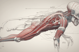
Layers of the Scalp
- The scalp consists of 5 layers, which can be remembered using the word "SCALP"
- S: Skin (thick, hairy, with sebaceous and sweat glands)
- C: Connective tissue (containing arteries, veins, nerves, and lymphatics)
- A: Aponeurosis (a thin, tendon-like structure connecting occipitalis and frontalis muscles)
- L: Loose areolar tissue (a thin connective tissue layer between the periosteum and epicranial aponeurosis)
- P: Pericranium (the outer layer of the skull bones)
Clinical Relevance of the Scalp
- Connective tissue layer:
- Dense, richly vascularized and innervated
- Contains septa that connect this layer with the layers above and below
- Wounds do not gape, but may cause profuse bleeding due to adherence of artery walls to connective tissue septa
- Aponeurosis:
- A strong, fibrous sheet
- Wounds reaching this layer gape due to muscle contraction
- Loose connective tissue layer:
- Continuous with the eyelids anteriorly
- Considered the "dangerous layer" due to emissary veins that can spread infection from the scalp to the cranial cavity
- May cause a "black eye" if infected
Muscles of the Face
- Occipito-frontalis muscle:
- Composed of frontal and occipital bellies
- Originates from the skin of the eyebrows (frontal bellies) and the highest nuchal line (occipital bellies)
- Inserts into the epicranial aponeurosis
- Actions: pulls the scalp forward and raises the eyebrows (frontal bellies), and pulls the scalp backward (occipital bellies)
- Nerve supply: temporal branch of the facial nerve (frontal bellies) and posterior auricular branch of the facial nerve (occipital bellies)
Boundaries of the Face
- Superiorly: the hair margin
- Inferiorly: the lower border of the mandible
- Sideways: from one auricle to the other
- The forehead is common to both scalp and face
- The face has no deep fascia except over the parotid and buccinator muscles
Muscles of Facial Expression
- Orbicularis oculi:
- Composed of orbital, palpebral, and lacrimal parts
- Actions: closes the eyelids tightly (orbital part), closes the eyelids gently (palpebral part), and dilates the lacrimal sac (lacrimal part)
- Supplied by temporal and zygomatic branches of the facial nerve (7th cranial nerve)
Lacrimal Apparatus
- Consists of:
- Lacrimal gland (secretes tears) and its excretory ducts
- Lacrimal punctum (opening) and canaliculi (convey tears to the surface of the eye)
- Located in the lacrimal fossa (superior lateral part of the orbital cavity)
Studying That Suits You
Use AI to generate personalized quizzes and flashcards to suit your learning preferences.