Podcast
Questions and Answers
What function does the myocardium serve in the heart?
What function does the myocardium serve in the heart?
- Responsible for contraction (correct)
- Lining of heart chambers
- Facilitates electrical conduction
- Supports vascular structure
Which type of blood vessel has thicker muscular walls and is primarily responsible for carrying blood away from the heart?
Which type of blood vessel has thicker muscular walls and is primarily responsible for carrying blood away from the heart?
- Arteries (correct)
- Sinusoids
- Veins
- Capillaries
How does atherosclerosis primarily affect the structure of arterial walls?
How does atherosclerosis primarily affect the structure of arterial walls?
- Thickening due to plaque buildup (correct)
- Increases elasticity and flexibility
- Enhances blood flow efficiency
- Reduces muscle fiber count
What is the main purpose of using immunohistochemistry in histological techniques?
What is the main purpose of using immunohistochemistry in histological techniques?
Which layer of blood vessels contains smooth muscle and is responsible for regulating vessel diameter?
Which layer of blood vessels contains smooth muscle and is responsible for regulating vessel diameter?
What is the characteristic histological feature of cardiac muscle tissue?
What is the characteristic histological feature of cardiac muscle tissue?
In which pathological condition is coagulative necrosis primarily observed?
In which pathological condition is coagulative necrosis primarily observed?
Which type of capillary is found in the kidneys and is characterized by small openings?
Which type of capillary is found in the kidneys and is characterized by small openings?
What distinguishes elastic arteries from muscular arteries?
What distinguishes elastic arteries from muscular arteries?
What common feature is shared by both veins and arteries?
What common feature is shared by both veins and arteries?
Flashcards are hidden until you start studying
Study Notes
Lab Histology for Cardiovascular System (CVS)
Key Components
-
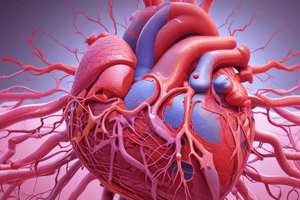
Heart
- Cardiac Muscle Tissue: Striated, involuntary, interconnected fibers (intercalated discs).
- Endocardium: Inner layer lining heart chambers, composed of endothelial cells.
- Myocardium: Thick middle layer of cardiac muscle, responsible for contraction.
- Epicardium: Outer layer, also part of the pericardium, contains connective tissue and blood vessels.
-
Blood Vessels
-
Arteries:
- Thick muscular walls with three layers: tunica intima (endothelium), tunica media (smooth muscle), tunica externa (connective tissue).
- Elastic arteries (e.g., aorta) have a significant elastic layer for recoil; muscular arteries have more smooth muscle.
-
Veins:
- Thinner walls than arteries, larger lumen.
- Valves present to prevent backflow.
- Three layers similar to arteries but less muscular and elastic.
-
Capillaries:
- Single layer of endothelial cells for efficient exchange of materials.
- Types: Continuous (most common), fenestrated (kidneys), and sinusoidal (liver, spleen).
-
Histological Techniques
-
Tissue Preparation:
- Fixation: Preserves tissue structure (common fixatives include formalin).
- Embedding: Infiltrating tissue with paraffin for sectioning.
- Sectioning: Thin slices for microscopic examination.
-
Staining Methods:
- Hematoxylin and Eosin (H&E): General stain for overall structure.
- Masson's Trichrome: Differentiates collagen from muscle.
- Immunohistochemistry: Uses antibodies to identify specific proteins.
Pathological Changes
- Atherosclerosis: Thickening of arterial walls due to plaque buildup; characterized by foam cells.
- Myocardial Infarction: Necrosis of cardiac tissue; histologically shows coagulative necrosis.
- Hypertrophy: Enlargement of cardiac muscle cells often due to pressure overload; noticeable in myocardium.
- Vascular Disorders: Changes in vessel walls, such as inflammation (vasculitis) or fibrous thickening.
Microscopic Features
- Cardiac Muscle: Central nuclei, striations, branching fibers.
- Endothelial Cells: Flat, elongated nuclei; form a continuous layer in vessels.
- Smooth Muscle Cells in Vessels: Spindle-shaped cells in tunica media, often arranged in circular layers.
Common Clinical Applications
- Histology is crucial for diagnosing cardiovascular diseases through biopsy samples.
- Understanding tissue architecture helps in assessing damage and guiding treatment plans.
Conclusion
Histological examination of the cardiovascular system provides vital insights into normal and pathological conditions. Key components include the heart and blood vessels, with various staining techniques enhancing the visualization of specific structures and abnormalities.
Key Components
-
Heart Anatomy:
- Composed of four main layers: endocardium (inner lining), myocardium (muscle layer), and epicardium (outer layer), with cardiac muscle being striated and involuntary.
- Cardiac muscle fibers interconnect through intercalated discs for synchronized contractions.
-
Blood Vessels:
- Arteries: Feature thick walls with three layers: tunica intima (endothelium), tunica media (smooth muscle), and tunica externa (connective tissue); elastic arteries aid in returning to shape after stretching.
- Veins: Have thinner walls and larger lumens compared to arteries; contain valves to prevent backflow, consisting of the same three layers but with less smooth muscle.
- Capillaries: Composed of a single layer of endothelial cells, facilitating efficient material exchange; types include continuous, fenestrated, and sinusoidal.
Histological Techniques
-
Tissue Preparation:
- Fixation: Essential for preserving tissue structure using agents like formalin.
- Embedding: Involves infiltrating tissues with paraffin to facilitate thin slicing for microscopy.
-
Staining Methods:
- Hematoxylin and Eosin (H&E): Standard stain for visualizing the overall structure of tissue.
- Masson's Trichrome: Distinguishes collagen fibers from muscle in tissue sections.
- Immunohistochemistry: Utilizes antibodies to visualize specific proteins within tissue samples.
Pathological Changes
- Atherosclerosis: Characterized by arterial wall thickening due to plaque and foam cell accumulation.
- Myocardial Infarction: Necrosis of heart tissue; histologically identifiable by coagulative necrosis.
- Hypertrophy: Enlargement of cardiac muscle cells, commonly due to sustained pressure overload; most evident in the myocardium.
- Vascular Disorders: Includes inflammation (vasculitis) and fibrous thickening of vessel walls.
Microscopic Features
- Cardiac Muscle: Distinctive central nuclei, visible striations, and branching fibers that contribute to contraction.
- Endothelial Cells: Exhibit flat, elongated nuclei; form a continuous layer lining blood vessels.
- Smooth Muscle Cells: Spindle-shaped in the tunica media, typically organized in circular layers to regulate vessel diameter.
Common Clinical Applications
- Histological evaluations are fundamental for diagnosing cardiovascular diseases using biopsy samples.
- Knowledge of tissue architecture is critical for assessing damage and developing appropriate treatment strategies.
Conclusion
- Histological examination of the cardiovascular system reveals important insights into both normal and diseased states, emphasizing the role of staining techniques in identifying specific structures and abnormalities.
Studying That Suits You
Use AI to generate personalized quizzes and flashcards to suit your learning preferences.


