Podcast
Questions and Answers
Which layer of the heart is primarily responsible for contracting and pumping blood?
Which layer of the heart is primarily responsible for contracting and pumping blood?
- Pericardium
- Epicardium
- Myocardium (correct)
- Endocardium
What structure is located between the atria and ventricles to prevent backflow of blood?
What structure is located between the atria and ventricles to prevent backflow of blood?
- Myocardial valves
- Atrioventricular valves (correct)
- Semilunar valves
- Pulmonary valves
What type of muscle makes up the majority of the heart wall?
What type of muscle makes up the majority of the heart wall?
- Skeletal muscle
- Adipose tissue
- Cardiac muscle (correct)
- Smooth muscle
Which layer of the heart directly contacts the blood within the chambers?
Which layer of the heart directly contacts the blood within the chambers?
What is the primary function of the Purkinje fibers in the heart?
What is the primary function of the Purkinje fibers in the heart?
Which statement about the epicardium is accurate?
Which statement about the epicardium is accurate?
What physiological condition is primarily indicated by hypertrophic cardiomyopathy?
What physiological condition is primarily indicated by hypertrophic cardiomyopathy?
Which atrioventricular valve is present on the right side of the heart?
Which atrioventricular valve is present on the right side of the heart?
What is the primary function of Purkinje fibers in the heart?
What is the primary function of Purkinje fibers in the heart?
Which layer of the cardiac valves directly faces the ventricle?
Which layer of the cardiac valves directly faces the ventricle?
Which statement about the endocardium is accurate?
Which statement about the endocardium is accurate?
What is the primary role of the atrioventricular valves?
What is the primary role of the atrioventricular valves?
The conducting system of the heart primarily involves which of the following components?
The conducting system of the heart primarily involves which of the following components?
What are the three layers of the cardiac valves?
What are the three layers of the cardiac valves?
Which of the following valves is classified as a semilunar valve?
Which of the following valves is classified as a semilunar valve?
What role does the subendocardium play within the heart's structure?
What role does the subendocardium play within the heart's structure?
What is a primary characteristic of Purkinje fibers?
What is a primary characteristic of Purkinje fibers?
Which structure allows the heart to contract as a single coordinated unit?
Which structure allows the heart to contract as a single coordinated unit?
How does atrial natriuretic factor (ANF) affect the kidneys?
How does atrial natriuretic factor (ANF) affect the kidneys?
What is the role of gap junctions in cardiac muscle tissue?
What is the role of gap junctions in cardiac muscle tissue?
What differentiates the sarcomeres in cardiac muscle compared to skeletal muscle?
What differentiates the sarcomeres in cardiac muscle compared to skeletal muscle?
In which part of the heart does the conduction system begin?
In which part of the heart does the conduction system begin?
What is the primary function of the AV bundle in the heart's conduction system?
What is the primary function of the AV bundle in the heart's conduction system?
Which of these structures are found in the endocardium?
Which of these structures are found in the endocardium?
The conduction pathway through the heart includes all of the following in correct sequence except:
The conduction pathway through the heart includes all of the following in correct sequence except:
What type of muscle contraction is primarily exhibited by atrial cardiomyocytes?
What type of muscle contraction is primarily exhibited by atrial cardiomyocytes?
Flashcards are hidden until you start studying
Study Notes
Endocardium
- Inner layer of heart, analogous to intima of blood vessels
- Consists of endothelium and basal lamina resting on a thin layer of connective tissue
- Subendocardium is a loose connective tissue layer between endocardium and myocardium, containing nerves, blood vessels, and branches of the heart's conducting system
- Valves are part of the endocardium
Cardiac Valves
- Two types: atrioventricular (mitral or tricuspid) and semilunar (aortic or pulmonary)
- Ensure unidirectional blood flow
- The thin cusps are normally avascular
Atrioventricular Valves
- Tricuspid valve has three cusps
- Mitral valve has two cusps
Semilunar Valves
- Aortic and pulmonic valves have three cusps each
- They have a free edge, line of closure and annulus
### Histology of Cardiac Valves
- Layers: fibrosa, spongiosa and endocardium (with endothelial lining)
- Endocardial layers are named according to the chamber they face
- Atrioventricular valves: auricularis (faces atrium) and ventricularis (faces ventricle)
- Semilunar valves: arterialis (faces aorta or pulmonary artery) and ventricularis (faces ventricle)
Summary of Heart and Valves
- The heart beats independently of nervous stimuli, but has postsynaptic and sympathetic input
- Two circulatory systems: systemic and pulmonic
Heart Development
- Cardiac tube develops into a chambered heart via: linear heart, loop, crescent, twisting and septation
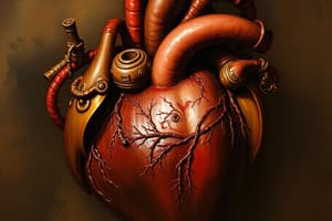
### Layers of Cardiac Chambers
- Composed of three layers: epicardium, myocardium, endocardium
- Surrounded by a pericardial cavity
Heart Structure and Blood Flow
- Normal heart: four chambers—left atrium (LA), right atrium (RA), right ventricle (RV), left ventricle (LV)
Clinical Correlation- Hypertrophic Cardiomyopathy
- Hypertrophy of cardiac muscle
- Contrast with normal septum and left ventricle (LV)
Clinical Correlation- Cardiac Hypertrophy
- Increase in LV wall thickness is hypertrophy
- Note: RV hypertrophy
### Heart Layers
- Epicardium
- Myocardium
- Endocardium
Endocardium
- Lots of blood vessels
- Step-like areas of interdigitation between adjacent sarcomeres
- Contractions are rhythmic, spontaneous, and involuntary
### Atrial Cardiomyocytes
- Large mitochondria and glycogen deposits
- ANF granules: released mostly by right atrial cardiomyocytes, decrease Na+ reabsorption by kidney tubules, lowering blood pressure; located in the atrium, not the ventricle
Myocardium: Cardiomyocytes
- Striated, short, branched, and interconnected
- One central nucleus (at most, two nuclei)
- Wider, but less numerous T tubules
- Less developed sarcoplasmic reticulum: diads
- Depolarization (Ca+2) occurs at T tubules, "voltage sensor."
Myocardium: Cardiomyocytes - Microscopic Anatomy of Cardiac Muscle
- Intercalated discs between adjacent cardiac cells
- Desmosomes
- Fascia adherens
- Gap junctions
- Keep cells together and electrically coupled, allowing heart to contract as a single unit
### Sarcomere
- Basic contractile unit of cardiac muscle
- Two sets of protein filaments: thick myosin filaments and thin actin filaments
- Each protein strand has a globular head at each end
- Sliding filament model: myosin heads bound to actin produce movement of thin filaments = sarcomere shortening
Cardiac Muscle Cells
- Sarcomere components visualized via electron microscope:
- Z (tubulin + a-actinin)
- I (thin actin + troponin + tropomyosin)
- A (thick myosin + actin)
### Clinical Correlation- Myocardial Infarction
- Myocyte injury and coagulation necrosis, edema
### Nervous System of the Heart
- SA node --> Atrium --> Internodal fibers --> AV node --> Bundle of His --> Purkinje fibers
Conducting System of the Heart
- Purkinje fibers found in sub-endocardium
- Larger than normal cardiac myofibers with specialized junctions
- Rich in glycogen
- Sparse and peripheral myofibrils
- Respond to hormones (epinephrine, norepinephrine) that increase force of contraction, parasympathetic decreases HR, sympathetic increases HR
Conducting System of the Heart, Pathway
- SA --> internodal --> AV --> bundle --> Purkinje
Studying That Suits You
Use AI to generate personalized quizzes and flashcards to suit your learning preferences.